Diagnostic method for proteinaceous binding pairs, cardiovascular conditions and preeclampsia

a proteinaceous binding and diagnostic method technology, applied in the field of placental growth factor detection, can solve the problems of a little discouraged use of modern immunodiagnostics

Inactive Publication Date: 2007-05-17
ABBOTT LAB INC
View PDF8 Cites 32 Cited by
  • Summary
  • Abstract
  • Description
  • Claims
  • Application Information

AI Technical Summary

Problems solved by technology

Other reagents are available for the detection of markers in biological samples, but the need to carefully characterize these...

Method used


Image

  • Diagnostic method for proteinaceous binding pairs, cardiovascular conditions and preeclampsia
  • Diagnostic method for proteinaceous binding pairs, cardiovascular conditions and preeclampsia
  • Diagnostic method for proteinaceous binding pairs, cardiovascular conditions and preeclampsia

Examples


examples

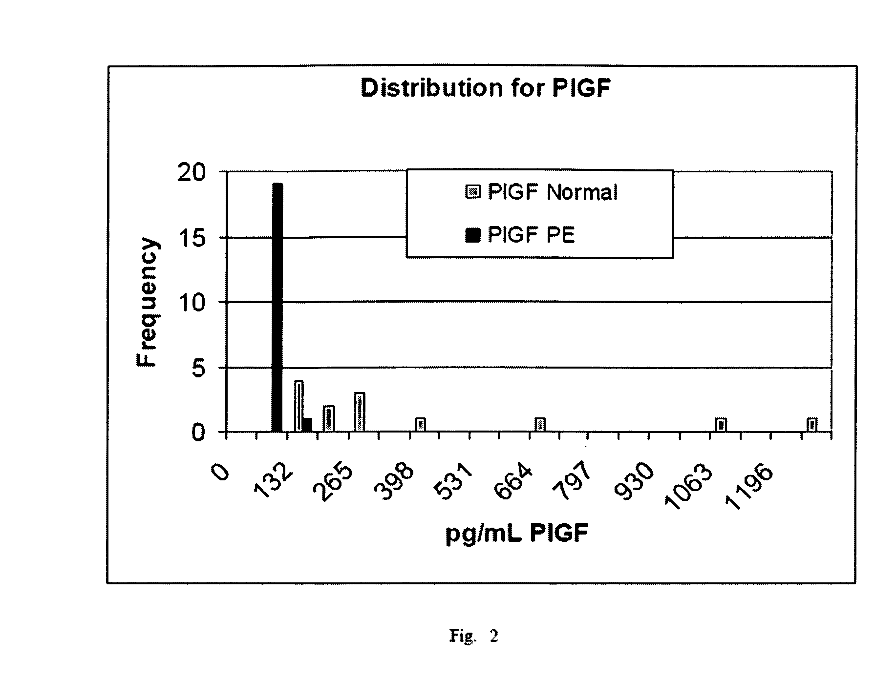
[0078] The invention is illustrated with data obtained from various immunoassays for total PlGF, free PlGF, total sFlt-1, and free sFlt-1. A selection of these data deemed to be most illustrative of the invention and inventive concepts are presented in the attached drawings and discussed briefly in the Brief Description of the Drawings. As is clear from the entirety of this description of the invention, the examples are meant to illustrate the claimed invention rather than to limit its scope.

Further Examples

[0079] The following additional examples provide more detail regarding two preferred embodiments of the present invention

example 2

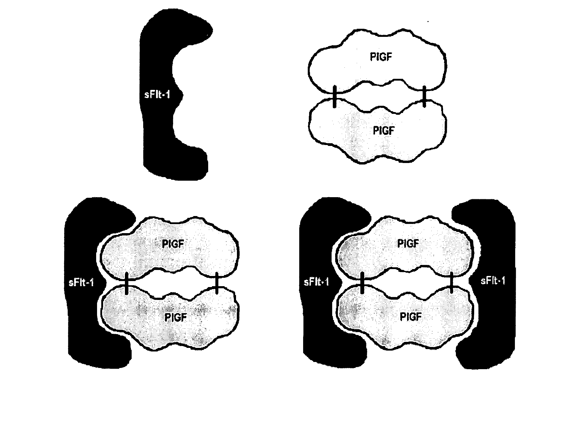
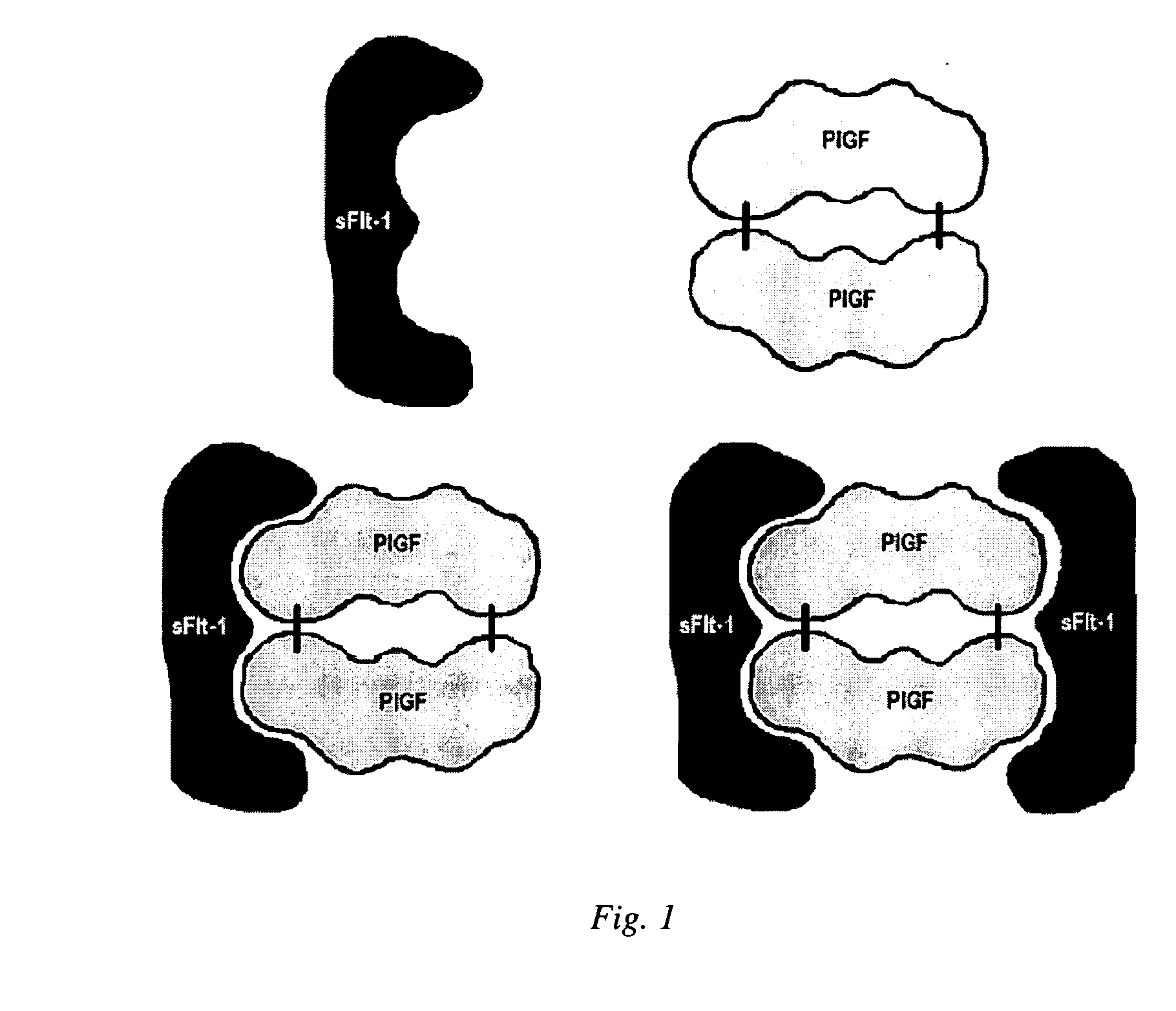
[0080] This example illustrates the inventive method in an assay used to detect free sFlt-1 in a biological sample using a portion of PlGF as a sbp for sFlt-1.

[0081] A monoclonal antibody against sFlt-1 was coated on magnetic carboxyl-latex microparticles (4.7 microns) at a protein concentration of 0.1 mg / nL of microparticles at a concentration of 1% by weight in 50 mM sodium MES (2-morpholinoethanesulfonic acid) at pH 6.0. After 10 minutes, EDAC, (ethyl-3-(-3-dimethylaminopropyl)carbodiimide) was added and allowed to react for one hour before washing the particles with phosphate buffered saline. The particles were then diluted to 0.1% in a buffer for use in an automated immunochemical analyzer.

[0082] Acridinylated PlGF was prepared by dissolving PlGF in phosphate buffered saline and incubation with an acridinium-ester at a mass ratio of PlGF to acridinium of 150,00 / 1. The conjugate was then purified by HPLC chromatography and diluted to a concentration of approximately 75 ng / mL. ...

example 3

[0085] This example illustrates the inventive method in an assay used to detect free PlGF in a biological sample using a portion of sFlt-1 as a sbp for PlGF.

[0086] Paramagnetic latex microparticles (4.7 microns), derivatized with carboxyl functional groups, was coated with anti-sFlt-1 antibody (containing domains 1-3 of the fms-like tyrosine kinase 1) at a protein concentration demonstrated to be sufficient to maximize the amount of protein absorbed to the surface area of the microparticles (2% solids by weight) in 50 mM MES, pH 5.5. In another embodiment, the sFlt-1 could be bound directly to a solid substrate. After 10 minutes, the non-absorbed sFlt-1 was removed by washing the particles multiple times with MES buffer. Following washing the particles, EDAC was added and allowed to react forming a covalent coupling of the sFlt-1 molecules to the particles. The particles were then washed with phosphate buffered saline to stop the reaction and remove unreacted EDAC. The particles ar...


PUM


Abstract

Method of measuring the quantity of a first proteinaceous specific binding partner (sbp) in a biological sample comprising detecting the binding of the first proteinaceous sbp with a labeled second proteinaceous sbp, wherein neither the first or second sbp is an antibody or fragment thereof, which is preferably a method of determining the amount of sFlt-1, particularly free sFlt-1, and the amount of PlGF, particularly free PlGF, in a sample, which is preferably used in a method of predicting risk of preeclampsia comprising comparing free PlGF to free sFlt-1.

Description

CROSS-REFERENCE TO RELATED APPLICATIONS [0001] In accordance with 35 U.S.C. § 119(e), this application claims the benefit of provisional application U.S. Ser. No. 60 / 736,659 filed Nov. 14, 2005, the disclosure of which is specifically incorporated by reference herein.TECHNICAL FIELD OF THE INVENTION [0002] The Field of the invention relates to the detection of placental growth factor (PlGF), soluble fms-like tyrosine kinase (sFlt-1), and related molecules in biological samples that are preferably obtained from patients. BACKGROUND OF THE INVENTION [0003] Immunodiagnostics enables the detection, diagnosis, prognosis of diseases, dysfunctions, and other conditions afflicting or affecting animals, including humans. It has become highly desirable to perform immunodiagnostics testing with the aid of automated testing equipment that minimizes the investigator's time handling samples and data. The rapid commercial growth of immunodiagnostics since 1980 has been made possible in part by tec...

Claims


Application Information

Patent Timeline
IPC IPC(8): G01N33/543
CPCG01N33/566G01N33/689G01N33/74G01N2800/368
Inventor SOGIN, DAVID C.LAIRD, DONALD M.YU, GEORGEDOSS, ROBERT C.
Owner ABBOTT LAB INC
Why Eureka
Try Eureka