

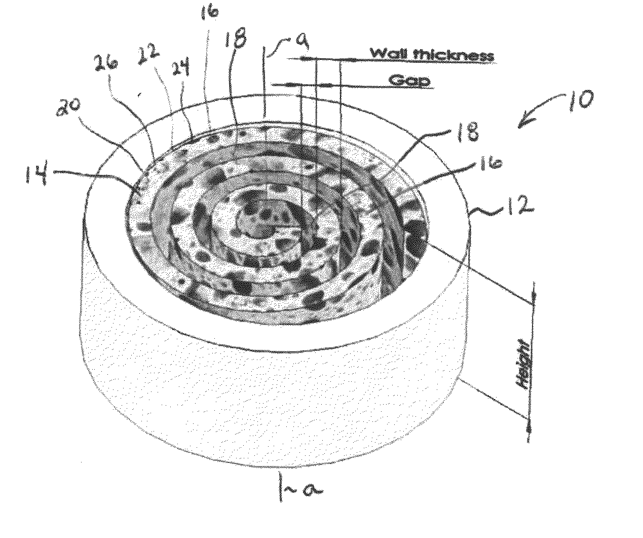
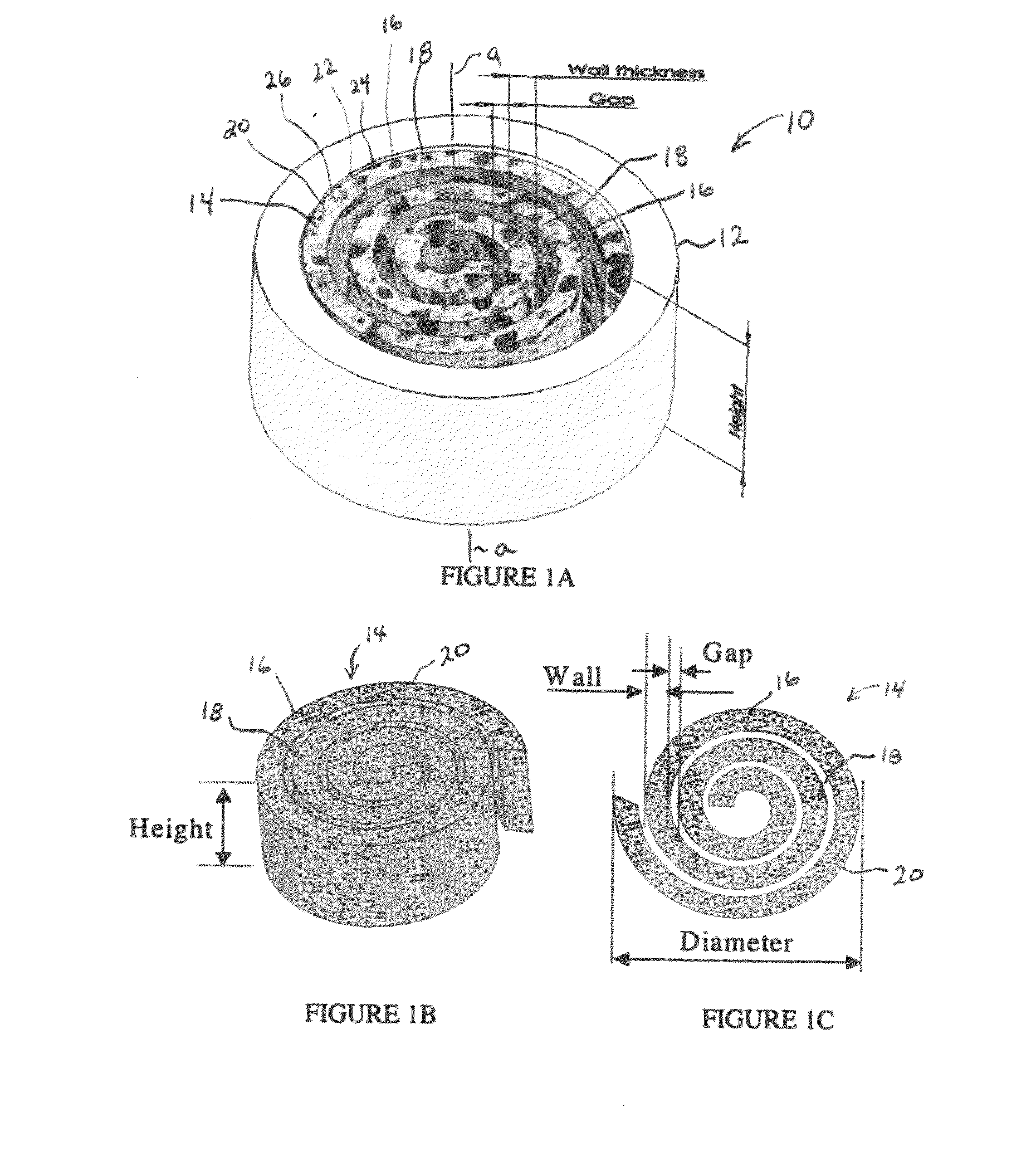
Synergetic functionalized spiral-in-tubular bone scaffolds
a functionalized, spiral-in-tubular bone technology, applied in the direction of prosthesis, peptide/protein ingredients, drug compositions, etc., can solve the problems of poor interconnection with non-uniform pores within the closed porous structure, poor biomechanical strength, poor biodegradability of bioceramics, etc., to achieve the highest amount of calcium deposition, the effect of high alp activity
- Summary
- Abstract
- Description
- Claims
- Application Information
AI Technical Summary
Benefits of technology
Problems solved by technology
Method used
Image

Examples
example 1
Fabrication of Spiral-in-Tubular Scaffolds
example 1.1
Low Porosity Poly(ε-Caprolactone) (PCL) Scaffolds
[0113]Poly(ε-caprolactone) (PCL) sheets (50-100 μm in thickness) were fabricated using a solvent evaporation method. Briefly, PCL in dichloromethane (DCM) (33% w / v) was spread on the surface of a glass Petri dish and the DCM was evaporated leaving a PCL sheet. The PCL sheet was then rolled along with a piece of copper sheet that acted as a mold to form a low-porosity PCL spiral scaffold. After incubation in an oven (45° C. for 30 minutes), the scaffold was immediately transferred to ice cold water for at least 24 hours to immobilize its shape. The copper mold was then removed from the low porosity scaffold prior to further experimentation.
example 1.2
High Porosity PCL Spiral Scaffolds
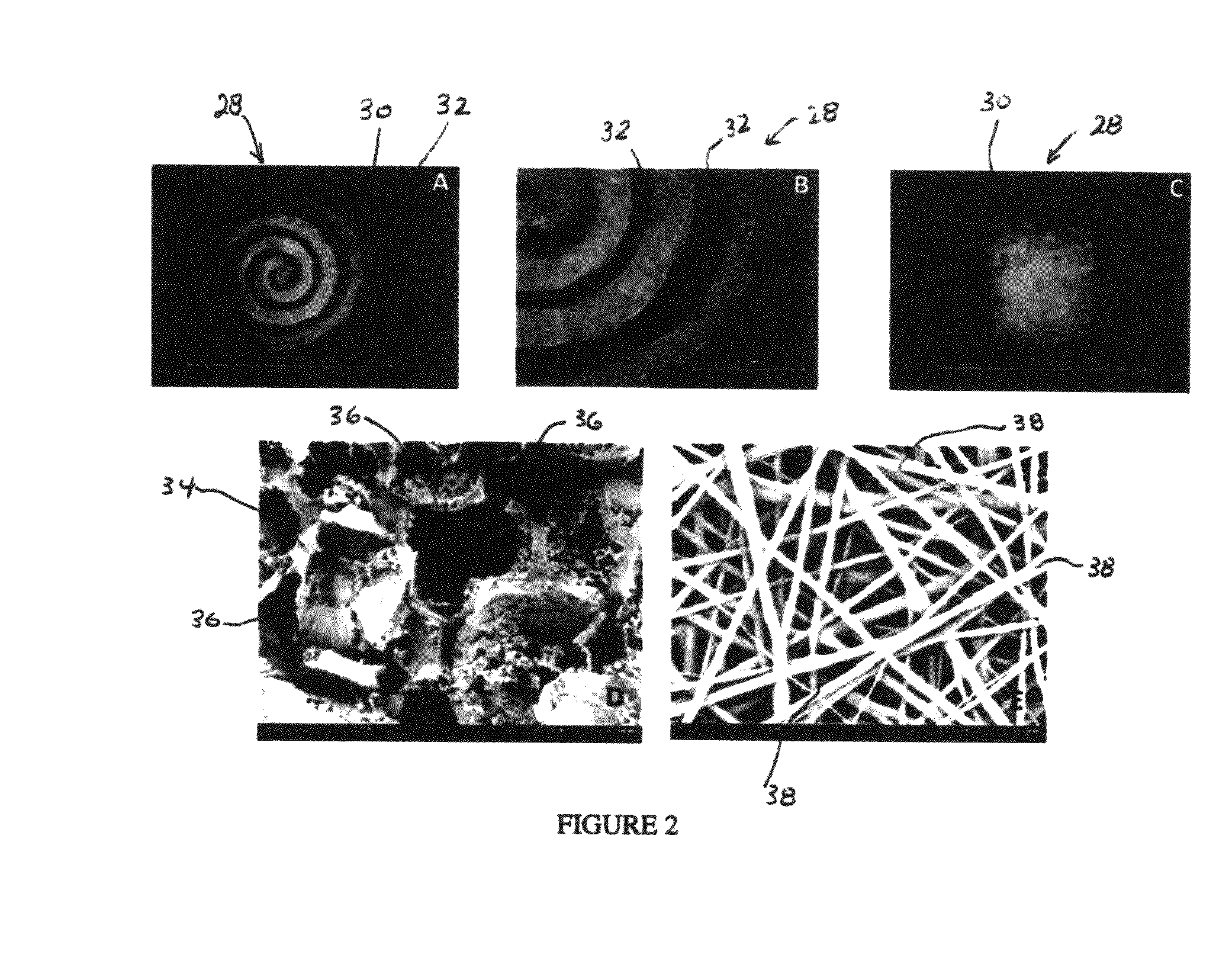
[0114]High porosity PCL spiral scaffolds were prepared using a salt-leaching method. Briefly, sodium chloride (NaCl) crystals (150-300 μm in size) and NaCl particles (200 μm in size) were added to a PCL / DCM solution (33% w / v) in a 1:1 (w / w) ratio. The mixture was spread onto a glass Petri dish and the spiral scaffolds were formed as described above. The scaffolds were then submerged into deionized water to remove the salt. The resulting spiral scaffolds proved to be highly porous.
PUM
| Property | Measurement | Unit |
|---|---|---|
| Depth | aaaaa | aaaaa |
| Stability | aaaaa | aaaaa |
| Modulus | aaaaa | aaaaa |
Abstract
Description
Claims
Application Information