
Image processing system and method of enhancing the quality of an ultrasound image

a processing system and ultrasound technology, applied in image enhancement, instruments, ultrasonic/sonic/infrasonic diagnostics, etc., can solve the problems of extensive increase of the time required to complete diagnosis and increase of the time incurred to adjust image parameters

Active Publication Date: 2007-07-19
KOREA ADVANCED INST OF SCI & TECH
View PDF3 Cites 19 Cited by
  • Summary
  • Abstract
  • Description
  • Claims
  • Application Information

AI Technical Summary

Problems solved by technology

However, since the slight adjustment of the image parameters is not conducted automatically but rather manually in the conventional ultrasound diagnostic system, an extensive amount of time is incurred to adjust the image parameters.
Therefore, there is a problem in that the amount of time needed to complete diagnosis is extensively increased.

Method used


Image

  • Image processing system and method of enhancing the quality of an ultrasound image
  • Image processing system and method of enhancing the quality of an ultrasound image
  • Image processing system and method of enhancing the quality of an ultrasound image

Examples


Embodiment Construction

[0014] A detailed description may be provided with reference to the accompanying drawings. One of ordinary skill in the art may realize that the following description is illustrative only and is not in any way limiting. Other embodiments of the present invention may readily suggest themselves to such skilled persons having the benefit of this disclosure.

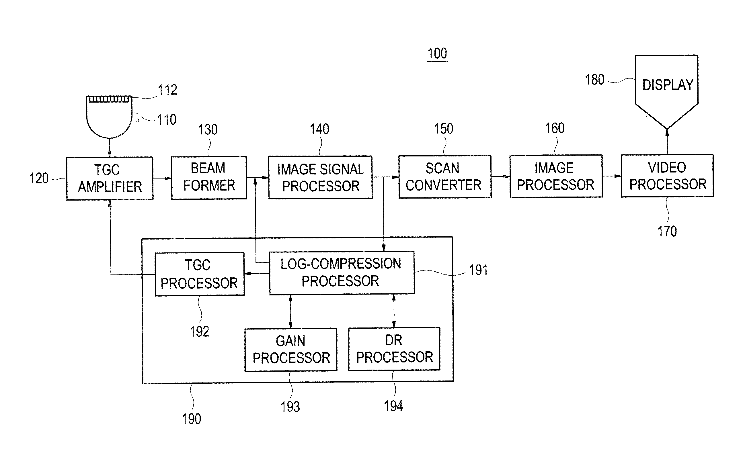
[0015]FIG. 1 is a block diagram illustrating an ultrasound diagnostic system 100, which is constructed in accordance with an embodiment of the present invention. The ultrasound diagnostic system 100 of the present invention includes a probe 110, a time gain compensation (TGC) amplifier 120, a beam former 130, an image signal processor 140, a scan converter 150, an image processor 160, a video processor 170, a display unit 180 and an image parameter processor 190. The image signal processor 140, the image processor 160, the video processor 170 and the image parameter processor 190 may be provided as one processor.

[0016] The probe 11...


PUM


Abstract

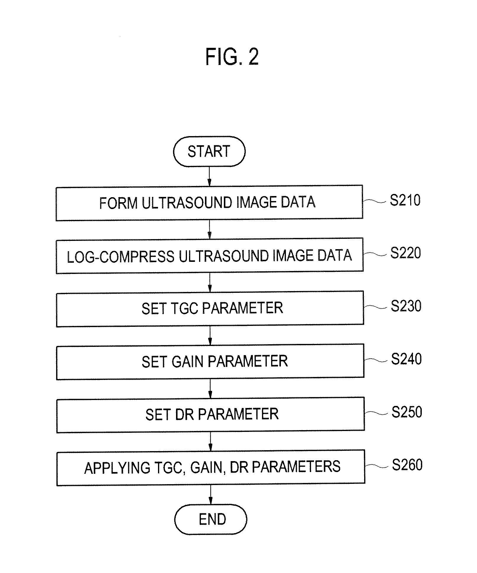
The present invention relates to an image processing system for enhancing the image quality of an ultrasound image. The image processing system may include: an image data forming unit for forming image data based on image signals acquired from a target object; a log-compression unit for log-compressing the image data to provide a log-compressed image; a TGC processor for analyzing a vertical profile of pixel intensities in the log-compressed image and automatically setting a TGC (time gain compensation) parameter based on the analysis result; a gain processor for calculating mean brightness of pixels included in the log-compressed image applying the set TGC parameter and comparing the calculated mean brightness with predetermined brightness, thereby automatically setting a gain parameter based on a comparison result; a DR processor for automatically setting a DR (dynamic range) parameter by using edge contrast and background roughness of the log-compressed image applying the set TGC and gain parameters; and an image forming unit for applying the TGC, gain and DR parameters to the image data.

Description

[0001] The present application claims priority from Korean Patent Application No. 10-2005-0112815 filed on Nov. 24, 2005, the subject matter of which is incorporated herein by reference. BACKGROUND [0002] 1. Field of the Invention [0003] The present invention generally relates to ultrasound image processing, and more particularly to an image processing system and a method of enhancing the quality of an ultrasound image. [0004] 2. Background of the Invention [0005] An ultrasound diagnostic system has become an important and popular diagnostic tool since it has a wide range of applications. Specifically, due to its non-invasive and non-destructive nature, the ultrasound diagnostic system has been extensively used in the medical profession. Modern high-performance ultrasound diagnostic systems and techniques are commonly used to produce two or three-dimensional diagnostic images of internal features of an object (e.g., human organs). [0006] The ultrasound diagnostic system generally us...

Claims


Application Information

Patent Timeline
IPC IPC(8): G06K9/00
CPCG01S7/52033G01S7/52063G06T2207/20021G06T2207/10132G06T5/009G06T5/92A61B8/00
Inventor AHN, CHI YOUNGRA, JONG BEOMKIM, YONG SUNLEE, DUHGOON
Owner KOREA ADVANCED INST OF SCI & TECH
Learn More