Method for forming and distributing a composite file including a dental image and associated diagnosis

a composite file and dental image technology, applied in dental surgery, applications, instruments, etc., can solve the problems of difficult to correlate the images with a separate image, unable to perform a diagnosis or otherwise make efficient use of the wealth of information provided, and implementing costly high-resolution printing on expensive film media

Inactive Publication Date: 2007-11-08
HOWERTON JR WILLIAM BRUCE
View PDF3 Cites 16 Cited by
  • Summary
  • Abstract
  • Description
  • Claims
  • Application Information

AI Technical Summary

Problems solved by technology

That is, any dental-related radiographic imaging likely produces a two-dimensional representation of the imaged area that includes distortion resulting from the attempt to image a three-dimensional subject.
However, since many dental practitioners are accustomed to viewing and performing a diagnosis through two-dimensional imaging of a three-dimensional subject, many such practitioners may be unable to perform a diagnosis or otherwise make efficient use of the wealth of information provided by images produced by three-dimensional imaging techniques.
However, such a method typically implements costly high-resolution printing on expensive film media.
Further, it may be difficult to correlate the images with a separate printed text (interpretations), delivery of this information through mail or other delivery channels is slow, and correcting images or typographical errors in the text often requires appropriately amended film or documentation to be prepared by the imaging provider and then re-forwarded to the referring dentist.
As such, the common thread between various DICOM viewers is generally limited to the capability of opening a DICOM format file.
Further, the entire DICOM data set is often very large in size.
Dentists, however, often do not have the time or skill to manipulate such large DICOM data sets and, since this method involves delivery to the referring dentist on hard media, such DICOM viewers and data may not be well-received by the referring dentist due to the time for delivery and / or the cumbersome and extensive nature of the provided data.
Another possible shortcoming in delivering to the referring dentist images produced from three-dimensional volume data sets is that the final radiographic study cannot be easily shared with more than one dentist that may be involved in treating the same patient.
That is, sharing such images between dentists, who may be remotely located, may be difficult and / or cumbersome, particularly where concurrent viewing of a particular image may be required for conferring between the remotely located dentists.
This is due, at least in part, to the size and complexity of the DICOM data set, the specialized nature of the DICOM viewer, and the separate file(s) for the accompanying description and / or interpretation of findings.

Method used


Image

  • Method for forming and distributing a composite file including a dental image and associated diagnosis
  • Method for forming and distributing a composite file including a dental image and associated diagnosis
  • Method for forming and distributing a composite file including a dental image and associated diagnosis

Examples


Embodiment Construction

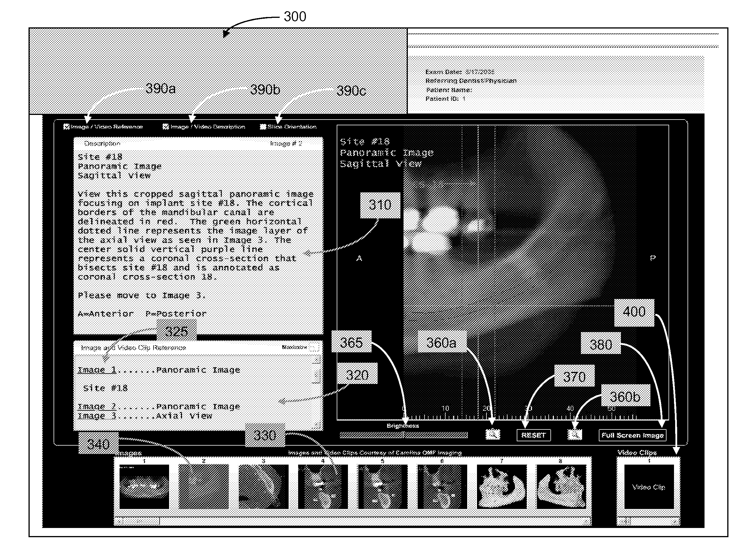
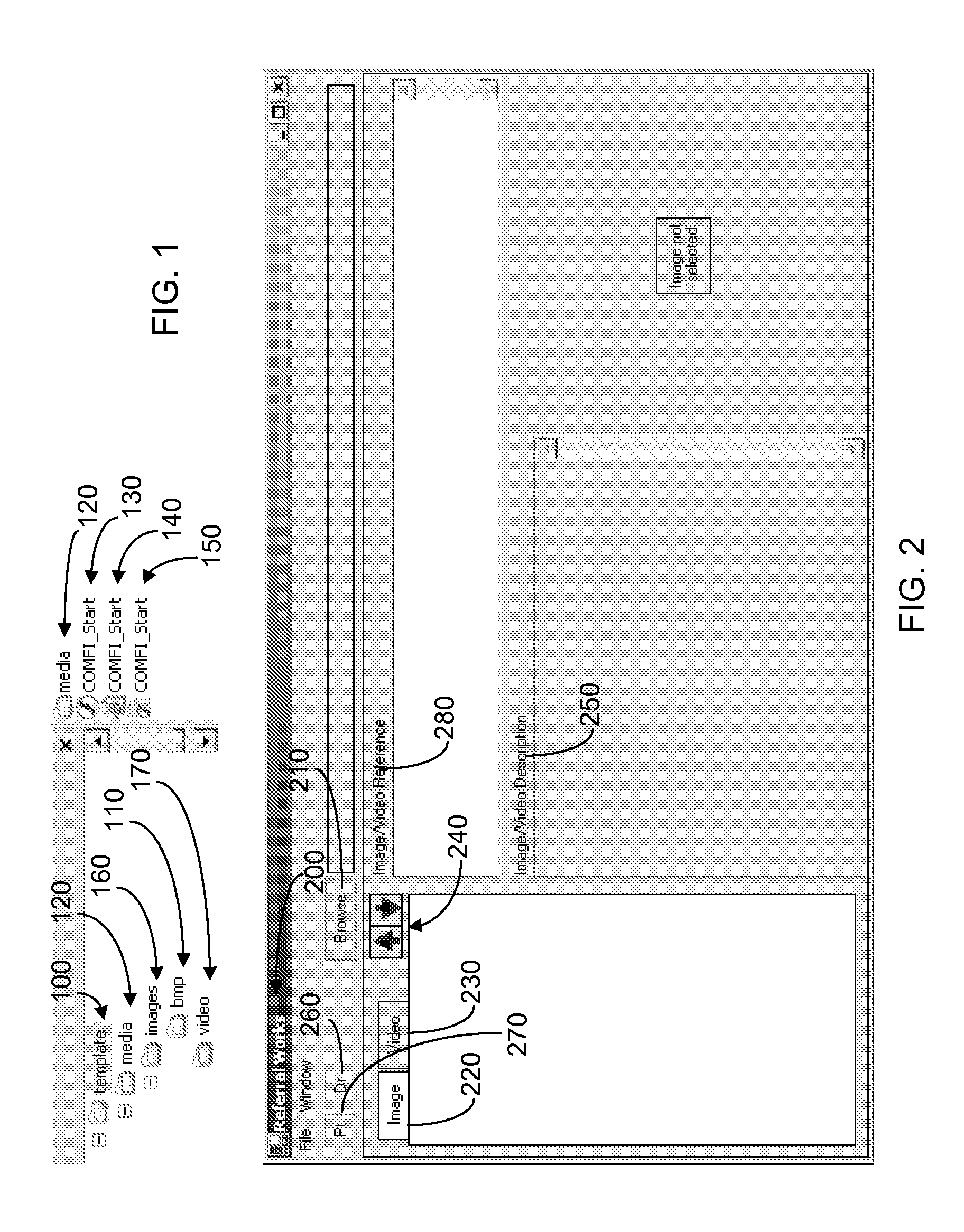
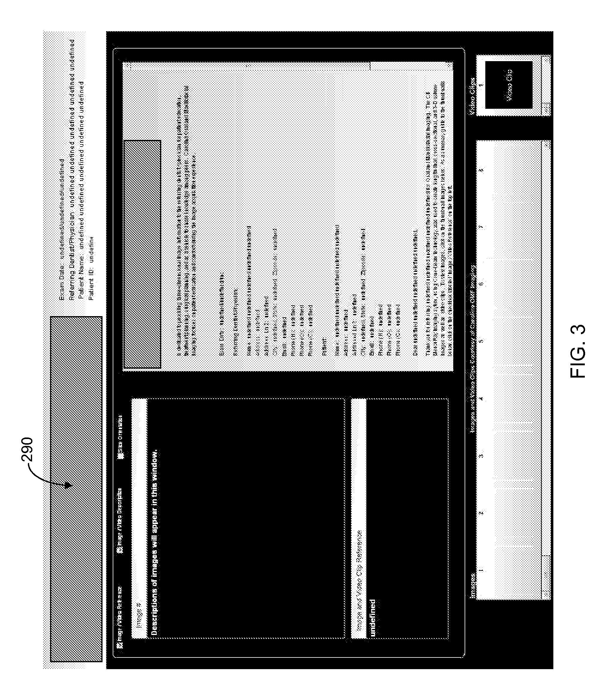
[0025]The present inventions now will be described more fully hereinafter with reference to the accompanying drawings, in which some, but not all embodiments of the inventions are shown. Indeed, these inventions may be embodied in many different forms and should not be construed as limited to the embodiments set forth herein; rather, these embodiments are provided so that this disclosure will satisfy applicable legal requirements. Like numbers refer to like elements throughout.

[0026]According to embodiments of the present invention, a patient is subjected to a three-dimensional imaging process in a dental imaging center using, for example, a conebeam technology imager or other suitable 3D imaging machine such as, for instance, a Model CB MercuRay for maxillofacial imaging, produced by Hitachi Medical Systems America of Twinsburg, OH. Such an imager generally includes a software package, or is otherwise configured to capture a comprehensive set of images of at least a portion of a ma...


PUM


Abstract

A method of providing dental-related imaging data of a patient to a dental practitioner is provided, the dental-related imaging data being obtained by a third party imaging provider at a third-party imaging provider site using a three-dimensional digital imaging device. Three-dimensional image data of the maxillofacial complex of the patient is obtained with the imaging device and analyzed to form a diagnosis. A two-dimensional image associated with the diagnosis is formed from the three-dimensional image data, and the two-dimensional image and diagnosis are combined into a composite file as part of the dental-related imaging data. The dental-related imaging data is stored in encrypted read-only format executable by a standard multi-media player and made available to the dental provider in a secure arrangement. The composite file is thereby selectively viewable by the dental provider for determining an appropriate treatment for the patient. Associated methods and apparatuses are also provided.

Description

CROSS-REFERENCE TO RELATED APPLICATION[0001]This application is a Continuation-in-Part of U.S. Utility application Ser. No. 11 / 417,381, filed May 4, 2006, which is incorporated by reference herein in its entirety.BACKGROUND OF THE INVENTION[0002]1. Field of the Invention[0003]The present invention relates to diagnostic imaging systems and, more particularly, to a method and apparatus for forming and distributing a composite file including a dental image and associated diagnosis.[0004]2. Description of Related Art[0005]Radiographic imaging in dentistry has historically been most commonly associated with two-dimensional images. That is, any dental-related radiographic imaging likely produces a two-dimensional representation of the imaged area that includes distortion resulting from the attempt to image a three-dimensional subject. Developments in three-dimensional imaging such as, for example, conebeam technology, has allowed three-dimensional imaging to be offered to private practiti...

Claims


Application Information

Patent Timeline
Patent Type & Authority Applications(United States)
IPC IPC(8): G06K9/00
CPCA61B6/14A61B6/463G06F19/321A61B6/5235A61B6/465G16H30/20G16H30/40G16H50/20
Inventor HOWERTON, JR., WILLIAM BRUCE
Owner HOWERTON JR WILLIAM BRUCE
Why Eureka
Try Eureka