Use of interleukin-18 inhibitors to inhibit tumor metastasis
a technology of interleukin-18 and tumor metastasis, which is applied in the field of tumor metastasis, can solve the problems of insufficient concentration of endotoxin or mannose receptor ligand, inability of most metastasizing cancer cells and the target tissues to produce these pro-inflammatory cytokines, and insufficient characterization of multiple mediators that evoke vcam-1 upregulation and its involvement during capillary transit of cancer cells
- Summary
- Abstract
- Description
- Claims
- Application Information
AI Technical Summary
Benefits of technology
Problems solved by technology
Method used
Image
Examples
example 1
Quantitative B16M Cell Adhesion to Primary HSE Cultures
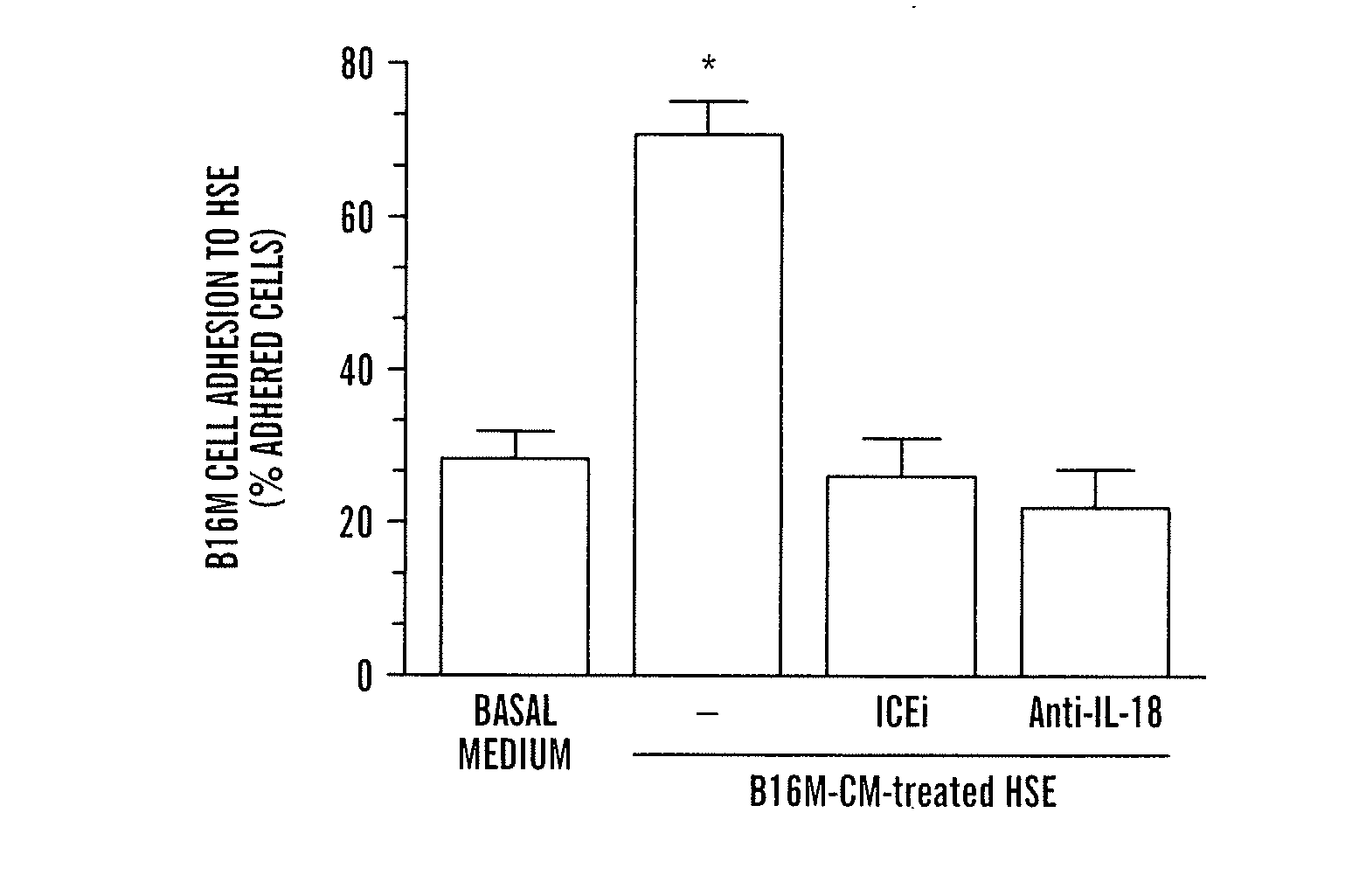
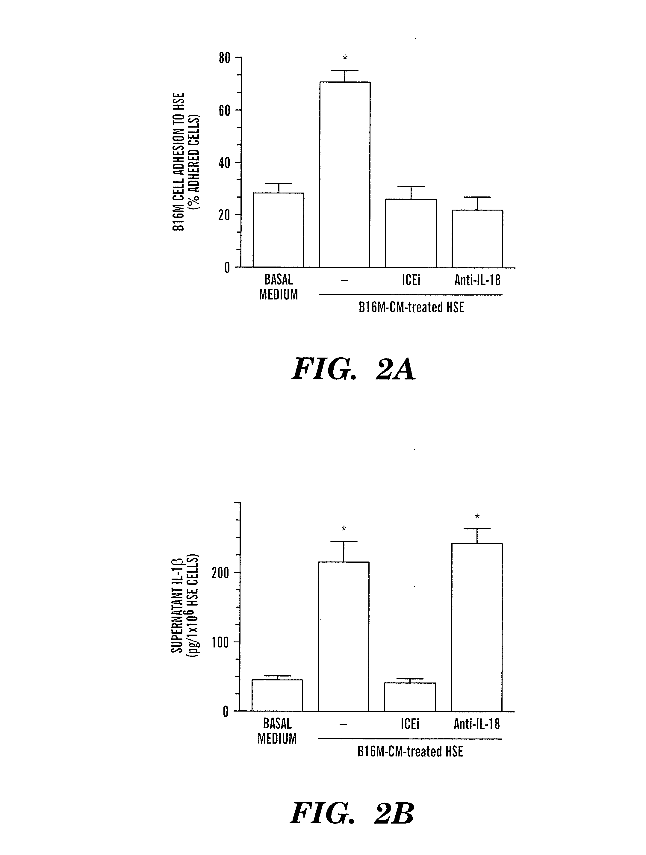
[0035] HSE was separated from syngeneic mice, identified and cultured as previously described (26). B16M cells were labeled with 2′,7′-bis-(2-carboxyethyl)-5,6-carboxyfluorescein-acetoxymethylester solution (BCECF-AM, Molecular Probes, Eugene, Oreg.) as reported (16). Then, 2×10⁵ cells / well were added to 24-well-plate cultured HSE and 8 min later, wells were washed three times with fresh medium. The number of adhering cells was determined using a quantitative method based on a previously described fluorescence measurement system (16). In some experiments, HSE cells were pre-incubated with B16M-CM for several hours before addition of B16M cells.
example 2
[0036] Wild-type, IL-1β− / − and ICE− / − male C57BL / 6J mice were generated as previously described (27). Six- to eight-week-old mice, housed five per cage, were used. Hepatic metastases were produced by the intrasplenic injection into anesthetized mice (Nembutal, 50 mg / kg intraperitoneal) of 3×10⁵ viable B16 melanoma cells suspended in 0.1 ml Hanks' balanced salt solution. Mice were killed under anesthesia on the 10th day after the injection of cancer cells. Liver tissues were processed for histology. Densitometric analysis of digitalized microscopic images was used to discriminate metastatic B16M from normal hepatic tissue and the liver metastasis density, which is the number of metastases per 100 mm3 of liver (based on the mean number of foci detected in fifteen 10×10 mm2 sections per liver), was calculated using previously described stereological procedures (17).
example 3
Reduced Metastasis and Growth of B16M Cells Injected Into IL-1β and ICE Deficient Mice
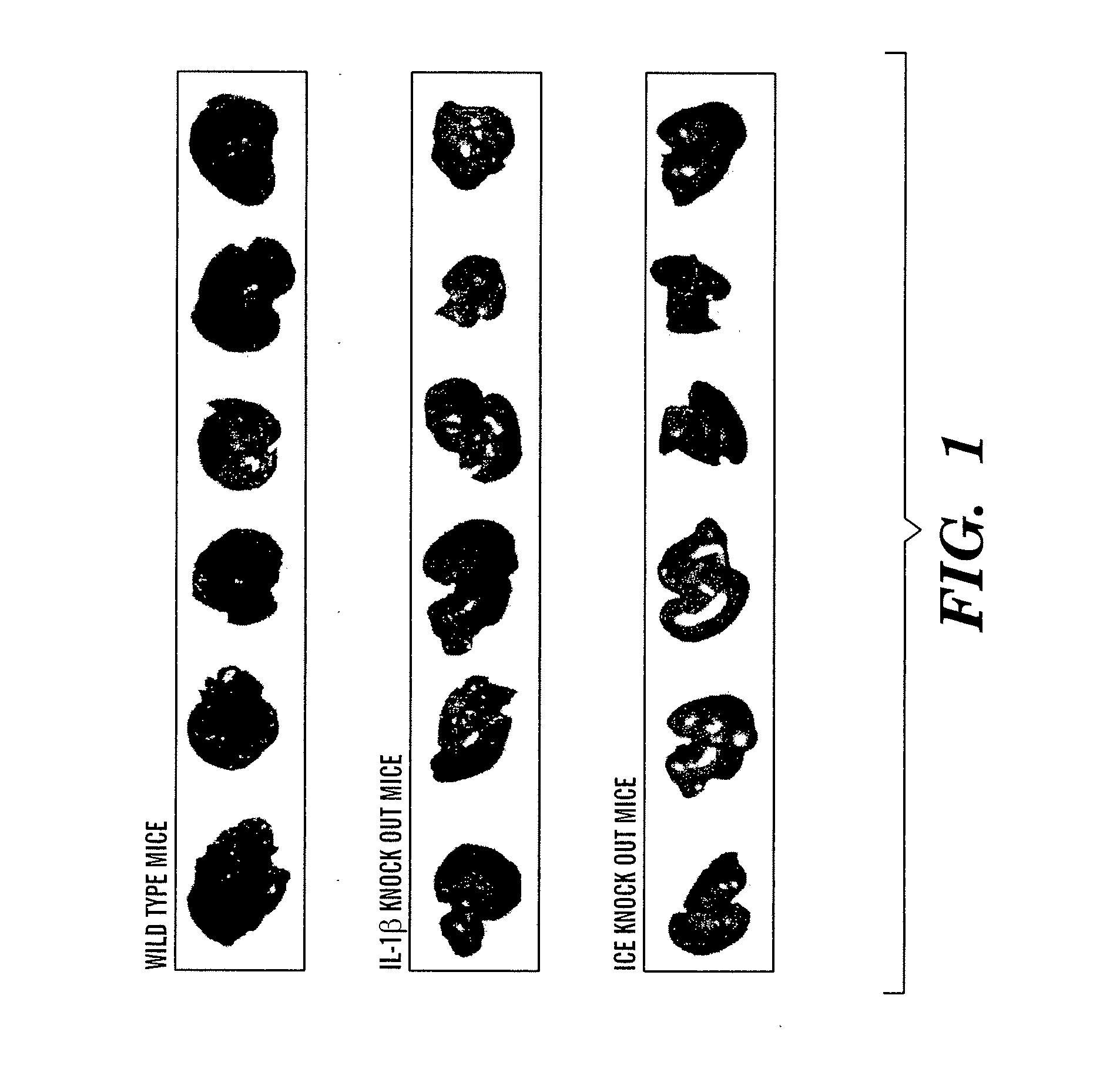
[0037] Two independent experiments, one year apart, were performed using two different batches of same B16M cells intrasplenically injected in adult C57Bl / 6J wild-type, ICE− / − and IL-1β− / − mice. Necropsic inspection demonstrated visible melanotic tumors in the spleen from all assayed mice, without significant differences in size as evaluated by splenic weight (Table 1). In contrast, a marked decrease in metastasis occurred in IL-1β− / − and, specially, ICE− / − mouse livers compared to wild-type mouse livers (FIG. 1). A quantitative histological analysis on number and size of metastatic foci was carried out to determine metastasis density (as no. foci / 100 mm3) and volume (percent organ occupancy) parameters in studied mouse livers. Compared to wild-type mice (Table 1), hepatic metastasis density significantly (P− / − and ICE− / − mouse livers by 84%-to-90%, indicating that most of injected B16M cells were...
PUM
| Property | Measurement | Unit |
|---|---|---|
| concentration | aaaaa | aaaaa |
| adhesion | aaaaa | aaaaa |
| adhesive function | aaaaa | aaaaa |
Abstract
Description
Claims
Application Information
 - R&D
- Intellectual Property
- Life Sciences
- Materials
- Tech Scout
- Unparalleled Data Quality
- Higher Quality Content
- 60% Fewer Hallucinations
Browse by: Latest US Patents, China's latest patents, Technical Efficacy Thesaurus, Application Domain, Technology Topic, Popular Technical Reports.
© 2025 PatSnap. All rights reserved.Legal|Privacy policy|Modern Slavery Act Transparency Statement|Sitemap|About US| Contact US: help@patsnap.com


