Determining mammographic image view and laterality

a mammographic image and view type technology, applied in image enhancement, image analysis, instruments, etc., can solve the problems of not all films are correctly labeled or oriented for viewing, method requires operator interaction, and is subject to operator error

Inactive Publication Date: 2008-03-20
CARESTREAM HEALTH INC
View PDF6 Cites 43 Cited by
  • Summary
  • Abstract
  • Description
  • Claims
  • Application Information

AI Technical Summary

Benefits of technology

[0016]An advantage of the method of the present invention is that it does not require interpreting text characters from a

Problems solved by technology

As can be expected, not all films are correctly labeled or oriented properly for viewing, due to technician inexperience or error.
While such

Method used


Image

  • Determining mammographic image view and laterality
  • Determining mammographic image view and laterality
  • Determining mammographic image view and laterality

Examples


Embodiment Construction

[0026]The present description is directed to elements forming part of, or cooperating more directly with, apparatus in accordance with the invention. It is to be understood that elements not specifically shown or described may take various forms well known to those skilled in the art.

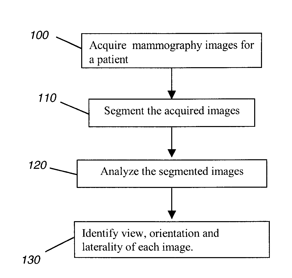
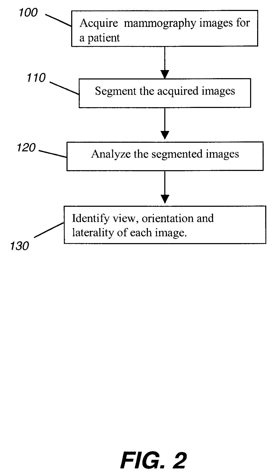
[0027]The method of the present invention uses analysis of segmented images and probability logic to identify type and laterality for mammography images. Using the method of the present invention, a system can accept, as input image data, the standard set of mammography images for a patient and can identify the image view type (MLO or CC) and laterality (R or L). These images can then be provided to a system for display using an appropriate hanging protocol or pattern that meets the needs of the radiology practitioner.

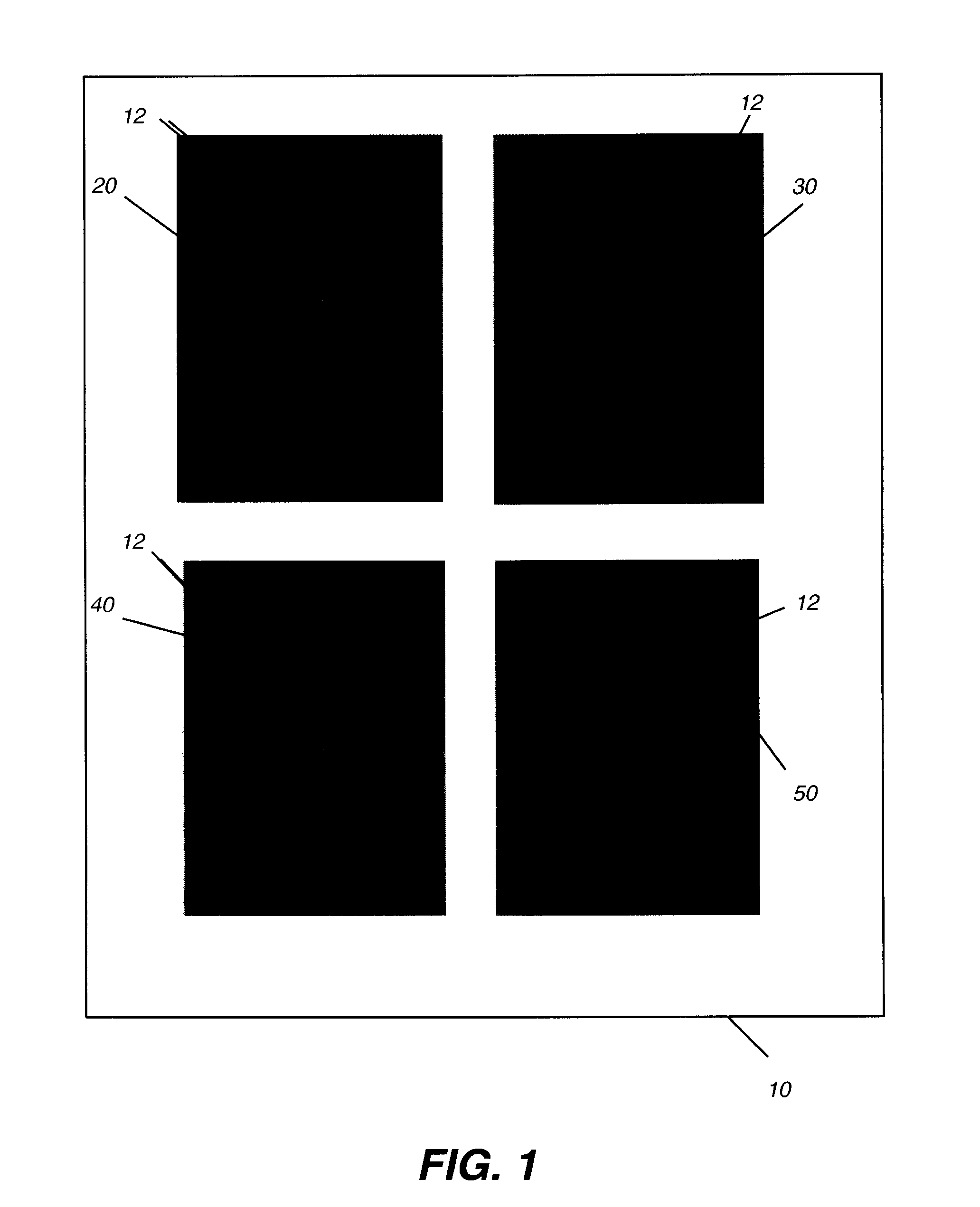
[0028]The method of the present invention makes some assumptions about digital mammography images. For example:[0029](i) The images are of the standard image set with four views (RCC, LCC,...


PUM


Abstract

A method for displaying a mammography image. Digital data of the mammography image is obtained. The mammography image is segmented to identify at least a first diagnostically relevant region comprising an image of the breast tissue and a second diagnostically relevant region. A view type is assigned for the image, either cranio-caudal or medio-lateral oblique view, according to a symmetry index calculated from the segmented first diagnostically relevant region. Right or left laterality is assigned to the image according to a laterality feature calculated according to the relative position of at least the second diagnostically relevant region within the image.

Description

FIELD OF THE INVENTION[0001]This invention generally relates to techniques for processing mammogram images, and more particularly to techniques for automatic detection of view type and laterality for digital mammographic images.BACKGROUND OF THE INVENTION[0002]Screening mammography is a method for detecting signs of breast cancer in otherwise asymptomatic women. The American College of Radiology recommends annual mammography for women over forty. Women undergo an X-ray exam in which X-ray films of the breast are exposed and then developed for later review. A radiologist reads the films and assesses the likelihood of the presence of signs of breast cancer. If a suspicious finding is present, the woman may be invited for additional, more detailed diagnostic X-ray exams, ultrasonic exams, and / or biopsy.[0003]In a typical screening exam in the United States, four X-rays of the breast are obtained. In conventional practice, two mammographic views are obtained for each breast: a cranio-ca...

Claims


Application Information

Patent Timeline
IPC IPC(8): G06K9/00
CPCG06T2207/30068G06T7/0042G06T7/73
Inventor LUO, HUI
Owner CARESTREAM HEALTH INC
Why Eureka
Try Eureka