
Visceral Anchors For Purse-String Closure of Perforations

a technology of perforation and visceral anchors, which is applied in the field of visceral anchors, can solve the problems of difficult to properly tension each individual suture, unwanted and sometimes deadly infection, and not without their drawbacks, and achieve the effect of increasing versatility and controlling the closure of perforations

Active Publication Date: 2008-06-05
WILSONCOOK MEDICAL +1
View PDF45 Cites 187 Cited by
  • Summary
  • Abstract
  • Description
  • Claims
  • Application Information

AI Technical Summary

Benefits of technology

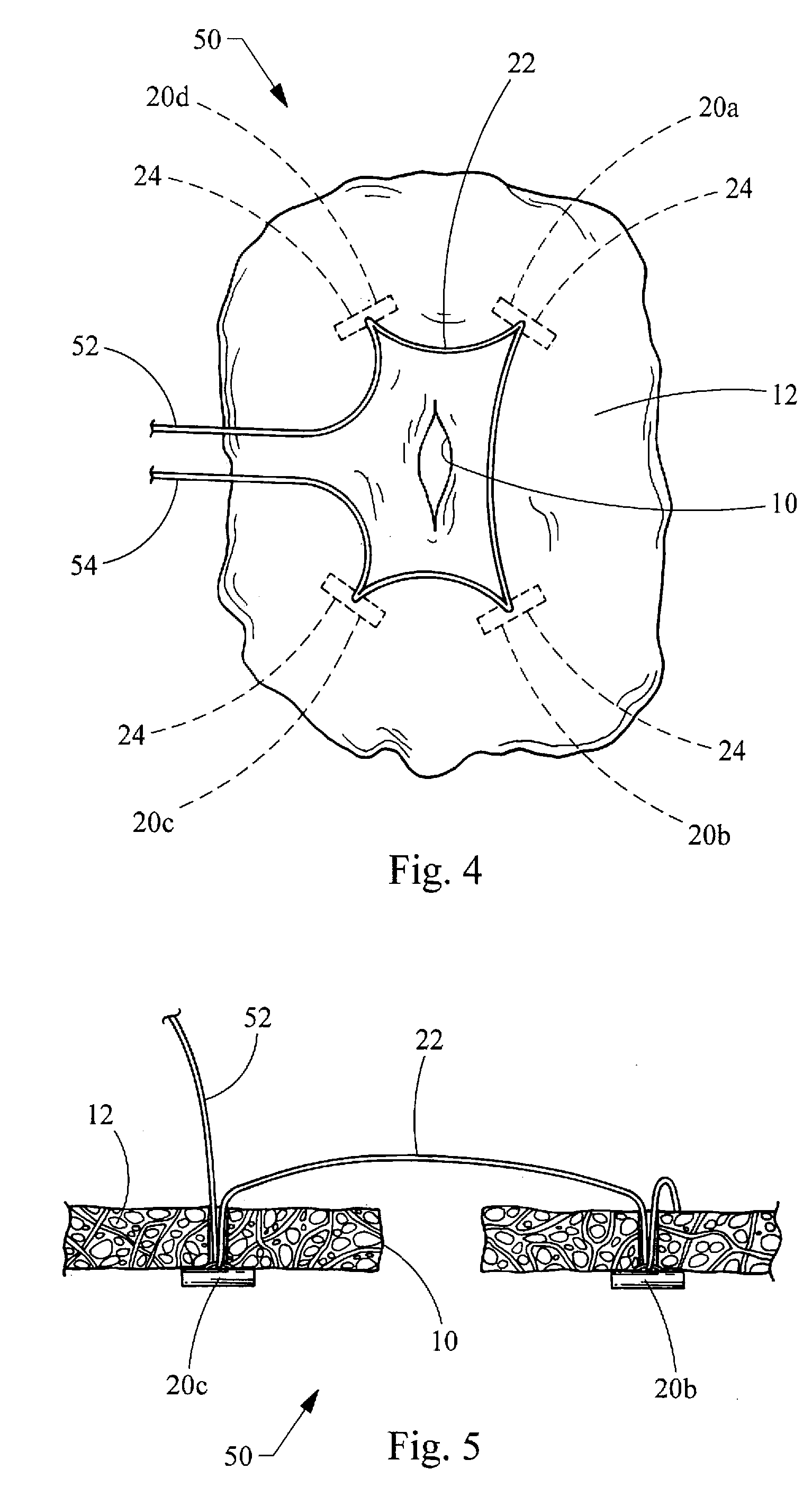
[0005]The present invention provides medical devices and related methods for closing a perforation in a bodily wall that provides reliable and complete closure of perforations, while at the same time gives increased versatility and control over perforation closure. One embodiment of a medical device, constructed in accordance with the teachings of the present invention, generally comprises a suture having opposing first and second ends and a set of visceral anchors. Each visceral anchor includes a crossbar having opposing ends, and a suture is slidably attached to the crossbar. The first and second ends of the suture are capable of being independently tensioned for translating the suture relative to each of the visceral anchors and closing the perforation.
[0006]According to more detailed aspects, the tensioning of the suture reduces the distance between the visceral anchors to close the perforation in a purse-string fashion. The tensioning of the suture causes compression of the bodily wall around the perforation. Each visceral anchor is positioned on one side of the bodily wall, and first and second ends of the suture are positioned on the opposite side of the bodily wall. The medical device may further include an overtube for delivery of the set of visceral anchors. The overtube has a set of lumens, each lumen receiving a visceral anchor. Portions of the suture located between the visceral anchors extend outside the lumens at a distal end of the overtube. The medical device may also include a set of puncture needles slidably received within the set of lumens, the set of puncture needles receiving the set of visceral anchors therein.
[0007]One embodiment of a visceral anchor, constructed in accordance with the teachings of the present invention, generally comprises a crossbar having a passageway. The crossbar has first and second opposing ends and defines a longitudinal axis. The passageway is located between the opposing ends of the crossbar and slidably receives the suture therein. The passageway has an entrance and an exit through which the suture passes. The entrance and exit are spaced apart a distance greater than 0.001 mm and less than 1.0 mm. By providing the entrance and exit of the passageway in close proximity, the leading and trailing portions of the suture may be kept closer together, and the visceral anchor pulled into closer contact with the tissue, while still retaining the ability to translate the suture relative to the anchor. The passageway is also structured to reduce friction between the suture and crossbar.
[0009]According to another aspect of the present invention, a method for closing a perforation in a bodily wall is provided in accordance with the teachings of the present invention. The method utilizes a set of visceral anchors connected to a suture as previously described. Each visceral anchor is passed through the bodily wall adjacent the periphery of the perforation. Each visceral anchor is on a distal side of the bodily wall, while the ends of the suture on a proximal side of the bodily wall. The ends of the suture are tensioned to reduce the distance between the visceral anchors and compress the bodily wall around the perforation. The ends of the suture are secured to maintain the compression of the bodily wall and close the perforation.

Problems solved by technology

Visceral anchors have also been successfully used in closing perforations, but are not without their drawbacks.
It can often be difficult to properly tension each of the individual sutures to ensure proper approximation of the tissue around the perforation and complete closure thereof.
This is especially critical within the gastrointestinal tract, where the travel of bacteria laden fluids outside of the tract may cause unwanted and sometimes deadly infection.

Method used


Image

  • Visceral Anchors For Purse-String Closure of Perforations
  • Visceral Anchors For Purse-String Closure of Perforations
  • Visceral Anchors For Purse-String Closure of Perforations

Examples


Embodiment Construction

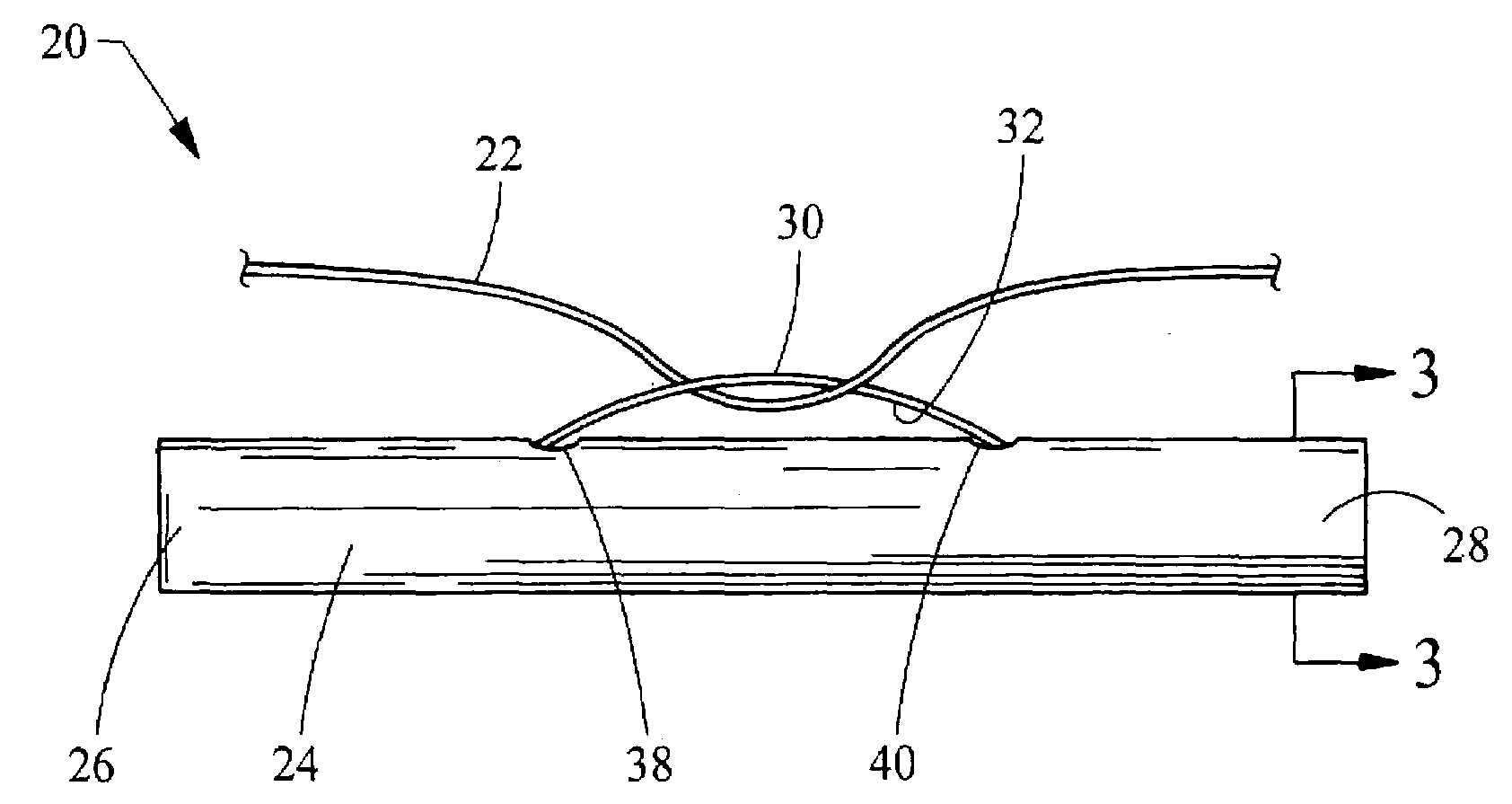
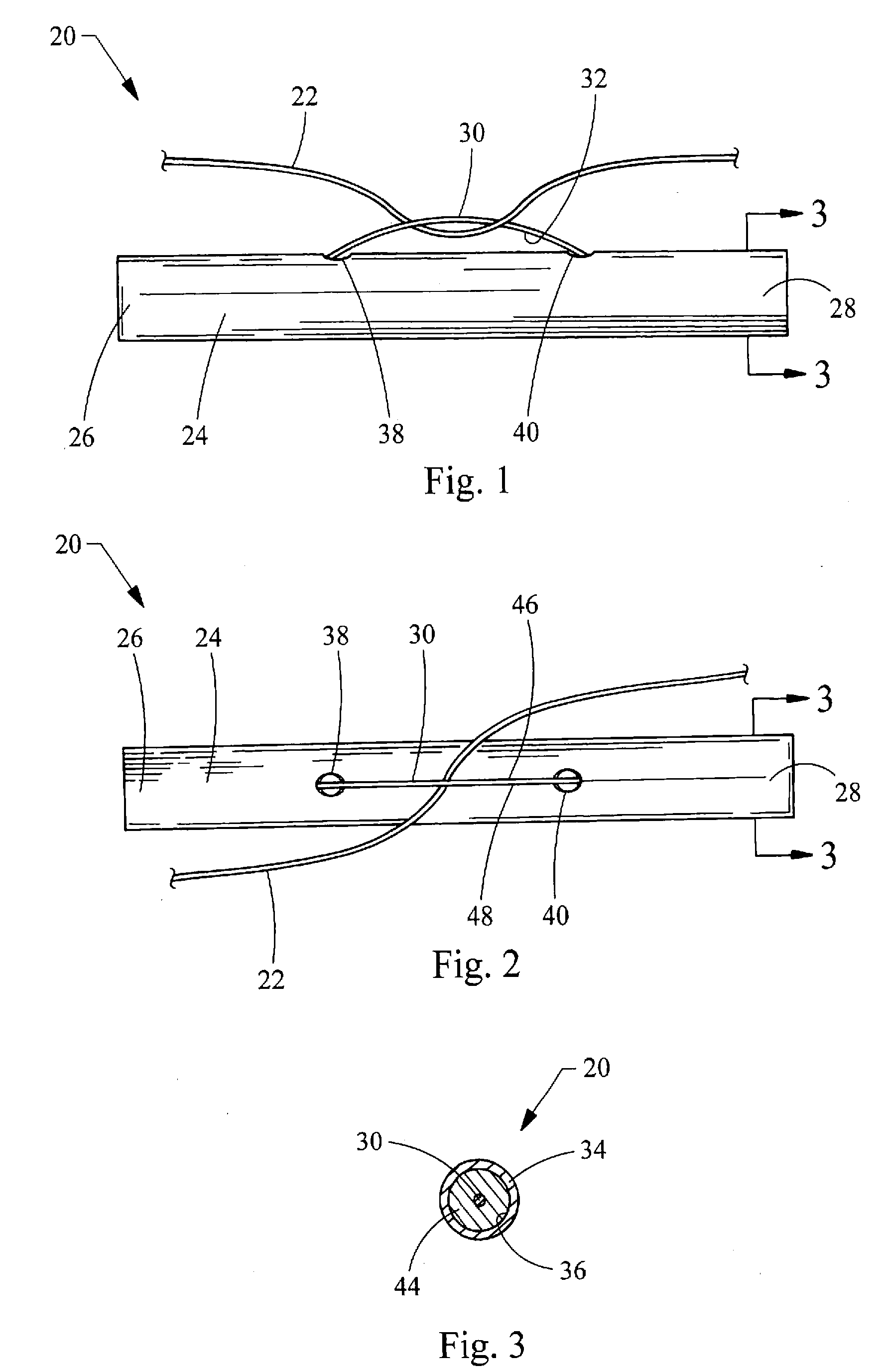
[0027]Turning now to the figures, FIGS. 1-3 depict a visceral anchor 20 constructed in accordance with the teachings of the present invention. The anchor 20 is utilized to connect a suture 22 to tissue for closing a perforation 10 in a bodily wall 12 (FIGS. 4 and 5). The anchor 20 generally includes a crossbar 24 having opposing ends 26 and 28. The crossbar 24 is preferably elongated, but may take any form suitable for connecting the suture 22 to the bodily wall 12. A wire 30 connected to the crossbar 24 to form a loop defining a passageway 32 between the wire 30 and crossbar 24. As best seen in FIG. 3, the crossbar 24 is constructed of a cannula having a tubular wall 34 defining a lumen 36. First and second apertures 38, 40 are formed in the tubular wall 34, and the wire 30 passes through the apertures 38, 40. The ends of wire 30 are secured within the lumen 36 of the cannula by welds 44. It will be recognized by those skilled in the art that the wire 30 may be secured to the cross...


PUM


Abstract

Medical devices and related methods for closing a perforation in a bodily wall The medical device generally includes a suture having opposing first and second ends and a set of visceral anchors. Each visceral anchor includes a crossbar having opposing ends and a suture slidably attached thereto. Each visceral anchor is passed through the bodily wall adjacent the periphery of the perforation. The ends of the suture are tensioned to reduce the distance between the visceral anchors and compress the bodily wall around the perforation. The ends of the suture are secured to maintain the compression of the bodily wall and close the perforation.

Description

CROSS-REFERENCE TO RELATED APPLICATIONS[0001]This application claims the benefit of U.S. Provisional Application Ser. No. 60 / 872,023 filed on Nov. 30, 2006, entitled “VISCERAL ANCHORS FOR PURSE-STRING CLOSURE OF PERFORATIONS” the entire contents of which are incorporated herein by reference.FIELD OF THE INVENTION[0002]The present invention relates generally to visceral anchors for connecting a suture to tissue, and more particularly relates to using visceral anchors and suture to close perforations in tissue.BACKGROUND OF THE INVENTION[0003]Perforations in bodily walls may be naturally occurring, or formed intentionally or unintentionally. In order to permanently close these perforations and allow the tissue to properly heal, numerous medical devices and methods have been developed employing sutures, adhesives, clips, staples and the like. One class of such devices is commonly referred to as tissue anchors (T-anchors) or visceral anchors. An exemplary visceral anchor is disclosed in...

Claims


Application Information

Patent Timeline
Patent Type & Authority Applications(United States)
IPC IPC(8): A61B17/04A61B17/34
CPCA61B17/0401A61B2017/00637A61B2017/0409A61B2017/0414A61B2017/1142A61B2017/0454A61B2017/0458A61B2017/0464A61B2017/0417
Inventor SURTI, VIHAR C.DESILETS, DAVID
Owner WILSONCOOK MEDICAL
Learn More