Multifunctional magnetic nanoparticle probes for intracellular molecular imaging and monitoring

a magnetic nanoparticle and probe technology, applied in the field of multifunctional magnetic nanoparticle probes, can solve the problems of inability to quantify gene expression in living cells, lack of novel labeling reagents for in vivo protein expression and interaction studies, and no existing method that enables dynamics studies, etc., to achieve enhanced signal-to-noise ratio in molecular imaging, efficient internalization of magnetic nanoparticle probes, and high specificity and sensitivity

Inactive Publication Date: 2008-12-02
GEORGIA TECH RES CORP +1
View PDF1 Cites 139 Cited by
  • Summary
  • Abstract
  • Description
  • Claims
  • Application Information

AI Technical Summary

Benefits of technology

[0015]The present invention fulfills in part the need to develop multifunctional magnetic nanoparticle probes for intracellular molecular imaging and monitoring. The novel nanoprobe design and probe / target recognition and binding strategies provide high specificity and sensitivity, and enhanced signal-to-noise ratio in molecular imaging. The peptide-based intracellular delivery of probes and specific cell-type targeting provide the vehicle for efficient internalization of the magnetic nanoparticle probes into selected cells and organs. In addition, the nanoparticle probes of the present invention provide the capability of combining MRI and FRET-based optical imaging, providing for improved spatial resolution and versatility. These innovations and their integration will provide a platform for a new generation of biomagnetic contrast agents for deep tissue molecular imaging and monitoring. The multifunctional magnetic nanoprobes offer a novel transduction mechanism for the in vivo detection of molecular markers for disease, infection, and injury.
[0016]Accordingly, the present invention provides improved compositions and methods of use for more sensitive, specific, or rapid nucleic acid or polypeptide detection.
[0017]The present invention also provides improved methods for the detection of gene expression associated with a response to external stimuli, e.g. a therapeutic drug candidate. The present invention further provides improved methods for the detection of gene expression, including genetic expression associated with an infection or disease state.
[0019]The present invention provides nanoparticle probe compositions for facilitating molecular imaging, comprising: a nucleic acid or polypeptide probe, a delivery ligand, and a detectable moiety, wherein the detectable moiety comprises a magnetic nanoparticle having a biocompatible coating thereon. The present invention also provides that the delivery ligand is a ligand that is capable of facilitating the traverse of a cell membrane. In a preferred embodiment, the magnetic nanoparticle is monocrystalline iron oxide nanoparticle (MION). The present invention also provides that the nanoparticle probe compositions may further comprise a second ligand that is capable of interacting with a molecule located on the outer surface of a particular type of cell or tissue.
[0022]In certain other embodiments, the invention provides compositions for facilitating signal transduction in molecular imaging, comprising two magnetic nanoparticle probe compositions wherein the first and the second magnetic nanoparticle probe compositions comprise a first polypeptide probe and a second polypeptide probe, respectively. The present invention provides that such a composition is useful to facilitate the measurement of polypeptide expression, protein interactions, and protein clustering in both in vivo and in vitro systems using magnetic imaging / magnetic spectroscopic measurements. In a preferred embodiment, the present invention provides that the first and the second polypeptide probes bind to a first and a second target sequence on a single subject polypeptide. In another preferred embodiment, the present invention provides that the first polypeptide probe binds to a first subject polypeptide; the second polypeptide probe binds to a second subject polypeptide. When the first subject polypeptide interacts with the second subject polypeptide, the first and second magnetic nanoparticles are brought in close proximity (e.g. 2-50 nm), and the effect on water relaxation (change in T1, T2, or its combination for contrast detection) from interaction of the first and second magnetic nanoparticles can be detected to determine binding of both the first and the second polypeptide probes.
[0026]The invention further provides methods for producing a magnetic nanoparticle probe composition for facilitating molecular imaging, comprising combining a magnetic nanoparticle with a biocompatible coating; adding a ligand that is capable of facilitating the traverse of a cell membrane; and adding a nucleic acid probe or polypeptide probe. The invention also provides that the methods may comprise the further step of adding a second ligand that is capable of interacting with a molecule located on the outer surface of a particular type of cell or tissue. The invention further provides that these methods may further comprise the step of adding a resonance energy transfer moiety to the magnetic nanoparticle probe composition.

Problems solved by technology

Technologies currently available for analysis and quantification of gene expression such as real-time RT-PCR, Northern blotting, expressed sequence tag (EST), serial analysis of gene expression (SAGE) and DNA microarrays are powerful tools for in vitro studies; however, they are not capable of quantifying gene expression in living cells.
For example, there is a lack of novel labeling reagents in performing in vivo protein expression and interaction studies.
There is virtually no existing method that enables studies of the dynamics of different intra-cellular molecular processes in deep tissue.
This is a daunting challenge in that none of the many approaches under development to address these problems have yet demonstrated compelling promise—even as generally effective laboratory-scale small animal models.
However, false-positive signals due to protein-beacon interaction and nuclease-induced beacon degradation significantly limit the sensitivity of the in vivo applications (Mitchell, 2001).
Although molecular beacons, with the dual FRET (fluorescence resonance energy transfer) design (Tsourkas et al, 2003a) and backbone modifications (Tsourkas at el, 2002b), can be used for sensitive and quantitative detection of mRNA in living cells, they are optical-based probes and therefore cannot be used for deep tissue imaging of RNA expression due to light absorption and scattering.
These coatings tend to have limited colloidal stability due to weak steric hindrance in preventing aggregation and have limited opportunity for functionalization.
Residual surfactant on the surface of nanoparticles can result in inefficient or incomplete coatings with this approach.
The amphiphilic polymer-based or silica-based coating approach often results in multilayer coatings (Lu et al., 2002), making the coating process difficult to control and resulting in a heterogeneous sample, and sometimes in the encapsulation of dimers or trimers of nanoparticles in the same shell (Lu et al., 2002).
These coating processes also lead to multilayer coatings, which can result in a heterogeneous sample.
In situ synthesis conditions limit the amount of functionalization that can be achieved.
However, traditional MRI contrast agents, as well as the use of magnetic nanoparticles with MRI to target cell surface receptors, are not suitable for detecting differential levels of gene expression inside living cells.

Method used


Image

  • Multifunctional magnetic nanoparticle probes for intracellular molecular imaging and monitoring
  • Multifunctional magnetic nanoparticle probes for intracellular molecular imaging and monitoring
  • Multifunctional magnetic nanoparticle probes for intracellular molecular imaging and monitoring

Examples


example 1

[0166]Compared with micrometer sized magnetic particles and chelates of paramagnetic ions such as gadolinium diethylenetriaminopentaacetic acid (Gd-DTPA), magnetic nanoparticles are much more efficient as relaxation promoters, and their effect on the relaxivities of water is measurable even at nanomolar concentrations, as demonstrated theoretically and experimentally (Koenig et al., 1995; Bulte et al., 1992; Le Duc, 2001; Josephson et al., 2001; Perez et al., 2002). Specifically, the use of ferromagnetic and superparamagnetic nanoparticles as contrast agents can induce a more than 10 fold increase in proton relaxivities (Coroiu et al., 1999). Based on the enzyme-cleavable contrast agents reported by Meade and coworkers (Louie et al., 2000; Huber et al., 1998), magnetic relaxation by gadolinium atoms is a short-range effect. The presence of a carbohydrate cap (less than 1 nm thick) prevents water molecules from contacting the contrast agent Gd (HP-DO3A). In contrast, superparamagneti...

example 2

Encapsulation of the Magnetic Nanoparticles

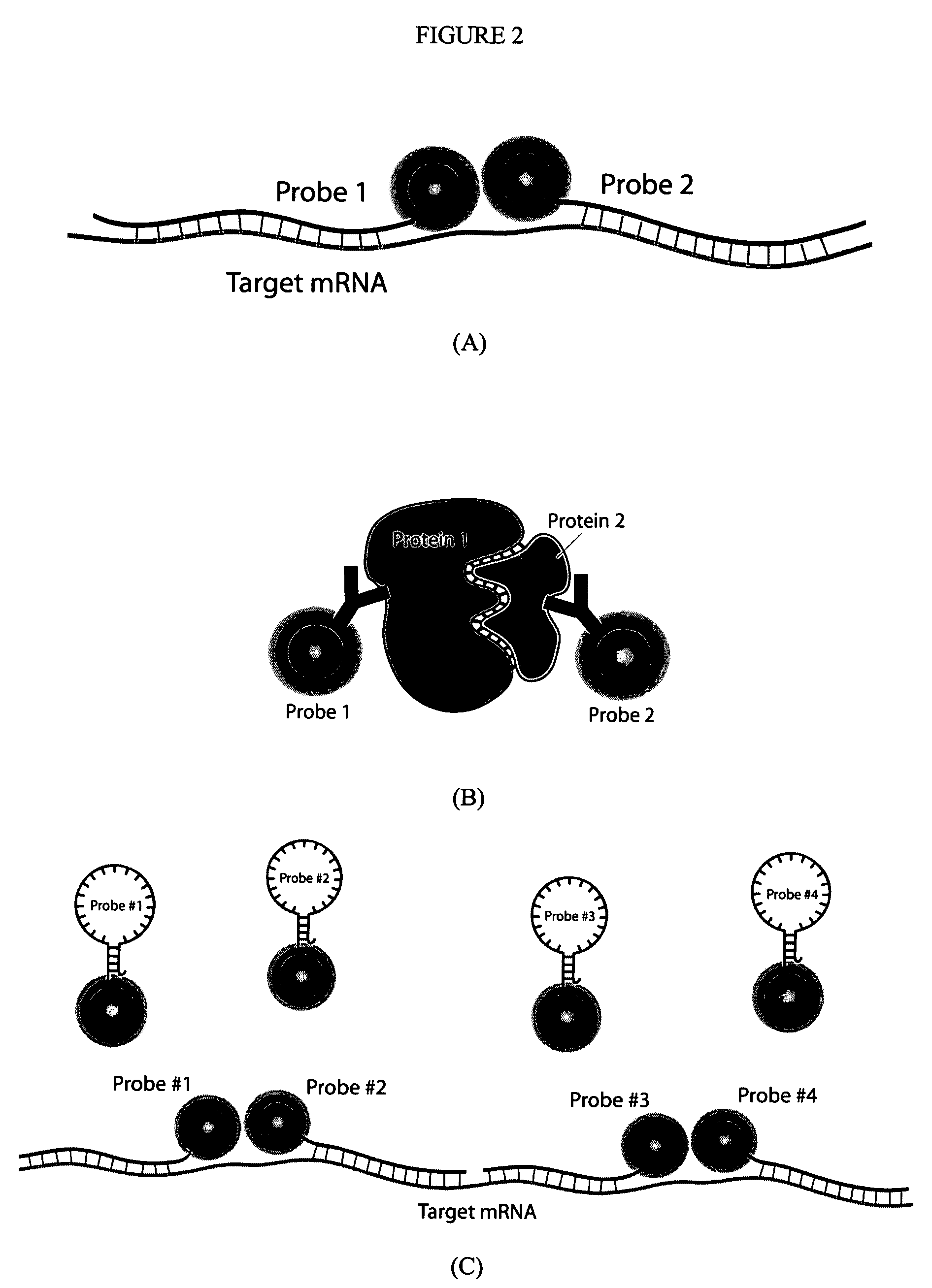
[0170]A critical component for the success of this invention is to make monocrystalline iron oxide nanoparticles (MIONs) water-soluble and create a stable suspension of MIONs that are biocompatible and surface-functionalized. A standard approach in the field is to use the polysaccharide dextran. A distinct disadvantage to this approach is the large thickness of the coating (approximately 15 nm). In order to minimize steric hindrance in hybridization between two probes, it is crucial to minimize the size of the coating. Therefore, efforts in the present invention were focused on developing alternative coatings. Two such post-synthesis coating processes will be described in detail here.

[0171]The first coating uses the monolayer ligand 16-mercaptohexadecanoic acid (Liu and Xu 1995). The MIONs were reacted with the organic surfactant 16-mercaptohexadecanoic acid (MHA). MHA has a carboxylic head group at one end of the molecule that reacts with ...

example 3

Bioconjugation of Magnetic Nanoparticles to Create Multifunctional Probes for Imaging and Delivery

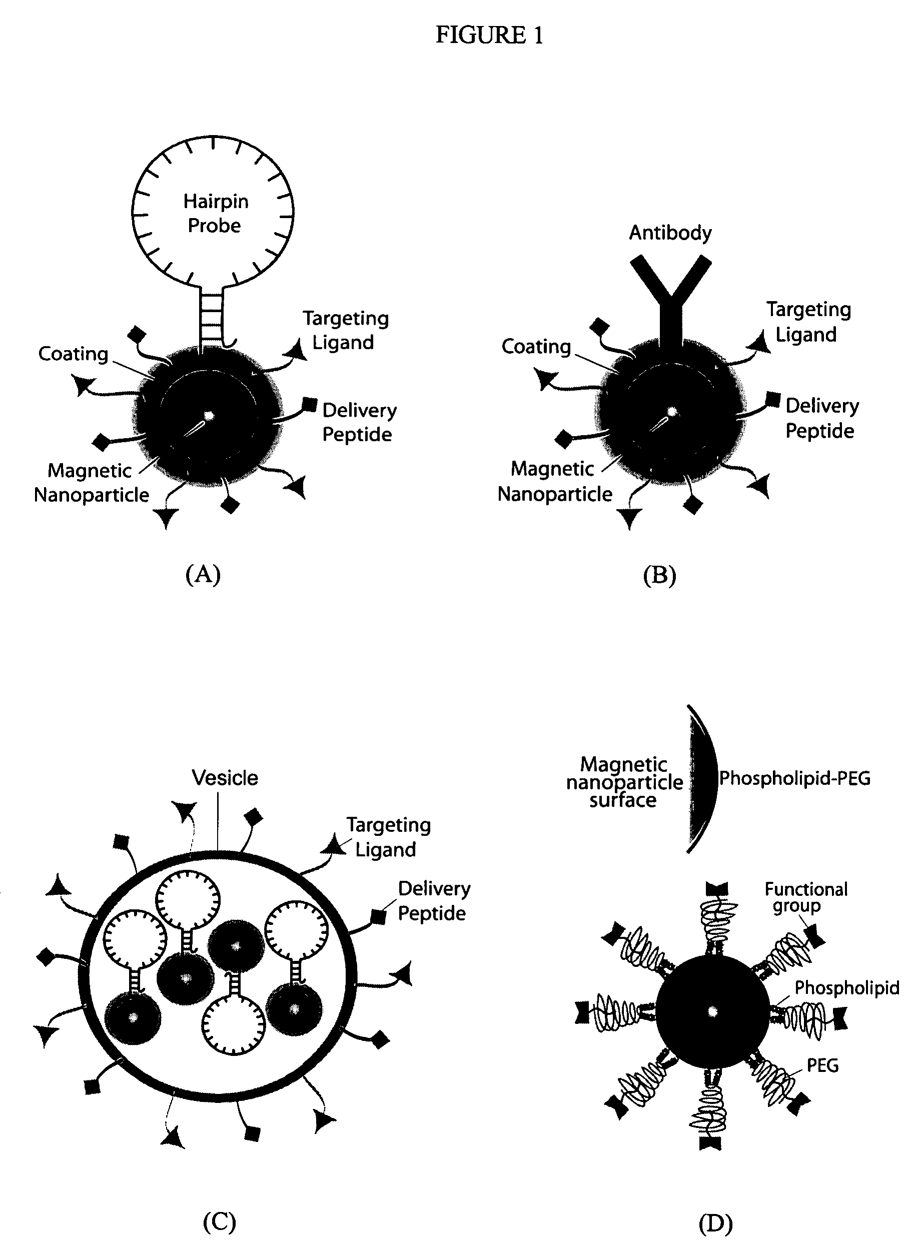
[0174]After encapsulation, the particles can be conjugated to peptides, proteins, fluorescent dye molecules, and hairpin oligonucleotide (ODN). Such conjugation is achieved by utilizing the functional group on the surface of the MIONs (in the specific cases described here, either reactive sulfhydryls or amines). These functional groups on the particle surface can be covalently conjugated to any of the above-described moieties by using standard cross-linking chemistry. Below will be described two specific examples of bioconjugation between MIONs and a) delivery peptide plus fluorescent dye and b) oligonucleotides. These examples use micelle-MIONs and MHA-MIONs, respectively; however, the coatings are versatile and either application is possible with both types of particles, with the micelle-MIONs being the preferred embodiment due to their superior solubility.

[0175]For optical imaging an...


PUM

PropertyMeasurementUnit
thickaaaaaaaaaa
core sizeaaaaaaaaaa
core sizeaaaaaaaaaa

Abstract

The present invention provides multifunctional magnetic nanoparticle probe compositions for molecular imaging and monitoring, comprising a nucleic acid or polypeptide probe, a delivery ligand, and a magnetic nanoparticle having a biocompatible coating thereon. The probe compositions may further comprise a fluorescent or luminescent resonance energy transfer moiety. Also provided are compositions comprising two or more such multifunctional magnetic nanoparticle probes for molecular imaging or monitoring. In particular, the nucleic acid or polypeptide probes bind to a target and generate an interaction observable with magnetic resonance imaging (MRI) or optical imaging. The invention thereby provides detectable signals for rapid, specific, and sensitive detection of nucleic acids, polypeptides, and interactions thereof in vivo.

Description

CROSS REFERENCE TO RELATED APPLICATIONS[0001]The present invention claims the priority benefit of U.S. Provisional Patent Application Ser. No. 60 / 421,361 filed Oct. 25, 2002, the entire contents of which are hereby incorporated by reference.ACKNOWLEDGEMENT OF FEDERAL RESEARCH SUPPORT[0002]This invention was made, at least in part, with funding from the Defense Advanced Research Project Agency (DARPA) of the US Department of Defense (DARPA-AFOSR Grant Number F49620-03-0320 to GB). Accordingly, the United States Government has certain rights in this invention.FIELD OF THE INVENTION[0003]This invention relates generally to multifunctional magnetic nanoparticle probes that may be used in both optical imaging and deep-tissue magnetic resonance imaging (MRI) and monitoring. More specifically, the present invention relates to magnetic nanoparticles that are coupled with ligand-based in vivo delivery, specific cell targeting, and a molecular probe approach for the detection of target nuclei...

Claims


Application Information

Patent Timeline
Patent Type & Authority Patents(United States)
IPC IPC(8): A61B5/055A61K51/00A61M36/14C12Q1/68G02B
CPCA61K49/0002A61K49/0041A61K49/0093A61K49/1836A61K49/186B82Y5/00B82Y30/00C12Q1/6818C12Q1/6834C12Q1/6841G01N33/5005G01N33/587C12Q2563/155C12Q2563/143C12Q2525/301C12Q2525/101
Inventor BAO, GANGNIE, SHUMINGNITIN, NITINLACONTE, LESLIE
Owner GEORGIA TECH RES CORP
Why Eureka
Try Eureka