Image registration of multiple medical imaging modalities using a multiple degree-of-freedom-encoded fiducial device

a fiducial device and degree of freedom technology, applied in the field of imaging registration of multiple medical imaging modalities, can solve the problems of not being used in quantitative intra-operative analysis, few attempts to relate fluoroscopic images to soft tissue anatomy, and little success, so as to improve the localization of surgically implanted objects, improve visualization and control, and facilitate dynamic dose calculation

Active Publication Date: 2008-10-23
THE JOHN HOPKINS UNIV SCHOOL OF MEDICINE +1
View PDF19 Cites 129 Cited by
  • Summary
  • Abstract
  • Description
  • Claims
  • Application Information

AI Technical Summary

Benefits of technology

[0016]An advantage of the present invention is to provide improved visualization and control during invasive surgical procedures.
[0017]Another advantage of the present invention is improved localization of surgically implanted objects in real time.
[0018]Another advantage of the present invention is to enable real time registration of multiple medical imaging modalities, whereby each modality provides images of different objects of distinct compositions in the same volume.
[0019]Another advantage of the present invention is to better enable dynamic dose calculation during radiation implant procedures, including the ability to adjust the planned implant locations to better optimize dosage as the implant progresses.

Problems solved by technology

While C-arm fluoroscopy has been used intra-operatively, it has not been utilized in quantitative intra-operative analysis.
Very few attempts have been made to relate fluoroscopic images to soft tissue anatomy, and with little success.
Here the fundamental problem is matching a large number of seeds with their projections in multiple X-ray images when some seeds obscure each other and solid objects also can get in the way.
Automated methods have been explored, but they also assume conditions that most likely cannot be met with a C-arm fluoroscope, particularly in a realistic intra-operative scenario.
Such assumptions include no extrinsic object in the field, optimal beam energy, arbitrary number and orientation of X-ray shots, or unlimited processing time and computational resources—none of which is realistic in real-time image-guided surgery.
In conclusion, “off-the-shelf” post-implant seed matching and reconstruction techniques of the related art cannot be expected to work in the operating room.
Many approaches have met with great difficulty due to the inability to accurately determine the imaging angles relative to the prostate for reconstruction of the multiple projection images.
This not only reduces accuracy, but makes the process too lengthy to be used intraoperatively.
The use of implanted needles as fiducial markers for registration of biplane TRUS data has been explored, but several key problems have been left unsolved.
First, the use of implanted needles as fiducials may not be practical, because most practitioners implant only one needle at a time and they do not use stabilizing needles.
Third, because X-ray and TRUS imaging are not simultaneous, it is imperative that the fiducials do not move relative to the prostate until both imaging sessions are complete.
Fourth, previous attempts at using implanted needles as fiducial markers did not account for the need to pre-operatively calibrate the C-arm fluoroscope, including removing image distortion and some form of intra-operative tracking to know where the multiple X-ray shots are coming with respect to one another.
While ultrasound is generally an excellent tool for guiding the implant needles with respect to prostate anatomy, it cannot reliably show the location of radioactive seeds after they are released in the prostate.
Intraoperative C-arm fluoroscopy does adequately show the location of implanted seeds, but it cannot differentiate soft tissue; thus, it cannot be used for visualizing prostate anatomy.
The problems and limitations apply to any procedure in which two imaging modalities are used, and in which there is a need to fuse or merge the products of the two imaging modalities into a single image space.

Method used


Image

  • Image registration of multiple medical imaging modalities using a multiple degree-of-freedom-encoded fiducial device
  • Image registration of multiple medical imaging modalities using a multiple degree-of-freedom-encoded fiducial device
  • Image registration of multiple medical imaging modalities using a multiple degree-of-freedom-encoded fiducial device

Examples


Embodiment Construction

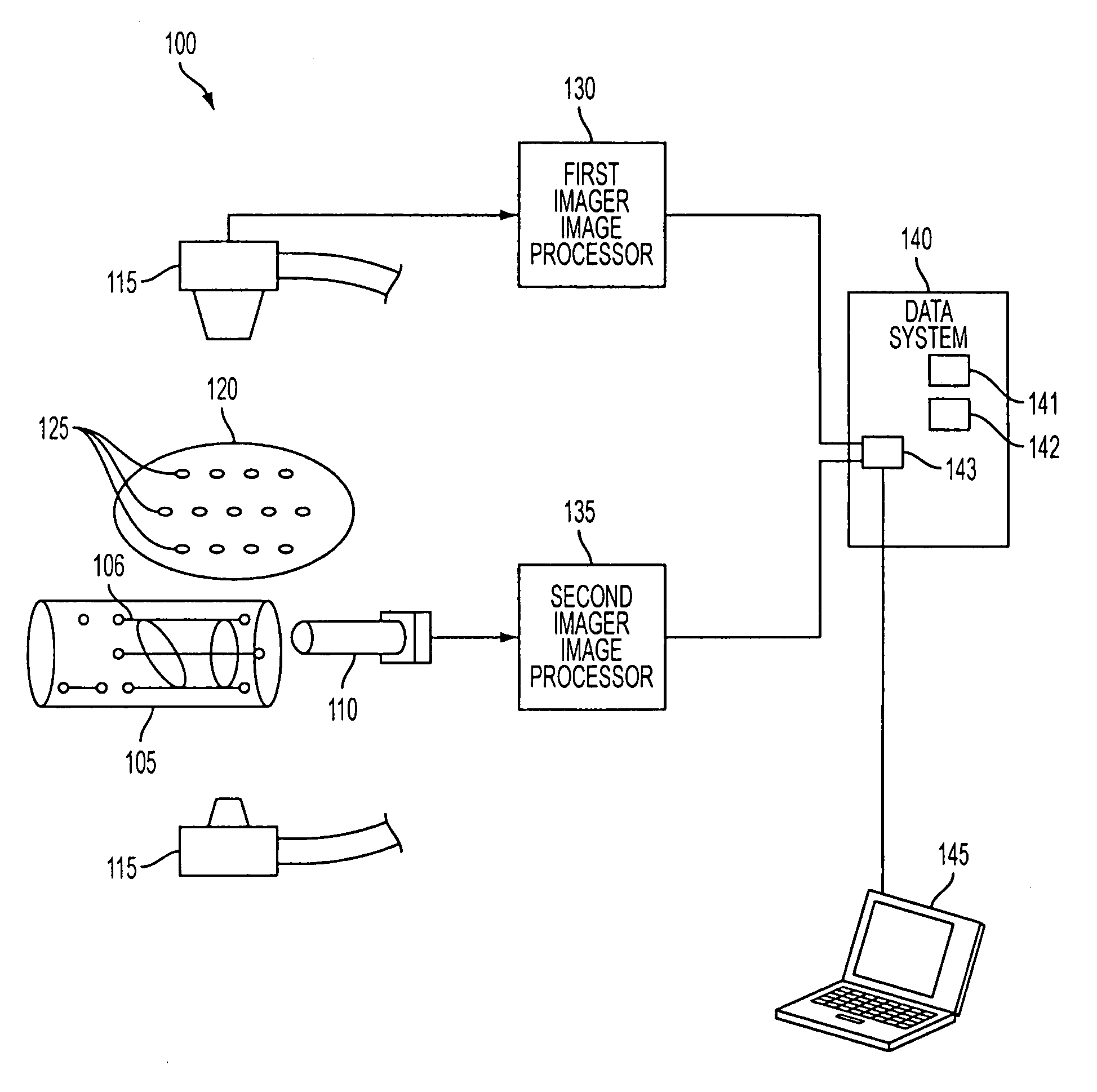
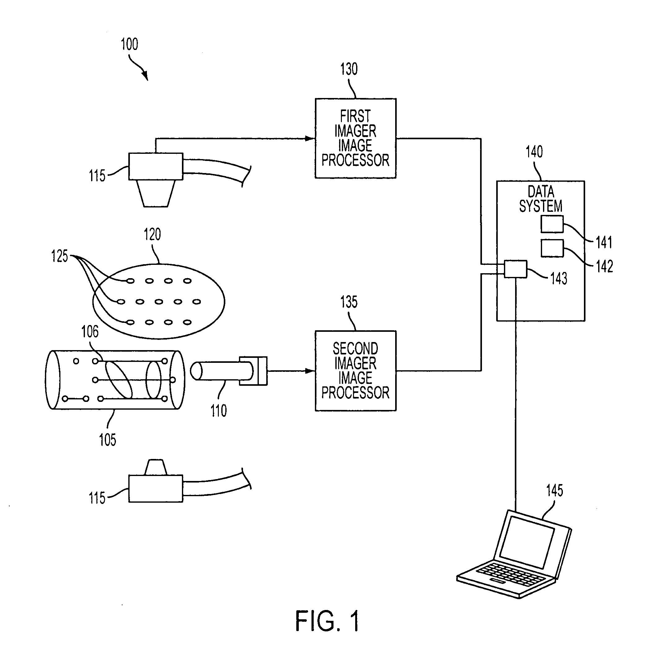
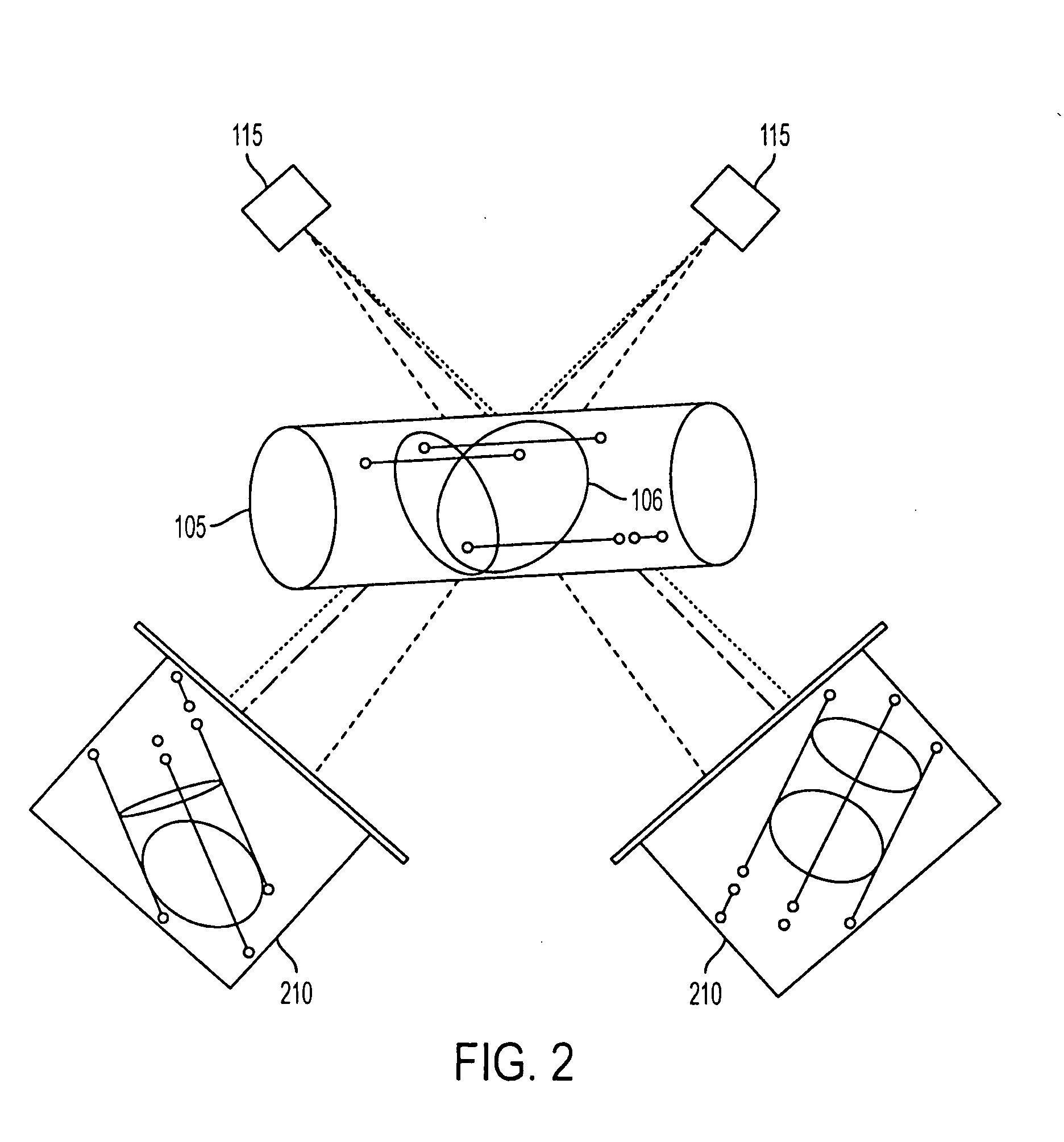
[0048]The invention provides registration of two medical imaging modalities by registering multiple images from a first imaging system, preferably at different angles, to a three-dimensional coordinate frame defined by the field of view of a second medical imaging system. The two medical imaging modalities preferably complement each other such that one imaging modality provides better quality images of certain features than the other, whereas the second imaging modality provides better quality images of other features. Registering the images from the two imaging modalities is done by determining a pose estimation, wherein the pose estimation is a six degree of freedom (six-DOF) coordinate transformation. The six-DOF transformation may be represented as a transformation matrix, or some other mathematical representation, from the coordinate frame of a fiducial, such as a fiducial device, to the coordinate frame of the field of view of the first imaging modality. The fiducial device is...


PUM


Abstract

Disclosed is a system and method for registering images from two medical imaging modalities in a six-degree-of-freedom coordinate space, for the purpose of providing real time registered imagery for optimization of medical procedures. The system and method uses fiducial that is visible to the first imager and in a fixed position and orientation relative to the second imager. The first imager may be an X-ray device such as a C-arm fluoroscope, and the second imager may be a Transrectal Ultrasound (TRUS) device.

Description

[0001]This application claims the benefit of U.S. Provisional Patent Application No. 60 / 488,965, filed on Jul. 21, 2003, and U.S. Non-provisional application Ser. No. 10 / 895,398, filed on Jul. 21, 2004, both of which are hereby incorporated by reference for all purposes as if fully set forth herein.[0002]Research and development efforts associated with the subject matter of this patent application was supported by the National Science Foundation under Grant No. ERC 9731478, and by the National Institutes of Health under Grant No. 1R43CA099374-01.BACKGROUND OF THE INVENTION[0003]1. Field of the Invention[0004]The present invention relates to the registration of multiple medical imaging modalities in support of real time optimization of medical procedures. In particular, the invention relates to the registration of ultrasound and C-arm fluoroscopy imagery for these purposes.[0005]Adenocarcinoma of the prostate is the most commonly diagnosed cancer in the U.S. male population. During t...

Claims


Application Information

Patent Timeline
Patent Type & Authority Applications(United States)
IPC IPC(8): A61B5/00A61BA61B5/05A61B6/00A61B8/00A61B8/12A61B17/00A61N5/00G21K4/00
CPCA61B6/504A61B8/12A61B6/583A61B6/5247A61B6/4417A61B2090/392A61B90/39A61B2090/3925A61B2090/3937A61B2090/3954A61B2090/3966A61N5/1014
Inventor FICHTINGER, GABORJAIN, AMEETMUSTUFA, TABISHWYROBEK, KEENANCHIRIKJIAN, GREGZHOU, YUBURDETTE, EVERETTE C.
Owner THE JOHN HOPKINS UNIV SCHOOL OF MEDICINE
Why Eureka
Try Eureka