Preoperative Surgical Simulation

a preoperative and surgical simulation technology, applied in the field of preoperative surgical simulation, can solve the problems of large amount of data, prohibitive cost of routine data analysis, and delicate and coordinated hand movements of procedures,

Active Publication Date: 2009-01-15
SIMBIONIX
View PDF106 Cites 107 Cited by
  • Summary
  • Abstract
  • Description
  • Claims
  • Application Information

AI Technical Summary

Problems solved by technology

These modalities provide noninvasive methods for studying internal organs in vivo, but the amount of data is relatively large and when presented as two dimensional (2D) images, it generally requires an anatomist/radiology specialist for interpretation.
Unfortunately, the cost incurred in manual interpretation of this data is prohibitive for routine data analysis.
However, these procedures involve delicate and coordinated hand movements, spatially unrelated to the view on a video monitor of the remotely controlled surgical instruments.
Depth perception is lacking on the flat video display and therefore it is not an easy task to learn to control the tools through the spati

Method used


Image

  • Preoperative Surgical Simulation
  • Preoperative Surgical Simulation
  • Preoperative Surgical Simulation

Examples


Embodiment Construction

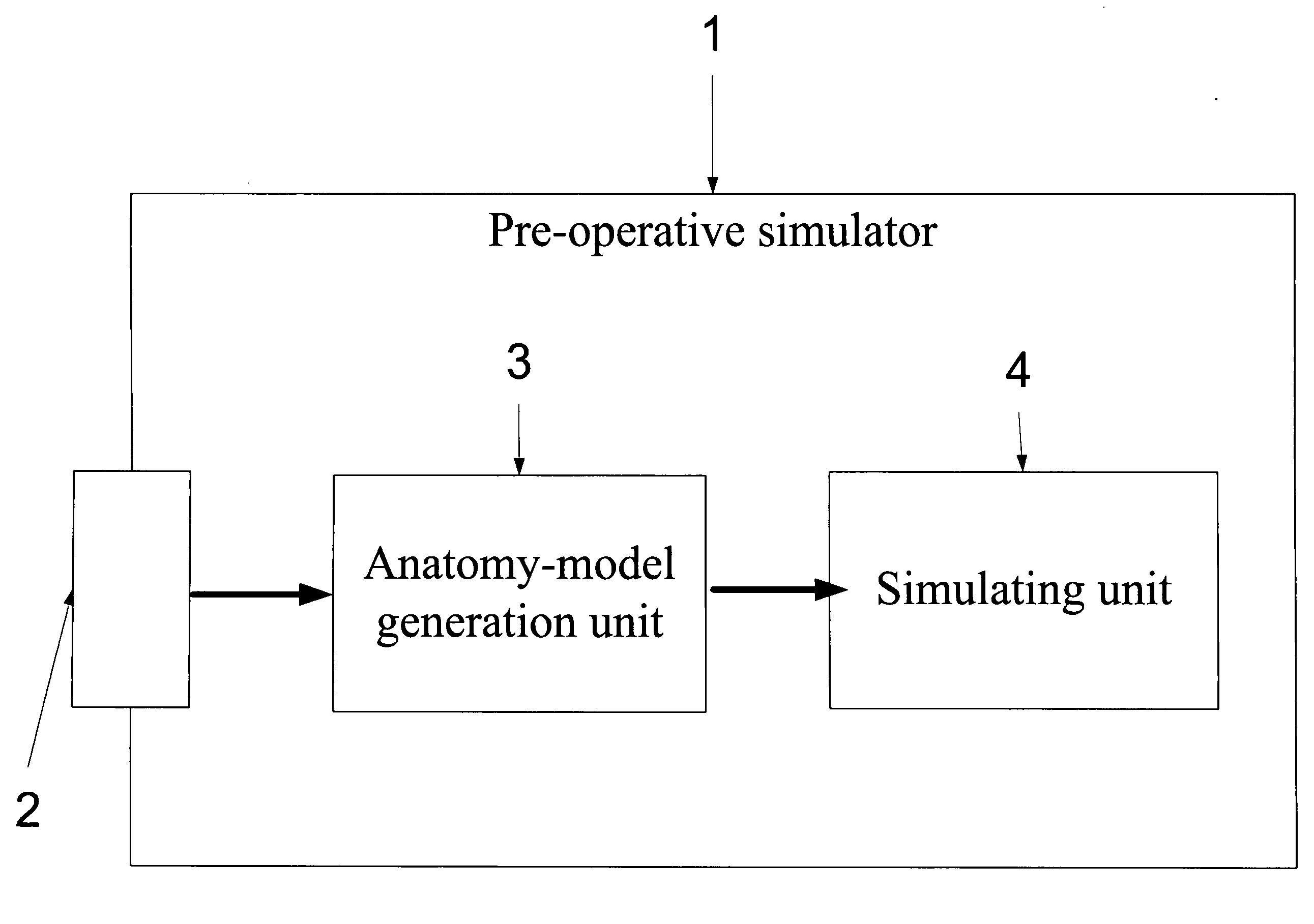
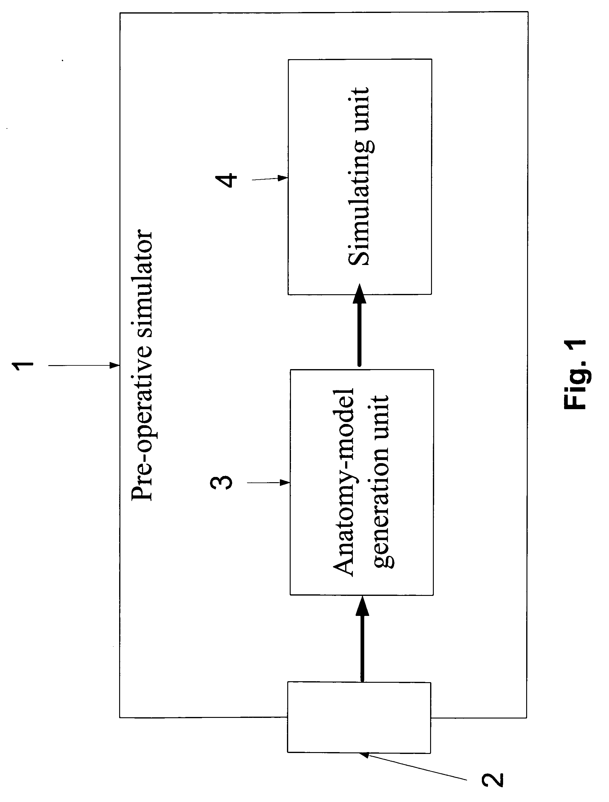
[0051]The present embodiments comprise a apparatus and a method for simulating an image-guided procedure. According to one embodiment of the present invention, the apparatus and the method allow a physician to set a pre-operative simulation of an image-guided procedure. The pre-operative simulation simulates an image-guided procedure that is about to be performed on a certain patient. In order to allow such a case-specific simulation, a 3D medical image that depicts an anatomical region of a certain patient who is about to be operated on is acquired and 3D anatomical models are generated based thereupon. Preferably, the 3D anatomical model defines the boundaries of a certain anatomy or an organ such as a vascular tract. During the pre-operative simulation, the 3D anatomical models are used for simulating an image-guided procedure on that region.

[0052]The principles and operation of an apparatus and method according to the present invention may be better understood with reference to ...


PUM


Abstract

An apparatus for simulating an image-guided procedure. The system comprises an input for receiving a three-dimensional (3D) medical image depicting an organ of a patient, a model generation unit for generating a 3D anatomical model of the organ according to the 3D medical image, and a simulating unit for simulating a planned image-guided procedure on the patient, according to the 3D anatomical model.

Description

FIELD AND BACKGROUND OF THE INVENTION[0001]The present invention relates to an apparatus and a method for performing a simulated image-guided medical procedure and, more particularly, but not exclusively to performing a simulated image-guided procedure according to a three-dimensional (3D) model of an organ that is based on a 3D medical image.[0002]Medical imaging is generally recognized as important for diagnosis and patient care with the goal of improving treatment outcomes. In recent years, medical imaging has experienced an explosive growth due to advances in imaging modalities such as x-rays, computed tomography (CT), magnetic resonance imaging (MRI) and ultrasound. These modalities provide noninvasive methods for studying internal organs in vivo, but the amount of data is relatively large and when presented as two dimensional (2D) images, it generally requires an anatomist / radiology specialist for interpretation. Unfortunately, the cost incurred in manual interpretation of thi...

Claims


Application Information

Patent Timeline
IPC IPC(8): G06G7/60
CPCG06T19/00G06T17/00G06T2210/41A61B19/50G06F19/3437G06F19/3481G06F19/321A61B34/10G16H50/50G16H30/20G16H30/40
Inventor BRONSTEIN, RANFISHER, NIVSHILON, OFEK
Owner SIMBIONIX
Why Eureka
Try Eureka