

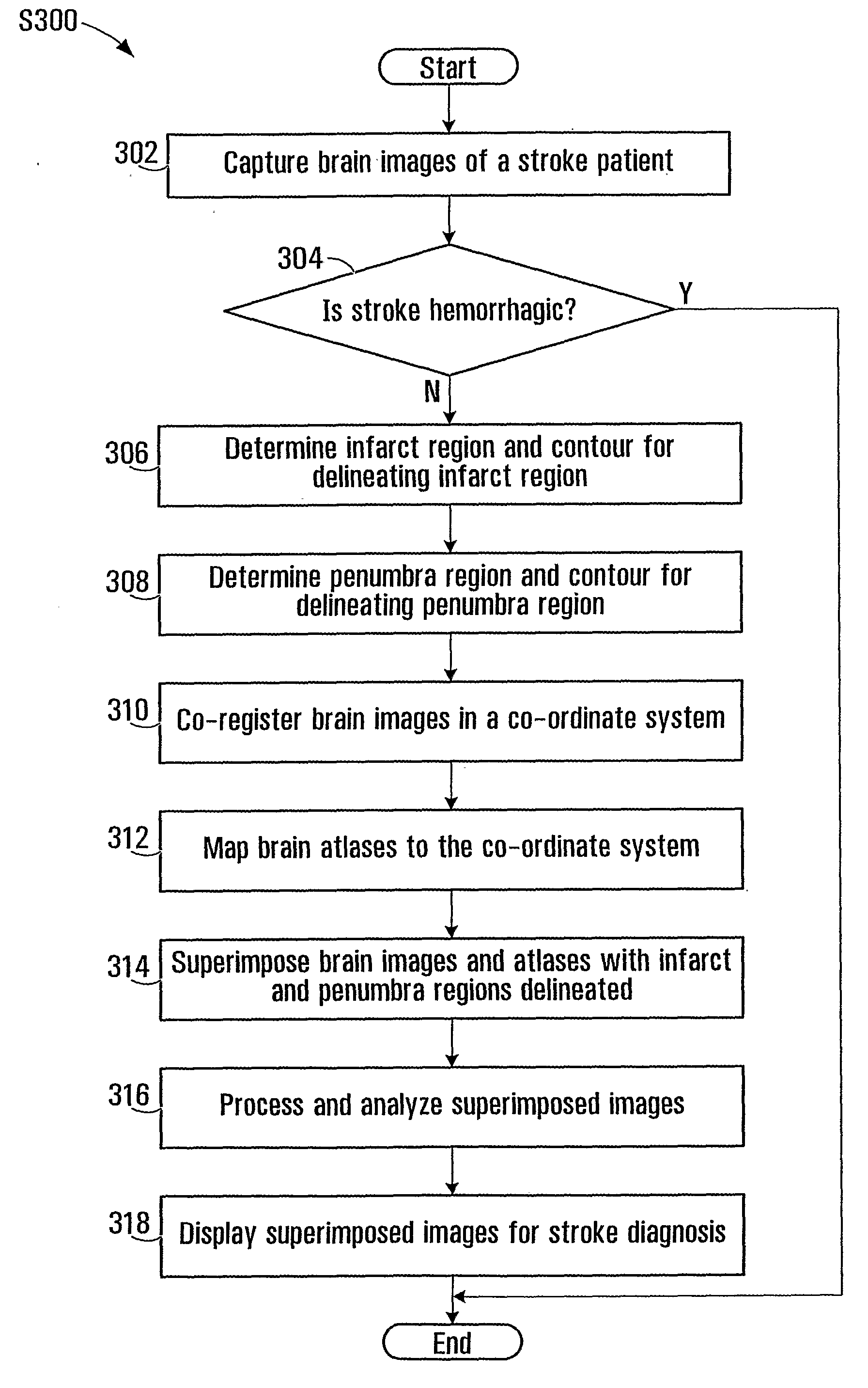
Superimposing brain atlas images and brain images with delineation of infarct and penumbra for stroke diagnosis
a brain atlas and brain image technology, applied in the field of presentation, processing and analysis of brain images for stroke diagnosis, can solve the problems of inconvenient stroke analysis, inability and death, inconsistent and inaccurate, etc., and achieve the effect of convenient stroke analysis and convenient identification and visualization
- Summary
- Abstract
- Description
- Claims
- Application Information
AI Technical Summary
Benefits of technology
Problems solved by technology
Method used
Image


Examples
Embodiment Construction
[0028]As used herein, an “image” may include a brain image, a map image, an atlas image, or a superposition of these images. The term “image” may refer to a displayed image or stored image data for displaying the image. The image data may be stored in digital form on a computer readable medium, such as in an electronic image file. An image may be two-dimensional (2D) or three-dimensional (3D). In some applications, 3D images may provide better views and additional information may be obtained from 3D images. In some applications, 2D images may be easier to process and display, and thus can be processed quicker with less consumption of computing power.
[0029]A “brain image” refers to an image of a brain that is captured using an imaging device. A brain image may be a CT image, an MR image or another scanned image, or the like.
[0030]Each of the terms “map” and “atlas” is used herein in a broad sense, but generally refers to a map image or an atlas image, respectively. A brain map may re...
PUM
 Abstract
Description
Claims
Application Information