Endoscope insertion support system and endoscope insertion support method

a technology of endoscope and support system, which is applied in the field of endoscope insertion support system and endoscope insertion support method, can solve the problems of difficulty in enabling and achieve the effect of facilitating the distal end portion of the endoscope to reach the target region

Active Publication Date: 2009-02-26
OLYMPUS CORP
View PDF2 Cites 53 Cited by
  • Summary
  • Abstract
  • Description
  • Claims
  • Application Information

AI Technical Summary

Benefits of technology

[0035]Such the endoscope insertion support system has an effect of enabling appropriate endoscopic diagnosis and treatment to be accomplished by presenting tubular information on any one of bronchus, blood vessel, large intestine, small intestine, pancreas duct, bile duct or lymphatic vessel.
[0036]The present invention has an effect of enabling a VOI (Volume of Interest) to be effectively set in a tubular organ having a strictured part and tubular path area information on a tubular organ to be correctly extracted.

Problems solved by technology

In a tubular path such as a bronchus having multi-step branches in the body, when the location of the disordered part is near the periphery of a branch, it is difficult to enable the distal end portion of the endoscope to reach the target region in a short period of time.

Method used


Image

  • Endoscope insertion support system and endoscope insertion support method
  • Endoscope insertion support system and endoscope insertion support method
  • Endoscope insertion support system and endoscope insertion support method

Examples


embodiment 1

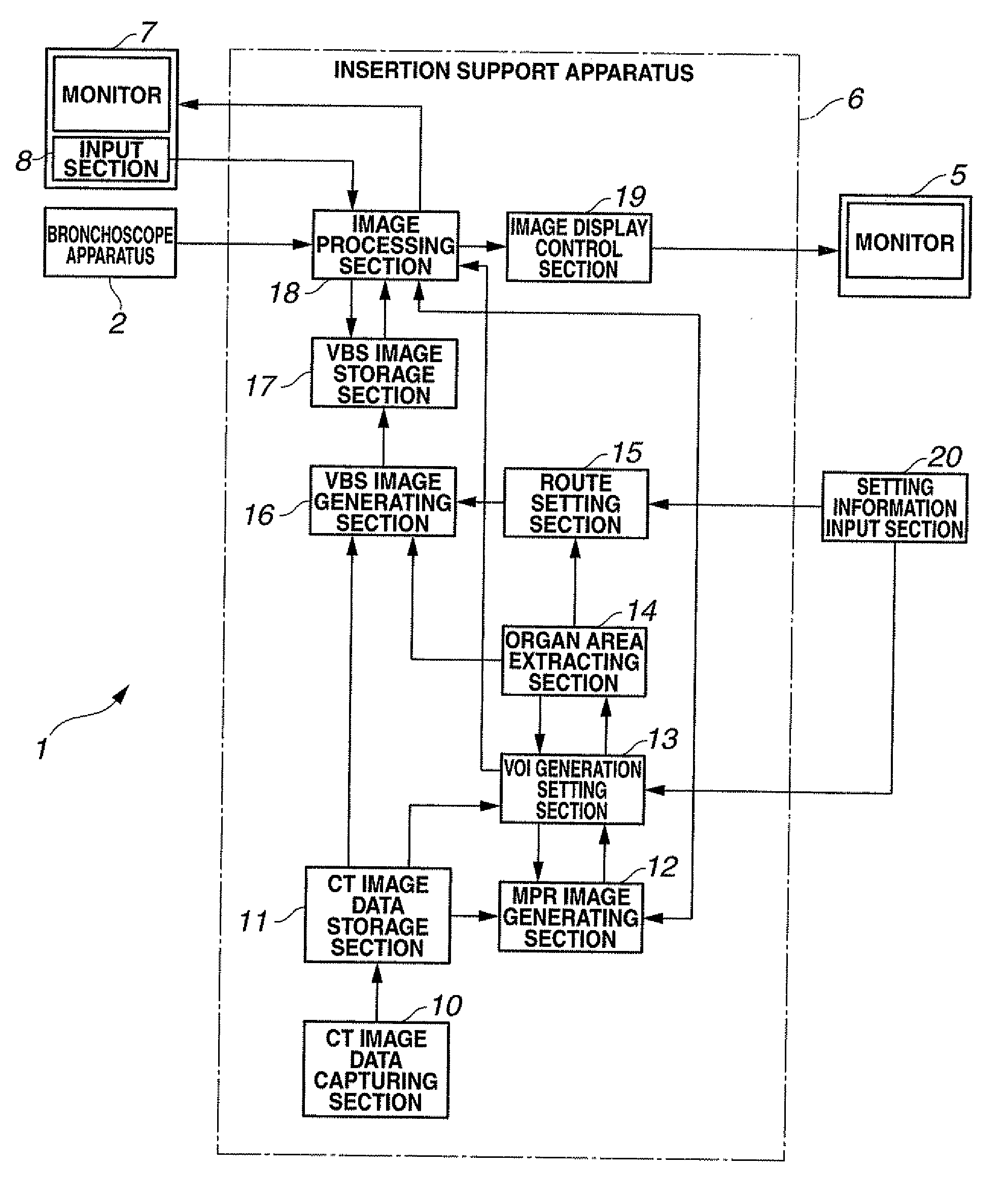
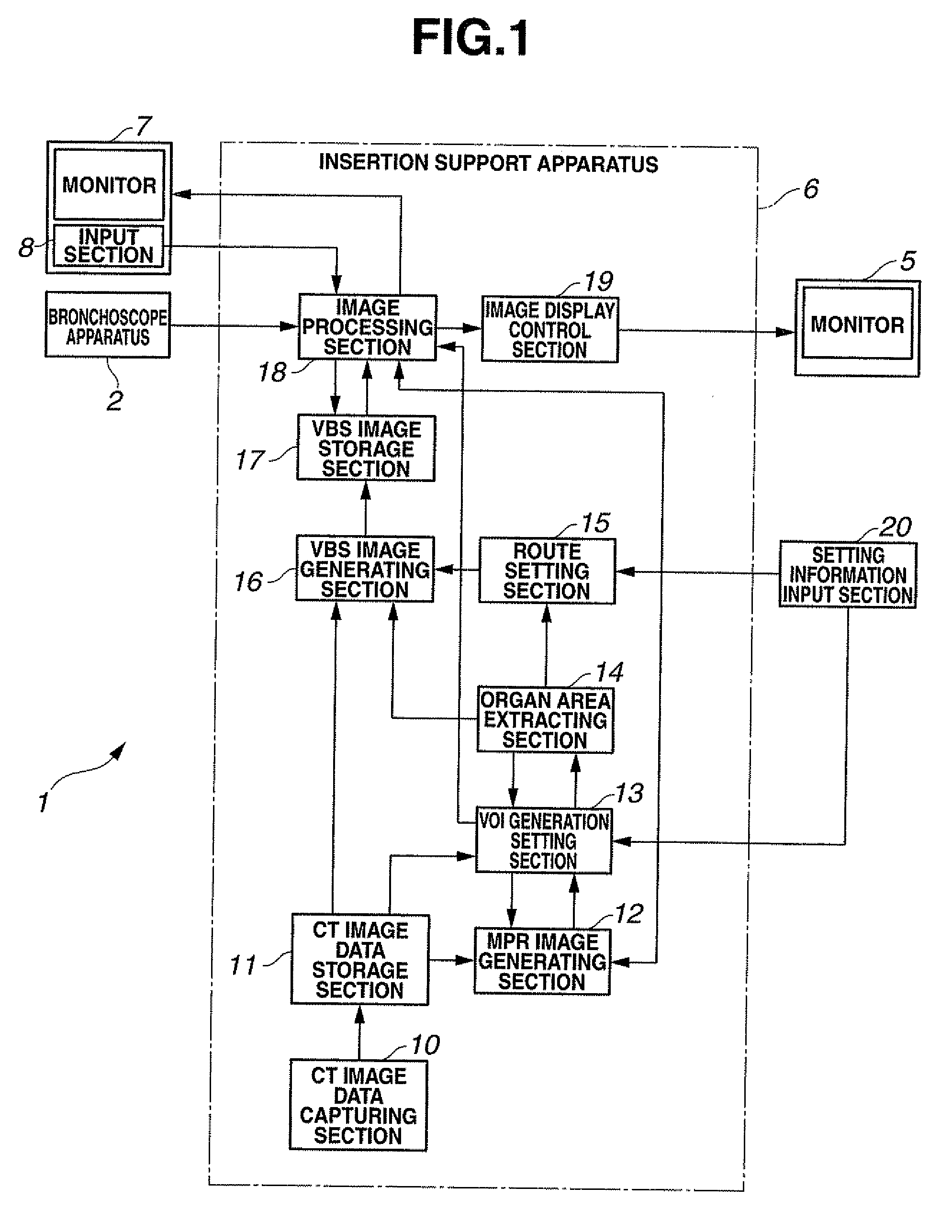
[0098]As shown in FIG. 1, a bronchoscope insertion support system 1 as the endoscope insertion support system of the present embodiment comprises a bronchoscope apparatus 2 and an endoscope insertion support apparatus 6 as an apparatus for extracting the bronchial shape.

[0099]The endoscope insertion support apparatus 6 extracts the bronchial shape on the basis of CT image data, and generates a virtual endoscopic image of the inside of the bronchus (hereinafter referred to as VBS image). Then, an endoscopic image of the inside of the bronchus obtained by the bronchoscope apparatus 2 (hereinafter referred to as live image) and the VBS image are synthesized to display a synthetic image on a monitor 5 to support insertion of the bronchoscope apparatus 2 into the bronchus.

[0100]Further the bronchoscope apparatus 2, though not shown comprises a bronchoscope having an image pickup section, a light source which supplies illuminating light to the bronchoscope, a camera control unit which sub...


PUM


Abstract

According to the invention, a VOI generation setting section disposed in an endoscope insertion support apparatus comprises a VOI setting function section, a VOI extending function section, a VOI direction determining function section, a VOI branch determining function section, a VOI resetting function section, a VOI information storage function section, a VOI size determining function section and a VOI branch extracting function section. This configuration enables a VOI (Volume of Interest) to be effectively set in a tubular organ having a strictured part and tubular path area information on the tubular organ to be extracted.

Description

CROSS REFERENCE TO RELATED APPLICATION[0001]This application is a continuation application of PCT / JP2007 / 059208 filed on Apr. 27, 2007 and claims benefit of Japanese Application No. 2006-128682 filed in Japan on May 2, 2006, the entire contents of which are incorporated herein by this reference.BACKGROUND OF THE INVENTION[0002]1. Field of the Invention[0003]The present invention relates to an endoscope insertion support system and an endoscope insertion support method for supporting the insertion of an endoscope into the tubular path of a tubular organ.[0004]2. Description of the Related Art[0005]In recent years, diagnosing by photographic images has come into extensive practice; for instance, three-dimensional image data within a subject are obtained by picking up tomograms of the subject with an X-ray CT (computed tomography) apparatus or the like, and the intended diagnosis is performed by using the three-dimensional image data.[0006]With a CT apparatus, by consecutively shifting...

Claims


Application Information

Patent Timeline
Patent Type & Authority Applications(United States)
IPC IPC(8): A61B1/00
CPCA61B1/00009A61B1/04A61B5/418A61B5/103A61B1/2676
Inventor MORI, KENSAKUKITASAKA, TAKAYUKIAKIMOTO, SHUNYAEBE, KOUHEIWADA, TAKESHI
Owner OLYMPUS CORP
Why Eureka
Try Eureka