Diagnostic imaging device having protective facade and method of cleaning and disinfecting same

a diagnostic imaging and protective technology, applied in diagnostics, medical science, applications, etc., can solve the problems of screen surface itself, damage to display screen or circuitry inside the device, increase the risk of contamination by infectious diseases and/or bacteria in the medical environment,

Active Publication Date: 2009-02-26
GENERAL ELECTRIC CO
View PDF13 Cites 9 Cited by
  • Summary
  • Abstract
  • Description
  • Claims
  • Application Information

AI Technical Summary

Problems solved by technology

The display screen surface itself may be damaged by repeated application of harsh chemicals and any fluid material that comes in contact with the display screen may seep into corners and edges of the frame, thereby potentially damaging the display screen or circuitry inside the device.
With known imaging devices using multiple components, there are part lines or seams where components join together, further increasing the risk of contamination by infectious diseases and / or bacteria in a medical environment in which a diagnostic imaging device may be employed.
A similar risk of contamination is posed around key pads, mechanical buttons, trackballs, touch pads, etc., that are part of the diagnostic imaging device.
However, there is a risk that the equipment may not be totally cleaned because small splatters of blood and other bodily fluids go unseen.
In some cases, ultrasound scanners are considered too contaminated to place close to a patient during an operating room procedure.
However, these drapes or covers tend to interfere with the visibility of images and the operation of the imaging devices and may not always be completely effective in eliminating contamination.
However, the user then may have to twist and strain just to see an image and an additional person may be required to operate the imaging device.

Method used


Image

  • Diagnostic imaging device having protective facade and method of cleaning and disinfecting  same
  • Diagnostic imaging device having protective facade and method of cleaning and disinfecting  same
  • Diagnostic imaging device having protective facade and method of cleaning and disinfecting  same

Examples


Embodiment Construction

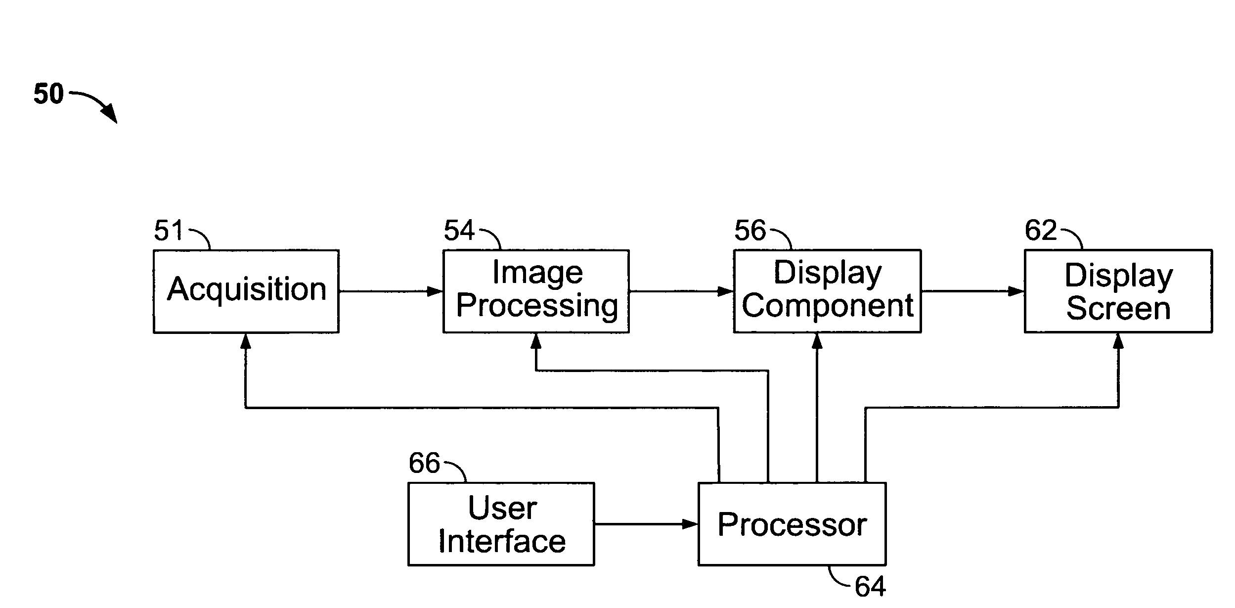
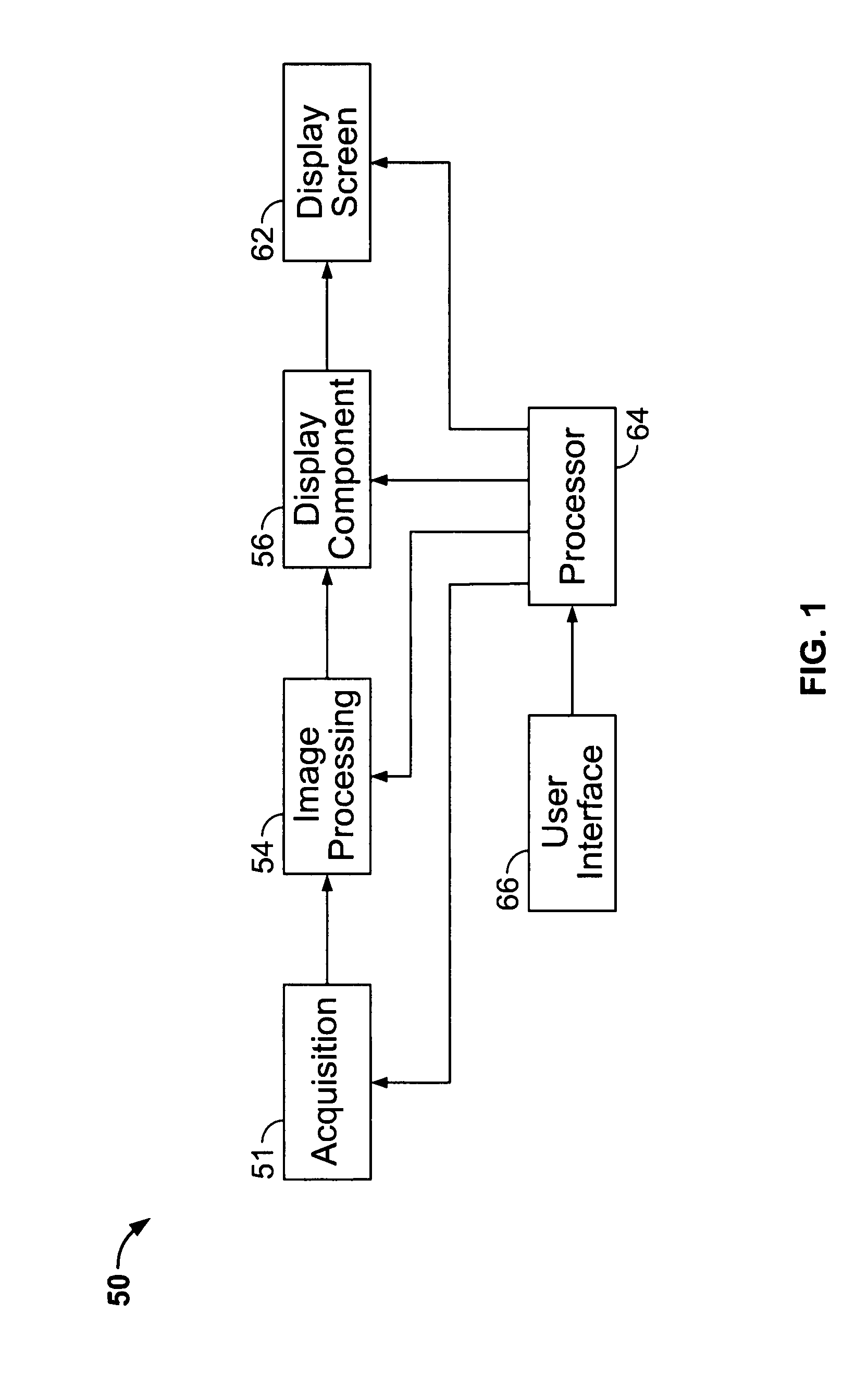
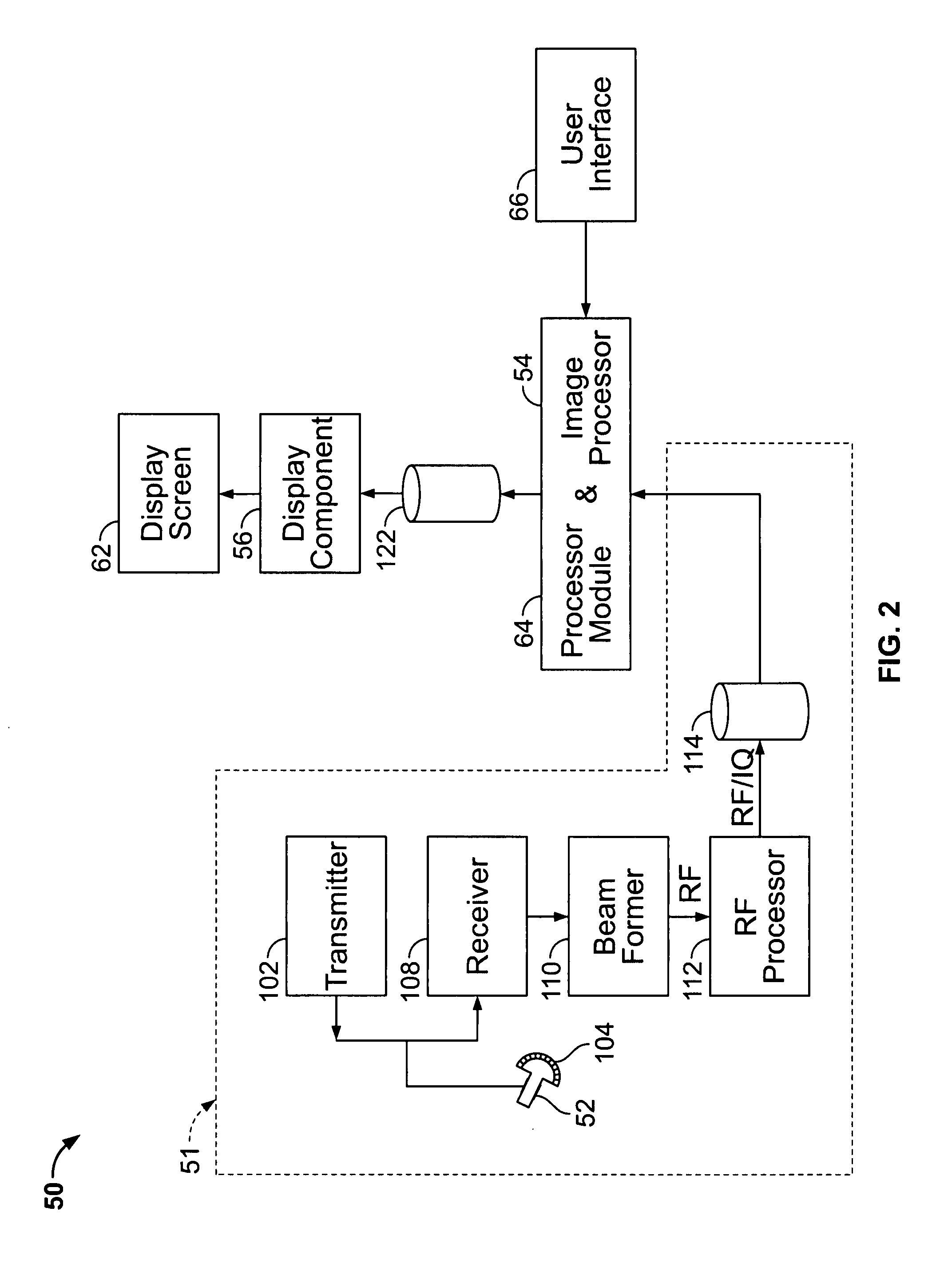
[0018]The foregoing summary, as well as the following detailed description of certain embodiments of the present invention, will be better understood when read in conjunction with the appended drawings. To the extent that the figures illustrate diagrams of the functional blocks of various embodiments, the functional blocks are not necessarily indicative of the division between hardware circuitry. Thus, for example, one or more of the functional blocks (e.g., processors or memories) may be implemented in a single piece of hardware (e.g., a general purpose signal processor or a block of random access memory, hard disk, or the like). Similarly, the programs may be stand alone programs, may be incorporated as subroutines in an operating system, may be functions in an installed software package, and the like. It should be understood that the various embodiments are not limited to the arrangements and instrumentality shown in the drawings.

[0019]As used herein, an element or step recited i...


PUM


Abstract

A diagnostic imaging device is provided that includes a display panel, a set of switches including at least one of a proximity switch and a capacitive switch, and a one-piece non-flexible facade covering the display panel and the set of switches. The facade is transparent over at least a portion of the display panel and configured such that the set of switches are operable by a user. A method for cleaning the imaging system includes operating the diagnostic imaging device, including the set of switches, by touching the one-piece non-flexible facade with human fingers or a stylus over selected switches, and, after said operating the diagnostic imaging device, disinfecting the diagnostic imaging device by cleaning the one-piece non-flexible facade with quaternary ammonium or isopropyl alcohol, or both.

Description

BACKGROUND OF THE INVENTION[0001]This invention relates generally to diagnostic imaging devices, and more particularly, to devices and apparatus that facilitate the cleaning and disinfecting of diagnostic imaging devices.[0002]Diagnostic imaging devices such as ultrasound imaging devices require a user interface to control scanning operation and a display screen to view images being scanned. Usually, these devices have a separate console and display screen, but some devices include a box or tablet shaped scanner, with buttons adjacent the display screen. In either embodiment, there are physically separate components joined together to form the device.[0003]In the case of an ultrasound imaging device, a display screen is used to view images produced by a probe and processed on electronic circuit boards of the device. In newer systems, the screen is often a flat panel framed in plastic without any other protection against chemicals or fluid splatter. The display screen surface itself ...

Claims


Application Information

Patent Timeline
Patent Type & Authority Applications(United States)
IPC IPC(8): A61B8/00
CPCA61B5/055A61B6/00A61B8/00A61B8/483A61B8/467A61B2562/247A61B6/4423A61B8/462A61B8/461A61B5/748
Inventor STONEFIELD, ANDREW DAVIDLANPING, LIURONCAGLIONI, AURELIE
Owner GENERAL ELECTRIC CO
Why Eureka
Try Eureka