System for 3-Dimensional Medical Image Data Acquisition

a technology of image data acquisition and x-ray imaging, applied in tomography, instruments, applications, etc., can solve the problems of adding noise to the x-ray image data, obscuring the surrounding anatomy, and beam hardening artifacts, etc., to achieve the effect of minimizing the introduction of metal artifacts

Active Publication Date: 2009-04-16
SIEMENS HEALTHCARE GMBH
View PDF11 Cites 18 Cited by
  • Summary
  • Abstract
  • Description
  • Claims
  • Application Information

AI Technical Summary

Benefits of technology

[0011]A system provides medical image processing and 3-dimensional image construction of an examination subject. An examination subject is placed on a support surface such as a bed. An x-ray imaging system acquires image data. The x-ray imaging system has a variable 3-dimensional acquisition axis relative to the examination subject on the support surface. A control processor, electrically coupled to the x-ray imaging system, enables a user to set a non-planar image data acquisition path for

Problems solved by technology

When the x-rays are deflected and strike the radiation detector, added noise is introduced into the x-ray image data.
While some absorption and scatter is expected, the increased or complete absorption due to the presence of highly dense objects in the subject being imaged will result in artifact(s) obscuring the surrounding anatomy that is of interest.
Beam hardening artifacts arise when x-rays are completely blocked from reaching the radiation detector due to the presence of highly dense objects.
The presence of beam hardening artifacts produces streaking in the x-ray images.
When the x-rays strike the radiation detector, artifacts will result which effect image quality of the x-ray images.
If the region of interest in the examination subject happens to lie adjacent to the radio-opaque object and in a direction perpendicular to the 3-dimensional acquisition axis, the metal artifacts in the image significantly degrade and even preclude an accurate diagnosis of the region of interest

Method used


Image

  • System for 3-Dimensional Medical Image Data Acquisition
  • System for 3-Dimensional Medical Image Data Acquisition
  • System for 3-Dimensional Medical Image Data Acquisition

Examples


Embodiment Construction

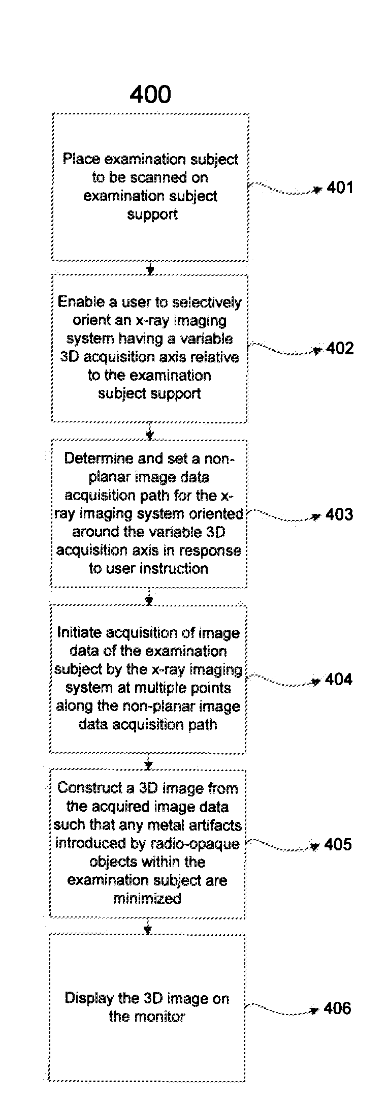
[0032]Known x-ray imaging systems for acquiring images of an examination subject containing a radio-opaque object produce poor quality images because of the metal artifacts introduced in the image. Radio-opaque objects are objects that do not allow x-rays or other types of radiation penetrate. Since radio-opaque objects block radiation, it is difficult to x-ray an examination subject that contains a radio-opaque object inside his / her body. Additionally, these radio-opaque objects create metal artifacts caused by beam hardening or scatter artifacts that can degrade the quality of an x-ray image. Known systems shift the examination subject and / or the examination subject support to shift the primary effect of metal artifacts away from an area of interest to other areas in the image, improving image quality for a specific area. However, in these systems, the image data is acquired along planar paths. Therefore, the metal artifacts introduced in the 3-dimensional image by radio-opaque ob...


PUM


Abstract

A method and system provides medical image processing and 3-dimensional image construction of an examination subject. A user is enabled to selectively orient an x-ray imaging system having a variable 3-dimensional acquisition axis relative to an examination subject support for holding an examination subject. A non-planar image data acquisition path is set for the x-ray imaging system oriented around the variable 3-dimensional acquisition axis in response to user instruction. Acquisition of image data of the examination subject is initiated by the x-ray imaging system at a plurality of points along the non-planar image data acquisition path. A 3-dimensional image is constructed from the acquired image data such that any metal artifacts introduced by radio-opaque objects within the examination subject are minimized. The 3-dimensional image is displayed.

Description

[0001]This is a non-provisional application of U.S. Provisional Application Ser. No. 60 / 979,536 filed Oct. 12, 2007, by John Rauch.FIELD OF THE INVENTION[0002]This invention concerns non-planar imaging data acquisition in an x-ray imaging system that improves image quality by diffusing beam hardening and scatter artifacts that arise from the presence of highly dense objects (e.g. metal) adjacent to or within less dense material.BACKGROUND OF THE INVENTION[0003]Conventional C-arm X-ray imaging systems acquire images along planar paths having a 3-dimensional acquisition axis. The 3-dimensional acquisition axis is the axis about which the x-ray source and radiation detector, held in fixed geometry by the C-arm, rotate. An examination subject being x-rayed by a conventional imaging system may have highly dense, radio-opaque objects (e.g. dental fillings, aneurysm clips or stents, screws, plates, etc.) disposed at various points through his / her body. These radio-opaque objects may includ...

Claims


Application Information

Patent Timeline
IPC IPC(8): H05G1/00
CPCA61B6/032A61B6/466Y10S378/901A61B6/4458A61B6/4464A61B6/5252A61B6/5258A61B6/03
Inventor RAUCH, JOHN CHRISTOPHER
Owner SIEMENS HEALTHCARE GMBH
Why Eureka
Try Eureka