Method for reducing metal artifact in computed tomography

a computed tomography and metal artifact technology, applied in tomography, image enhancement, instruments, etc., can solve the problems of deteriorating the quality of a reconstructed ct image, severe photon starvation of the measured attenuation of the metal given x-ray beam on the detector, and inaccurate after-image projection data, etc., to achieve the effect of minimizing metal artifacts

Active Publication Date: 2016-12-06
INST FOR BASIC SCI
View PDF4 Cites 0 Cited by
  • Summary
  • Abstract
  • Description
  • Claims
  • Application Information

AI Technical Summary

Benefits of technology

[0012]The present invention has been conceived in consideration of the problems as set forth above and is aimed at providing a method for reducing metal artifacts in computed tomography (CT), which enables an image closest to an original image to be obtained by minimizing metal artifacts in CT through introduction of a novel metal artifact reduction (MAR) technique referred to as an algebraic correction technique (ACT) using an intermediate image of an attenuation coefficient of an outside of a metal area.
[0024]According to the present invention, through introduction of the novel metal artifact reduction (MAR) technique referred to as the algebraic correction technique (ACT) using the intermediate image of the attenuation coefficient of the outside of the metal area, the image closest to the original image can be obtained by minimizing metal artifacts in CT.

Problems solved by technology

A metal object transplanted into the human body can cause serious side effects in a CT image and can deteriorate quality of a reconstructed CT image.
Generally, metals strongly attenuate an X-ray beam and measured attenuation of the metal given X-ray beam on the detector suffer from severe photon starvation.
Thus, after-image projection data becomes inaccurate.
As compared with the projection completion methods, model-based iterative methods are computationally intensive and have a limitation in clinical application.
Therefore, existing filling methods can cause inaccurate information on the projections passing through the red point.
As a result, a CT image reconstructed from such a corrected sinogram by a filtered back projection (FBP) method can be less efficient in restoring details of a true image of the phantom model.
Efficiency of the projection completion method has a high dependency on accuracy of synthesized data.

Method used


Image

  • Method for reducing metal artifact in computed tomography
  • Method for reducing metal artifact in computed tomography
  • Method for reducing metal artifact in computed tomography

Examples


Embodiment Construction

[0030]Prior to a detailed description of embodiments of the present invention, it is assumed that an image processing system (for example, computer system), which can be provided with CT image data scanned in a CT scanner and can process the provided CT image data, is established to realize a method according to the present invention. In addition, such an image processing system (computer system) includes: a controller provided with a processor processing image data or information; a display configured to display data (information) input from the outside and a result processed by the controller; and a memory storing data processed by the controller and various applications necessary for operation of the image processing system (computer system).

[0031]Hereinafter, embodiments of the present invention will be described in detail with reference to the accompanying drawings.

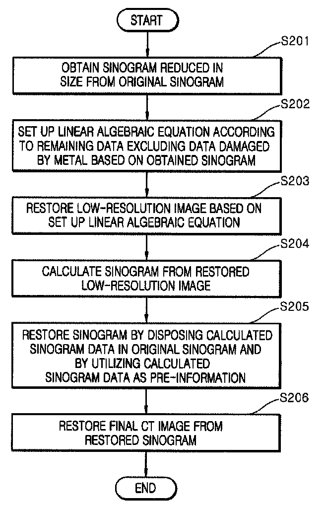
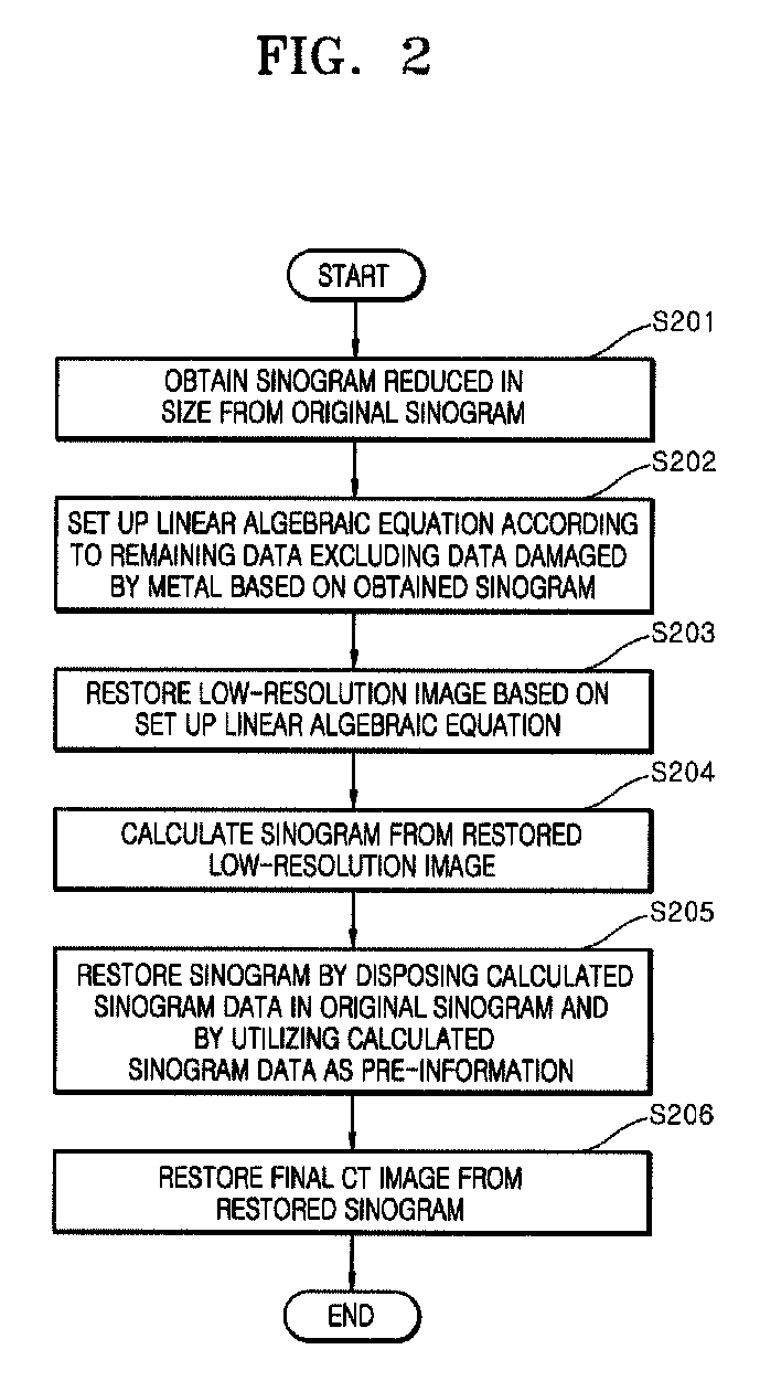
[0032]FIG. 2 is a flowchart showing processes of performing a method for reducing metal artifacts in CT according ...


PUM


Abstract

A method for reducing metal artifacts in computed tomography (CT) is disclosed. The method for reducing metal artifacts in CT includes: obtaining a sinogram reduced in size from an original sinogram; setting up a linear algebraic equation according to remaining data excluding data damaged by a metal based on the obtained sinogram; restoring a low-resolution image based on the set up linear algebraic equation; calculating a sinogram from the restored low-resolution image; restoring a sinogram by disposing the calculated sinogram data in the original sinogram and by utilizing the calculated sinogram data as pre-information; and restoring a final CT image from the restored sinogram. Through introduction of a novel metal artifact reduction (MAR) technique referred to as an algebraic correction technique (ACT) using an intermediate image of an attenuation coefficient of an outside of a metal area, an image closest to an original image can be obtained by minimizing metal artifacts in CT.

Description

CROSS-REFERENCE TO RELATED APPLICATIONS[0001]This application claims priority of Korean Patent Application No. 10-2014-0148888, filed Oct. 30, 2014, the contents of which is hereby incorporated by reference in its entirety.BACKGROUND[0002]1. Technical Field[0003]The present invention relates to a method for reducing metal artifacts in computed tomography (CT), and more particularly, to a method for reducing metal artifacts in CT, which restores an original image of an area generated by a metal in an X-ray sinogram by extracting pre-information from an image restored to a small size.[0004]2. Description of the Related Art[0005]Computed tomography (CT) provides 2-dimensional and 3-dimensional high-resolution tomographic images of a human body from a measured X-ray projection image.[0006]Although there is a controversy over dangers related to exposure to X-ray emission, CT has been widely used for the purpose of diagnosis and treatment as a powerful tool for investigating an inside of ...

Claims


Application Information

Patent Timeline
Patent Type & Authority Patents(United States)
IPC IPC(8): G06K9/00A61B6/03G06T11/00A61B6/00G06T5/00G06T7/00
CPCG06T11/003A61B6/032A61B6/5205A61B6/5258A61B6/5264G06T5/001G06T7/0012G06T2207/10081G06T2207/30004G06T2210/41G06T2211/421G06T11/005
Inventor KIM, SUNG WHANJEON, KI WANAHN, CHI YOUNG
Owner INST FOR BASIC SCI
Why Eureka
Try Eureka