Method and system of coregistrating optical coherence tomography (OCT) with other clinical tests

a co-registration and optical coherence tomography technology, applied in the field of image processing and analysis, can solve the problems of lack of focal pathology annotation, inability to show relevant pathologies in svp technique, and inability to meet the requirements of a satisfactory method or system for localizing focal in vivo pathology

Active Publication Date: 2009-09-22
DUKE UNIV
View PDF14 Cites 106 Cited by
  • Summary
  • Abstract
  • Description
  • Claims
  • Application Information

AI Technical Summary

Benefits of technology

[0005]Spectral domain optical coherence tomography (SD-OCT), also known as Fourier domain OCT, is a relatively new imaging technique that utilizes the Fourier transform function to gather depth data from the spectra of the OCT signal and thus eliminates the need to mechanically move the scanning mirror to obtain depth information as is required for commercially available time-domain systems. (See Huang D, Swanson E A, Lin C P, et al., “Optical coherence tomography,” Science 1991;254:1178-81; Puliafito C A, Hee M R, Lin C P, et al., “Imaging of macular diseases with optical coherence tomography,” Ophthalmology 1995;102:217-29; and Hee M R, Izatt J A, Swanson E A, et al., “Optical coherence tomography of the human retina,” Arch Ophthalmol 1995;113:325-32.) The SD-OCT technique significantly increases signal-to-noise ratio and increases the speed of data collection by a factor of 50 (conventional time-domain OCT functions at 400 A-scan / sec, while the SD-OCT system scans at 20,000 A-scan / sec). (See Wojtkowski M, Bajraszewski T, Gorczyńska I, et al., “Ophthalmic imaging by spectral optical coherence tomography,” Am J Ophthalmol 2004;138:412-9; Wojtkowski M, Leitgeb R, Kowalczyk A, et al., “In vivo human retinal imaging by Fourier domain optical coherence tomography,” J Biomed Opt 2002;7:457-63; and Wojtkowski M, Bajraszewski T, Targowski P, Kowalczyk A., “Real-time in vivo imaging by high-speed spectral optical coherence tomography,” Opt Lett 2003;28:1745-7.) Because of the increase in speed, a single cross-sectional scan of 1000 A-scans can be captured, processed, streamed to disk, and displayed in 60 ms (or 1 / 42 of the time required for a time-domain scan). Because of this speed, there is less eye movement during the SD-OCT scan and thus a more stable image with a significant decrease in artifact of the image caused by patient motion. Also because of this speed, a stack of 100 cross-sectional scans can be acquired in the time normally used to gather 6 low resolution cross-sectional scans of the macula on a time-domain system. The image stack across the macula can be processed to produce a three dimensional representation of structures. (See Wojtkowski M, Srinivasan V, Fujimoto J G, et al., “Three-dimensional retinal imaging with high-speed ultrahigh-resolution optical coherence tomography,” Ophthalmology 2005;112:1734-46.)
[0009]Present exemplary embodiments provide a method / system to annotate, extract or preserve different pathological conditions and / or changes that are recognized on cross-sections within a three dimensional volume so that the findings are maintained (preserved as visible) in an en face projection produced with a SVP technique. Furthermore, present exemplary embodiment(s) make it possible to coregister marked changes with other types of two dimensional en face images such as images from other ophthalmic devices (e.g., angiography device, microperimetry device, autofluorescence device, fundal photography device, etc.). The findings are maintained in an image resulting from a coregistration of the projection produced with the SVP technique and the other types of two dimensional en face images.
[0010]In more detail, present exemplary embodiments delineate, extract and preserve different pathological conditions and / or changes that are recognized on retinal cross-sections obtained from patients with retinal disease. The patients may have, for example, neovascular and non neovascular age related macular degeneration (AMD). With present exemplary embodiments, the delineated pathology (e.g., pathology delineated, via color-coded markings or sets of numbers, by a user and / or automatically by an image processing, rendering and interpolation algorithm) remains visible through the SVP and coregistration process. Thus the lateral extent and location of pathology (as well as other features of the pathology such as thickness, volume, size and / or severity) is precisely maintained relative to retinal vasculature on fundus images produced with the SVP technique.

Problems solved by technology

There currently exists no satisfactory method or system to localize focal in vivo pathology that correlates with function.
Although clinicians are able to define pathologies on retinal OCT cross-sections based on previous clinicopathologic correlation, this cross-sectional information is viewed separately and not integrated with conventional fundus imaging such as color photography and angiography.
Consequently, they lack annotation of focal pathology.
Even though some pathological structures can be observed on a two dimensional en face image produced with SVP, this technique may not show all the changes that might be relevant for the diagnosis because some information is lost.
In particular, the SVP technique may not show relevant pathologies because much information is lost in the summing of the pixels in the collapsing process.

Method used


Image

  • Method and system of coregistrating optical coherence tomography (OCT) with other clinical tests
  • Method and system of coregistrating optical coherence tomography (OCT) with other clinical tests
  • Method and system of coregistrating optical coherence tomography (OCT) with other clinical tests

Examples


Embodiment Construction

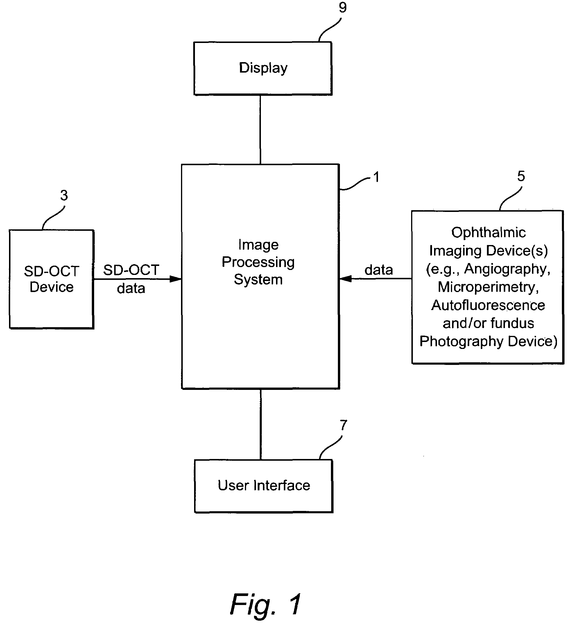
[0025]FIG. 1 illustrates an exemplary system having an image processing system 1. The image processing system 1 comprises a computer having processing components, storage memories, etc. The image processing system 1 receives imaging data from an SD-OCT scanning device 3 and from one or more ophthalmic device(s) such as an angiography, autofluorescence, fundal photography and / or microperimetry device 5. A user (e.g., ophthalmologist, radiologist, etc.) may provide input to the image processing system 1 through a user interface 7 (e.g., a keyboard and / or mouse). An output of the image processing system 1 is stored in for example a memory of the system 1 and may be displayed on display 9 or on a hard copy through operation of a printer (not shown). The processing performed by the image processing system may be partially or fully automated.

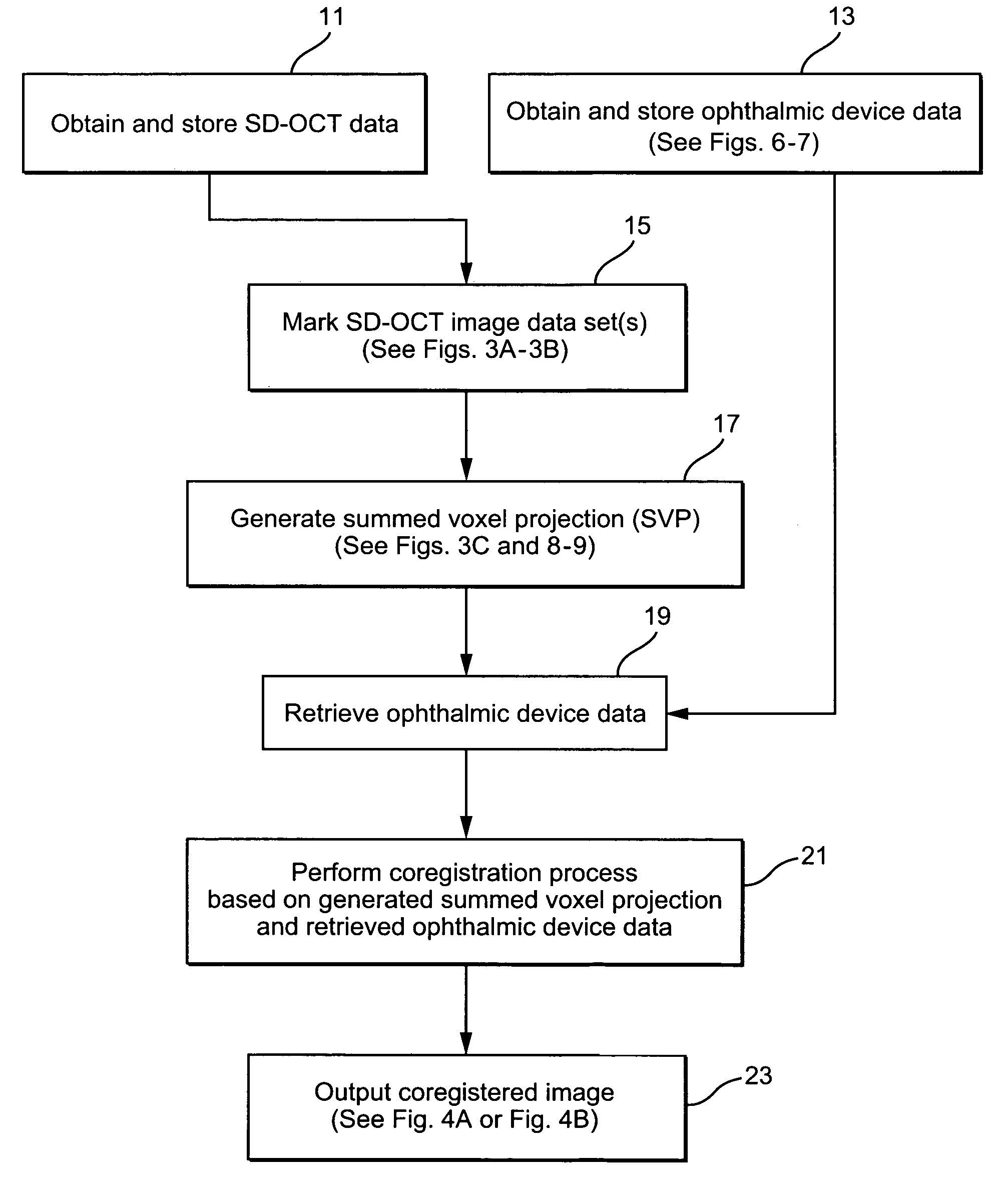
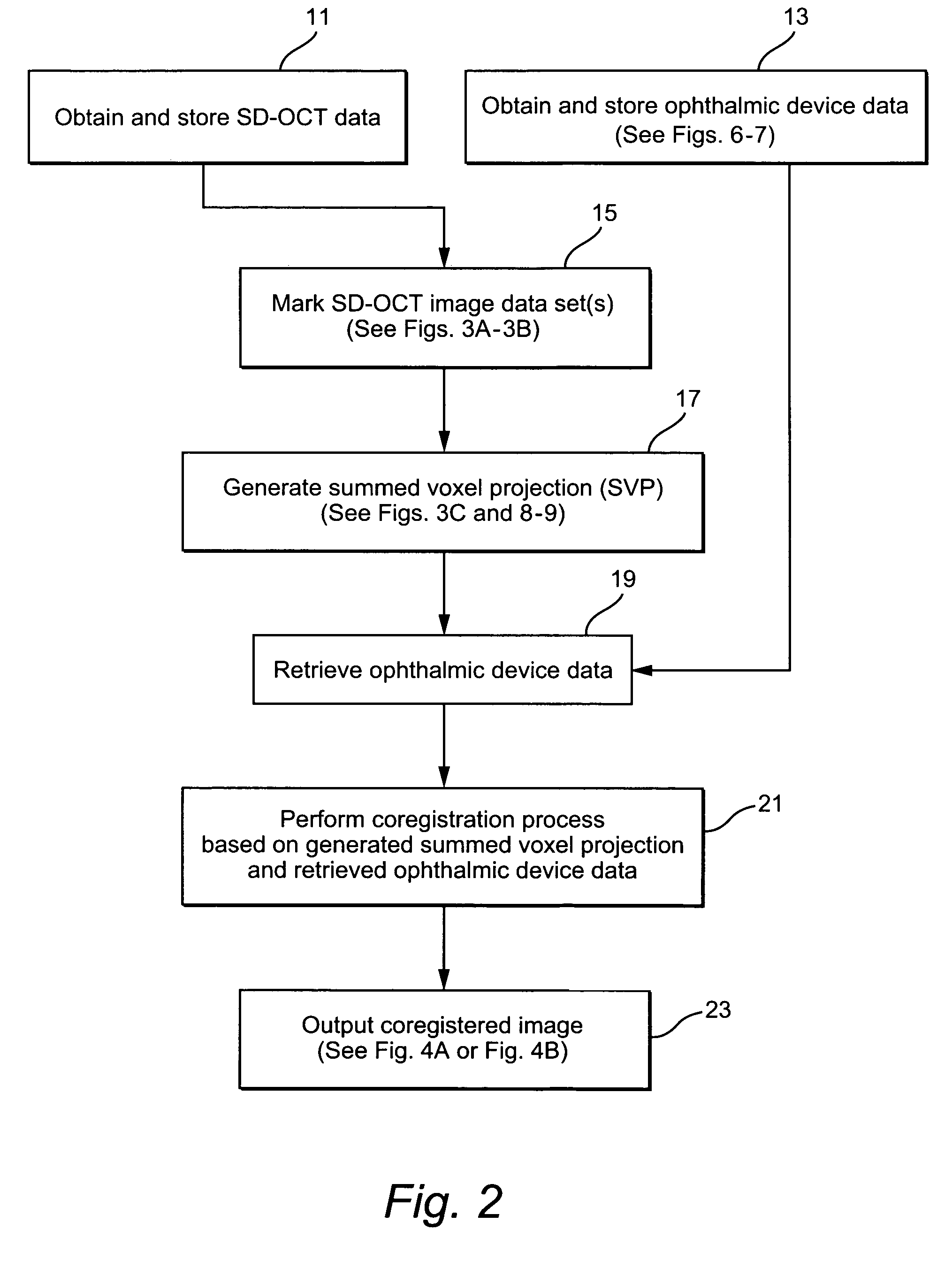
[0026]FIG. 2 illustrates an exemplary method which may be performed using the system illustrated in FIG. 1 to annotate, extract and / or preserve diffe...


PUM


Abstract

A method / system preserves annotations of different pathological conditions or changes that are recognized on cross-sections within a three dimensional volume of a patient's eye so that the annotations are maintained in a visible state in an en face projection produced with a SVP technique. it is thus possible to coregister the annotated conditions or changes with other types of two dimensional en face images such as images from other ophthalmic devices (e.g., angiography device, microperimetry device, autofluorescence device, fundal photography device.). The annotations are also maintained in a visible state in the coregistered image.

Description

CROSS REFERENCE TO RELATED APPLICATION[0001]This application claims priority from U.S. Provisional Application 60 / 737,776 filed Nov. 18, 2005, the entire content of which is incorporated herein by reference.STATEMENT OF FEDERAL SPONSORED RESEARCH[0002]This invention was made with government support under grants EB000243 and RR019769 awarded by the National Institutes of Health. The government has certain rights in the invention.TECHNICAL FIELD[0003]Exemplary embodiments of the present technology relate to a method and system of image processing and analysis. In particular, the present method / system preserves an annotation of pathology in a summed voxel projection (SVP) of a series of scans (for instance, from optical coherence tomography) for use in coregistration with data from other technologies such as ophthalmic imaging.BACKGROUND AND SUMMARY[0004]Clinicians and researchers continue to need better methods to gather biologically incisive data on retinal disease and in vivo pathol...

Claims


Application Information

Patent Timeline
Patent Type & Authority Patents(United States)
IPC IPC(8): G06K9/00
CPCA61B3/0025G01N21/4795G01N21/6456Y10S128/922G01N2021/8887G01N2021/1787
Inventor TOTH, CYNTHIA A.STOPA, MARCINBOWER, BRADLEY A.
Owner DUKE UNIV
Why Eureka
Try Eureka