Methods and Compositions for Identifying a Cell Phenotype
a cell phenotype and cell technology, applied in the field of compounds, can solve the problems of not being able to provide a pan-malignant tool for oncodiagnosis, not being able to express tinctorial selectivity for malignant cells in the method, and not being able to accurately identify cancer cells among the normal cellular population
- Summary
- Abstract
- Description
- Claims
- Application Information
AI Technical Summary
Benefits of technology
Problems solved by technology
Method used
Image
Examples
example 1
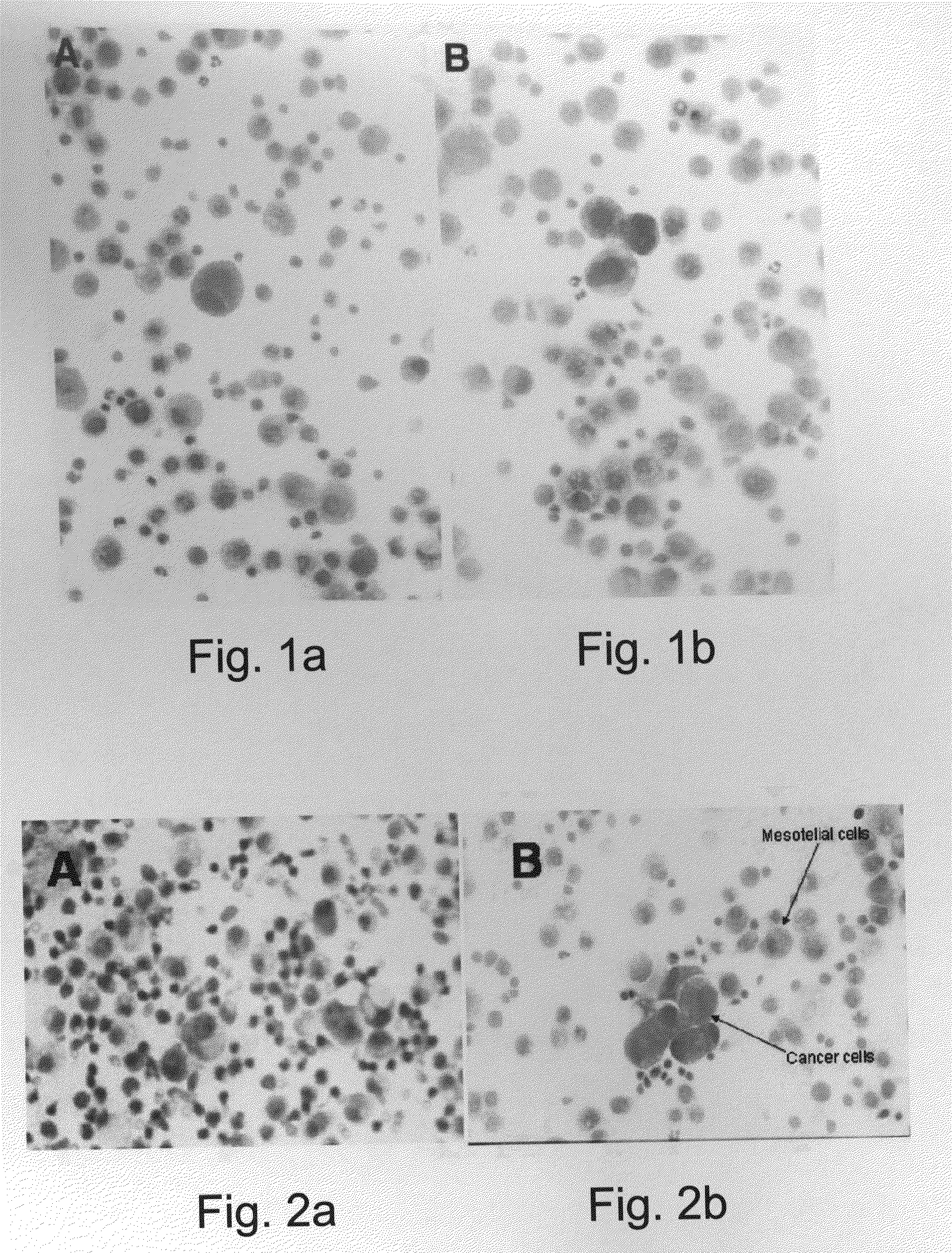
Differential Staining of Malignant and Non-Malignant Cells of Non-Cervical Biopsies
[0277]Materials and Experimental Procedures
[0278]The following materials and experimental procedures section relates to Examples 1 through Example 5 detailed hereinbelow.
[0279]Staining Procedures
[0280]Table 1b below provides step-by-step procedures for general staining according to the present invention. Please note that the air drying is preferably effected in accordance with the teachings of the present invention, improving the differential effect of the protocols. Multiwell plates could be stained following the drying: minimum 3 hours (hrs) with an optimum of 24 hours following staining.
TABLE 1bA - Protocol for non-cervical air-dried smears1.Air-dry slide or print2.Fixation: 2 minutes in Fix-13.Clean in running distilled water4.1 minute in Fix-25.Clean in running distilled water6.1 minute in Dye-17.Clean in running distilled water8.Ext-2 for 1 minute (min)9.Clean vigorously 5-7 seconds in distilled...
example 2
Differential Staining of Malignant and Non-Malignant Cells in Cervical Smears and Breast Cancer Smears
[0365]The procedures for cervical smears were developed in response to sub-optimal staining results of alcohol fixed smears by the protocol for non-cervical smears. These smears included both gynecological cervical pap-smears and non-cervical alcohol fixed breast cancer smears.
[0366]These staining procedures took advantage of the staining properties of conjugates of Bismark-Brown with Picric Acid, and Bismark Brown with Eosin, applying the effects of the plant extracts described in the protocol for non-cervical smears and the same fixation process. The protocols for cervical smears resulted in differential staining of normal cells in green tones and cancer cells in red tones (Flame red). In addition, both these staining procedures seemed capable of detecting most of the cellular changes, benign, malignant and pre-malignant, prevalent in cervical smears. These changes included: atypi...
example 3
Differential Staining of Malignant and Non-Malignant Cells in Paraffin-Embedded Histology Sections and Frozen Sections
[0374]The results obtained with histological sections are consistent with those obtained with cytological smears. However, it should be emphasized that while cytological smears usually contain a very small variety of cells and tissue types, a histology section represents the entire scope of tissue and cell types and distinguishing malignant from normal structures present a more difficult task. FIGS. 9A-D show some comparative results.
[0375]Similarly, in FIGS. 6A-D, the colon cancer cells adjacent to normal tissue were successfully differentiated by a clear and distinctive red cytoplasmic color as opposed to a clear and distinctive green cytoplasmic color indicating normal cells.
[0376]The staining protocol for histological sections yielded red staining of malignant neoplastic cells as well as some neoplasia. Hyperplasia, as well as dysplasia stained distinctly from no...
PUM
 Abstract
Description
Claims
Application Information
 - R&D Engineer
- R&D Manager
- IP Professional
- Industry Leading Data Capabilities
- Powerful AI technology
- Patent DNA Extraction
Browse by: Latest US Patents, China's latest patents, Technical Efficacy Thesaurus, Application Domain, Technology Topic.
© 2024 PatSnap. All rights reserved.Legal|Privacy policy|Modern Slavery Act Transparency Statement|Sitemap


