Method for selectively elevating and separating tissue layers and surgical instrument for performing the method

a surgical instrument and tissue layer technology, applied in the field of selectively elevating and separating tissue layers and surgical instruments for performing the method, can solve the problems of incomplete perforation of the target tissue, rapid fluid escape, and difficulty in maintaining fluid retention within the tissue, and achieves selective elevation of the mucosa, simple and economical solution, and significant reduction of the probe diameter

Active Publication Date: 2009-06-18
ERBE ELEKTROMEDIZIN GMBH
View PDF20 Cites 52 Cited by
  • Summary
  • Abstract
  • Description
  • Claims
  • Application Information

AI Technical Summary

Benefits of technology

[0013]Embodiments of the invention provide a surgical instrument that includes a device for feeding in at least one liquid, in particular for introduction of a liquid resulting in selective tissue elevation or separation, and / or dissection. The device may also include a device for high frequency surgery, wherein the device for high frequency surgery may be combined with the device for feeding in the at least one liquid, in order to achieve uniform handling. A multifunction device may thereby be achieved, which combines the advantages of water jet surgery (including introduction of liquid for selective tissue elevation or separation and / or dissection by means of liquid) with the advantages of high frequency surgery. The surgical instrument according to the disclosed embodiments makes it possible to selectively introduce a liquid under the mucosal layer into the submucosa to thereby selectively lift and / or separate selected tissue layers.
[0017]In any of the embodiments, the surgical instrument may further comprise a handle which also has a switch, in particular a sliding switch, for switching on the device for high frequency surgery and is connectable to a separate switch, in particular a foot switch, for activating the device for high frequency surgery. Two spatially separate switches must thereby be actuated, so that the device for high frequency surgery is switched on by the sliding switch and then activated by actuating the separate foot switch. It is thereby ensured that the device for high frequency surgery is not accidentally activated during a procedure. A separate foot switch can be adapted both for activating the device for high frequency surgery and for activating the feeding-in of the at least one liquid, so that by means of the separate foot switch, both the feeding-in of the liquid and the activation of the high frequency electrode can be actuated.
[0018]The probe may be comprised of a plastic hose, which results in a particularly simple and economical solution for designing a flexible probe. Alternatively, the probe may be comprised of a flexible metal tube, which is insulated on the outer periphery. By means of the metal inner core, the probe diameter can be significantly reduced without a severe loss of flexibility. Those skilled in the art can substitute a variety of suitable materials and activation devices, which can be used without deviating from the true scope and spirit of the disclosed invention.
[0019]The surgical instrument according to the disclosed embodiments allow for the selective elevation of the mucosa by introduction of a liquid under the mucosa at any angle, separation of the mucosal layer from an underlying tissue layer and fluid diffusion / penetration into the submucosal tissue matrix (which allows for improved fluid retention within the selected layer). With the aid of a single surgical device, changing instruments during the operation in order to re-inject liquid during resection is no longer required. Rather, when using the surgical instrument according to the invention, if the liquid begins to slowly diffuse or leak out of the submucosal tissue matrix, the device is simply switched over from cutting mode to liquid introduction mode and additional liquid is introduced into the submucosal tissue matrix.
[0020]By selectively targeting the submucosal tissue layer (comprised primarily of collagen) with a liquid delivered at a suitable pressure, the normal tissue properties of the submucosal tissue matrix can be altered, thereby addressing one of the major problems associated with the conventional “needle injection” technique, fluid retention within the tissue. Operating times can therefore be significantly reduced because the instruments do not have to be removed and the fluid retention of the tissue has been significantly improved. Another advantage of the surgical instrument according to the disclosed embodiments is that the function of introducing liquid under the mucosal layer for separation and selective tissue elevation of the tissue layers may be implemented without insertion of a needle or other mechanical instrument into the tissue. This is made possible by the tissue selective properties of the method and device. Advantages of this “needleless” tissue elevation method (as compared to other mechanically invasive methods) include automatically defining the appropriate tissue plane, improved fluid retention within the tissue, reliability, ease of use, faster and higher elevation and more predictability with respect to the introduction of the liquid.

Problems solved by technology

These, conventional “needle injection” techniques for delivery of fluidic agents (in order to allow the subsequent removal of selected tissues) have several problems associated with them.
One problem associated with these methods is that the user must rely on “tactile feel” and a constant delivery of fluid in order to precisely place the needle within the submucosal layer before additional liquid is injected under the mucosa via the needle.
This process is relatively “unselective” and often results in complete perforation of the target tissue and whereas the fluid accumulates under the mucosal layer without integrating with the submucosal tissue matrix, the fluid escapes rapidly (e.g., in less than a few minutes).
Additional problems associated with using conventional methods include difficulty in obtaining access to the delivery site (e.g., relative position of the device with respect to the tissue), necessity of repositioning the distal tip of the device in multiple locations, delivering the fluidic agent to the delivery site, accurately injecting the fluidic agent into the appropriate tissue layer or depth within the tissue, and problems maintaining fluid retention within the tissue, etc.
In addition, injection of the fluidic agent becomes extremely difficult when the target tissues are moving or are pliable.
This technique for resection is extremely difficult.
In particular, the difficulty of this type of intervention is that the injected liquid leaks out of the “fluid pocket” during the operation, causing the deeper layers to become thermally damaged by the needle knife.
This thermal damage can in turn lead to perforation of hollow organs (e.g., intestine) or undesired thermal damage to other tissue layers or structures.
The initial fluid retention issue is addressed using a self sealing micro-hole, however, after the first incision is made, the fluid escapes rapidly.
Therefore, continual re-introduction is still required as this device has not improved fluid retention within the tissue.
These attempts still do not offer a satisfactory solution because the escape of the liquid out of the submucosa is only slowed, but is not prevented.
Each of these patents discloses devices for submucosal needle injection, however, they fail to address all or the majority of the problems associated with this technically challenging and high risk procedure.
For example, these patents do not address the difficulties of “manually” finding the appropriate tissue plane without the risk of complete colonic transection or perforation with a needle device (layer thicknesses range from 0.5 mm to 1.5 mm), the limitations on the device approach to the target tissue, and problems with maintaining fluid retention within the tissue.

Method used


Image

  • Method for selectively elevating and separating tissue layers and surgical instrument for performing the method
  • Method for selectively elevating and separating tissue layers and surgical instrument for performing the method
  • Method for selectively elevating and separating tissue layers and surgical instrument for performing the method

Examples


Embodiment Construction

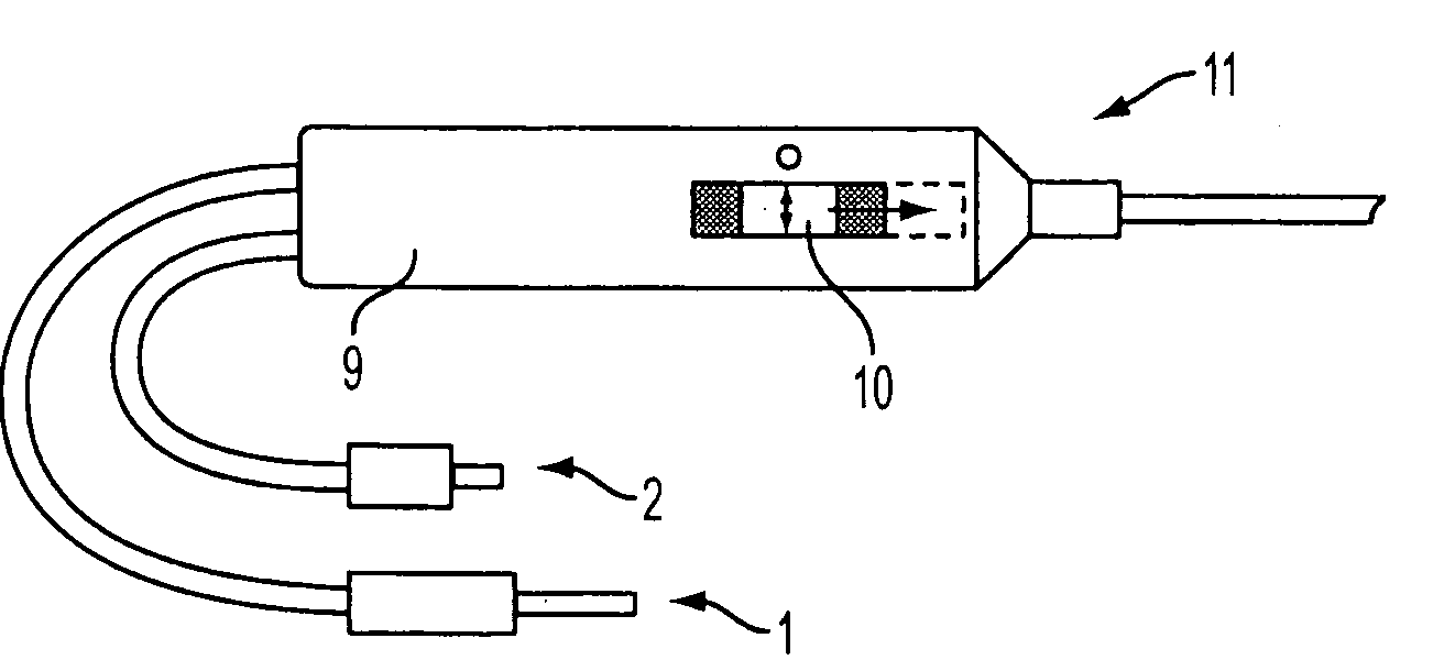
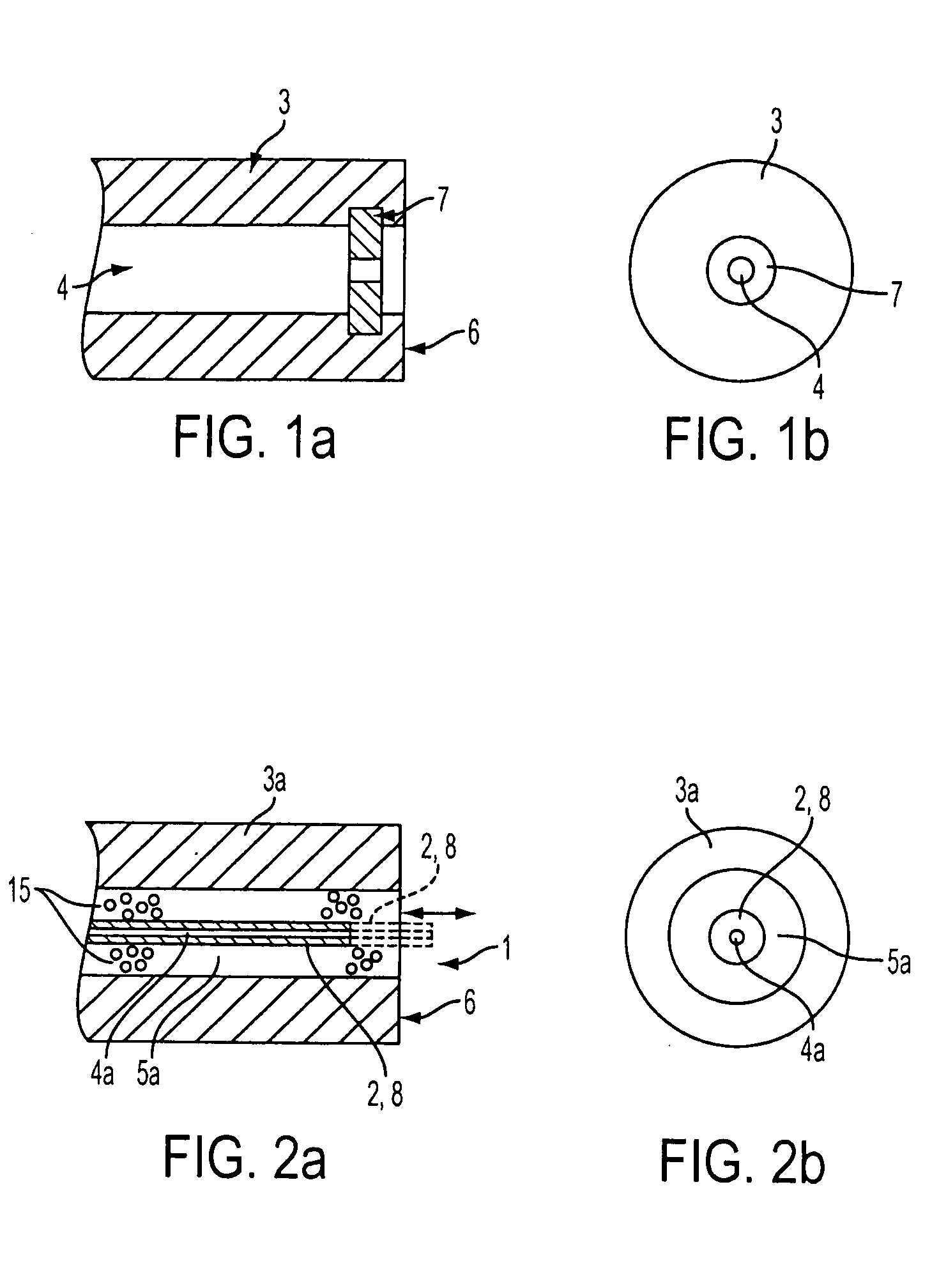
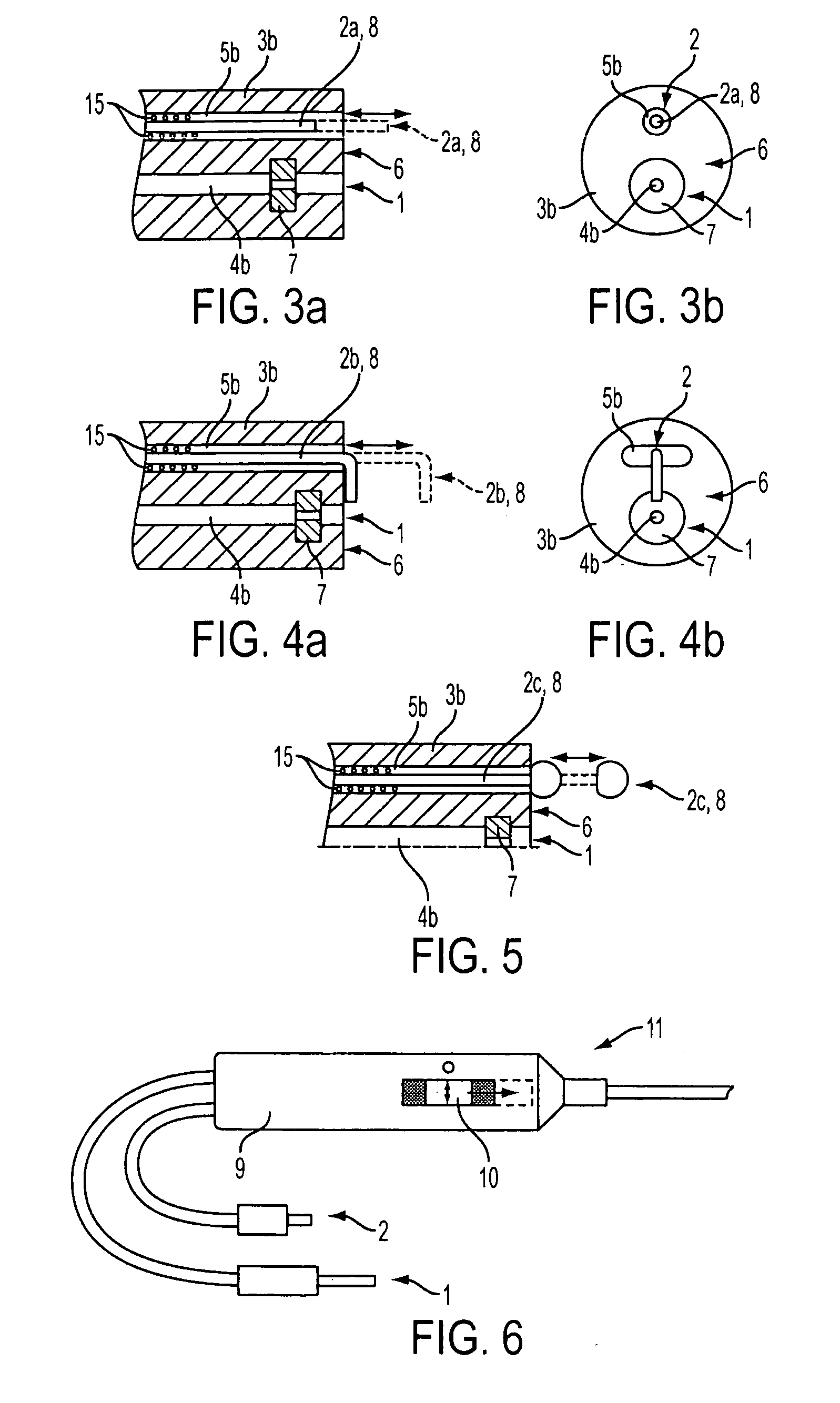
[0044]In one embodiment of the invention, a catheter device is provided and used for the delivery of a fluidic, therapeutic or diagnostic agent under a suitable pressure (selected on the basis of targeting a selected tissue layer) in order to selectively elevate, separate, and / or resect selected portions of tissues. As shown in FIGS. 1a and 1b, the distal end 6 of probe 3 comprises a channel 4 for feeding through at least one liquid through nozzle 7 at a suitable pressure. The suitable pressure can be changed according to the desired task. For selectively elevating and separating tissues, a certain target pressure is desired; for coagulating small vessels, a different target pressure may be desired; and for resecting or cutting selected tissues, a different target pressure may be desired. Probe 3 is preferably flexible in nature and can be comprised of a variety of different suitable materials. For example, the flexible probe 3 may be constructed from, nylon, PEEK, polyimide, stainl...


PUM


Abstract

A method for the selective elevation and separation of tissues comprised of multiple layers and a surgical instrument for performing the method. The method may be performed without the requirement of a solid mechanical device insertion or transection through the mucosal layer. In particular, the method and surgical instrument may be used for the selected separation and / or resection of selected portions of benign or malignant tissues (e.g. defining tissue planes, polyp elevation and removal, submucosal tissue tunneling, endoscopic mucosa resection, etc).

Description

CROSS-REFERENCE TO RELATED APPLICATIONS[0001]This application is a continuation-in-part of application Ser. No. 11 / 918,251, filed on Oct. 16, 2007, which is the U.S. national stage of International Application no. PCT / EP2006 / 002328, filed Mar. 14, 2006, the entire disclosures of both of which are incorporated by reference herein.FIELD OF THE INVENTION[0002]The invention relates to an improved method for the selective elevation and separation of tissues comprised of multiple layers using a tissue selective insertion method. In one embodiment, liquid is introduced without requiring a solid mechanical device insertion or transection through the mucosal layer and a surgical instrument for performing this method. In particular, the method and surgical instrument may be used for the tissue-selective separation and / or resection of selected portions of benign or malignant tissues (e.g., defining tissue planes, polyp elevation and removal, submucosal tissue tunneling, endoscopic mucosa resec...

Claims


Application Information

Patent Timeline
Patent Type & Authority Applications(United States)
IPC IPC(8): A61M29/00A61B17/00
CPCA61B17/00234A61B2018/00589A61B18/042A61B2017/00269A61B17/3203A61B17/3478A61B18/1482A61B2017/22074A61B2018/1407A61B17/32056A61B2018/00601
Inventor FISCHER, KLAUSMAURICE, DANDAY, JOHNENDERLE, MARKUS
Owner ERBE ELEKTROMEDIZIN GMBH
Why Eureka
Try Eureka