
Medical method and associated apparatus utilizable in accessing internal organs through skin surface

Inactive Publication Date: 2009-06-25
KANE SCOTT D +2
View PDF10 Cites 17 Cited by
  • Summary
  • Abstract
  • Description
  • Claims
  • Application Information

AI Technical Summary

Benefits of technology

[0007]It is another object of the present invention to provide such a technique and / or associated apparatus that is inexpensive and / or easy to use.
[0011]Pursuant to another feature of the present invention, an accurate positioning of the sheet or web on the skin surface of the patient is accomplished in part by aligning the sheet or web with reference marks on the patient. These reference marks are typically made during the scanning procedure and depend on the relative position of the scanner with respect to the patient. The printed sheet or web may also be provided with locators or reference marks that are alignable with the reference marks provided on the patient during the scanning procedure.
[0019]The image reproduction device may include a printer that reproduces the map or model on a print substrate. The computer may be programmed to provide locator reference marks on the print substrate for assisting in positioning the print substrate on the skin surface of the patient.

Problems solved by technology

A long standing difficulty in routine medical practice is that of accessing an internal tissue structure such as a blood vessel or the spinal column through the skin surface.
Needless to say, the conventional medical practice can be painful and traumatic to the patient.
It is generally impractical to use any of the several available scanning technologies to provide visual information as to internal tissue structures to guide a medical practitioner in accessing an internal organ or other tissue structure.
The currently available scanning devices must generally be placed in contact with or near the patient in a manner that blocks effective access to the scanned site.
In addition to being too cumbersome for many common patient access procedures, some of the currently available scanning technologies are simply too expensive to warrant widespread use for common needle directed or subdermal / hypodermic mediated operations.
The least expensive scanning technology, ultrasound, that is currently on the market generates images that for most purposes have too low a resolution to be of effective assistance in placing a needle tip at a desired target location.
There is however, an ultrasound technology not yet on the market that provides the requisite resolution in real time.

Method used


Image

  • Medical method and associated apparatus utilizable in accessing internal organs through skin surface
  • Medical method and associated apparatus utilizable in accessing internal organs through skin surface

Examples


Embodiment Construction

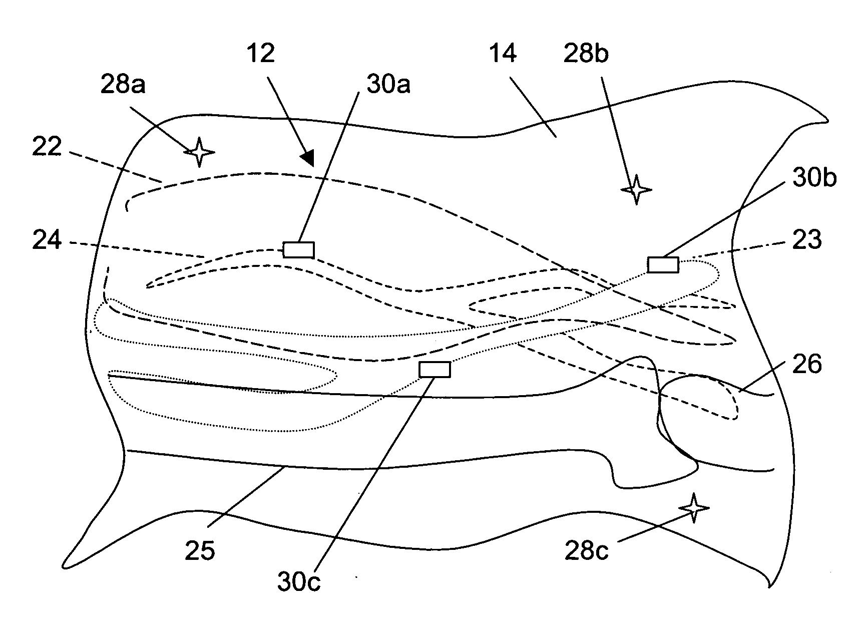
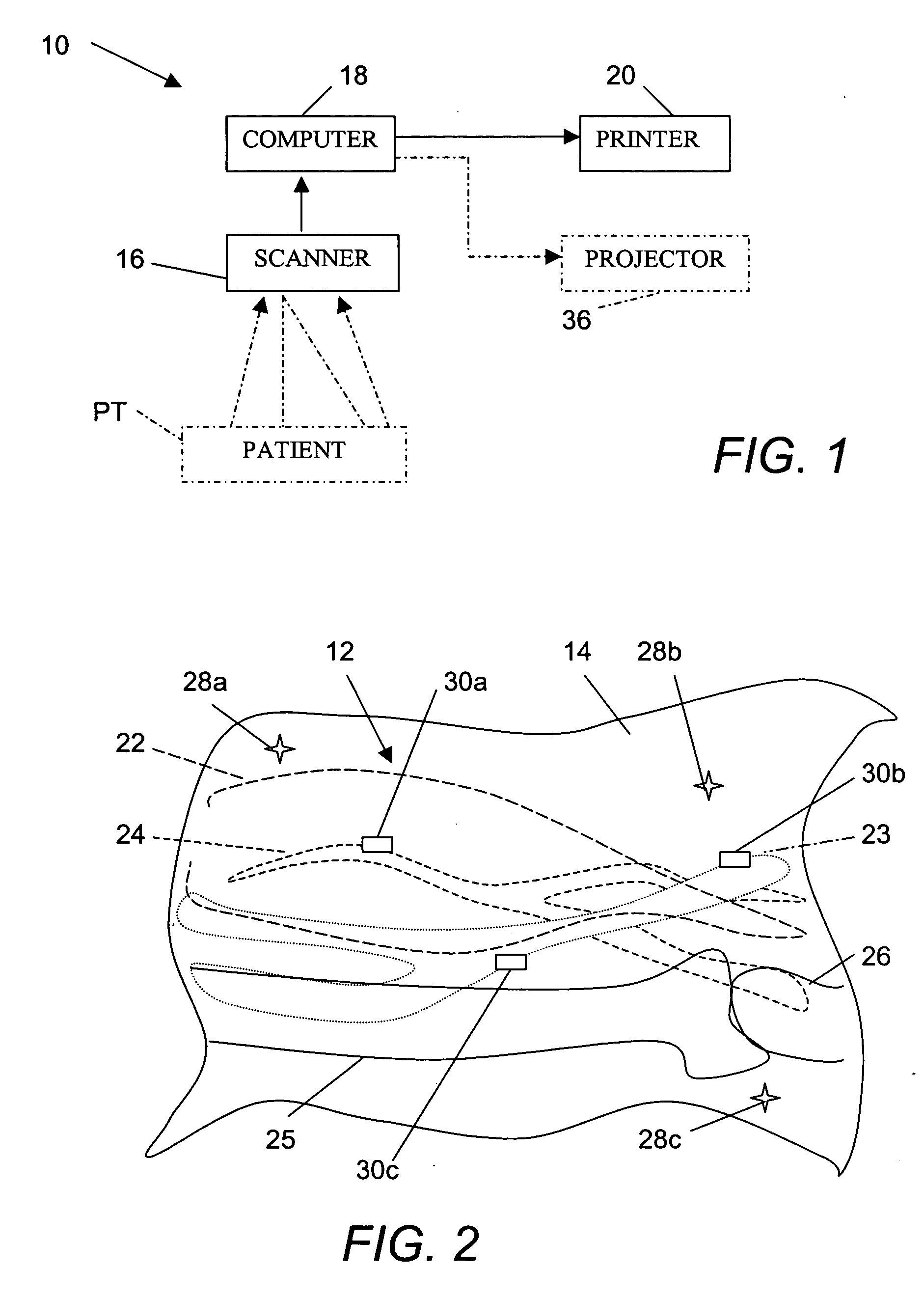
[0024]FIG. 1 schematically depicts an apparatus 10 for producing a visually readable map 12 (FIG. 2) of internal tissue structures of a patient PT exemplarily on a print substrate 14 such as a sheet, film, or web which is then placed over a patient at a desired location for assisting a medical practitioner in accessing a desired internal tissue structure of the patient. Apparatus 10 includes a scanner 16 such as an MRI apparatus, a CAT scanner, or an ultrasound scanner, for generating raw image data of internal tissue structures of a patient. A computer 18 is operatively connected to scanner 16 for deriving an electronic three-dimensional electronic map or model of the internal tissue structures from the raw data. Apparatus 10 further includes an image reproduction device 20 in the form of a printer that is operatively connected to computer 18 for reproducing the map or model in a visually readable format 12, as schematically depicted in FIG. 2. Computer 18 controls printer 20 to re...


PUM


Abstract

A medical apparatus includes a scanner for generating raw image data of internal tissue structures of a patient, and a computer operatively connected to the scanner and programmed to derive a three-dimensional electronic map or model of the internal tissue structures from the raw data. An image reproduction device is operatively connected to the computer for reproducing the map or model in a visually readable format. The computer controls the image reproduction device to reproduce the map or model. The visually readable format includes graphical representations of the tissue structures alignable on a skin surface of the patient with the tissue structures. In use, a medical practitioner inserts a sharp instrument into a patient through the reproduced map, using the map as a locator assist.

Description

BACKGROUND OF THE INVENTION[0001]This invention relates to a medical method and an associated apparatus utilizing a scanning device for obtaining three-dimensional structural data pertaining to internal tissue structures. The present method and apparatus utilize the three-dimensional data to provide a map of the internal tissue structures to assist the surgeon, anesthesiologist, radiologist, invasive cardiologist, or other medical personnel to access internal tissue structures at desired locations internal to a patient.[0002]A long standing difficulty in routine medical practice is that of accessing an internal tissue structure such as a blood vessel or the spinal column through the skin surface. Typically, a physician or medical assistant uses a needle to puncture the skin of a patient, for purposes of injecting a therapeutic or diagnostic composition into a target tissue structure or to extract a sample of tissue for diagnostic purposes. In nearly every case, the medical personnel...

Claims


Application Information

Patent Timeline
IPC IPC(8): A61B8/14G06F15/00A61B19/00
CPCA61B2019/5293A61B19/52A61B90/36A61B2090/366
Inventor KANE, SCOTT D.WILK, PETER J.NOHARA, TIMOTHY J.
Owner KANE SCOTT D
Learn More