Method for in vivo, ex vivo and in vitro repair and regeneration of cartilage and collagen and bone remodeling

a technology applied in the field of in vivo, ex vivo and in vitro repair and regeneration of cartilage and collagen and bone remodeling, can solve the problems of reducing personal productivity and quality of life, increasing morbidity and mortality for men and women, and the need for revision arthroplasty

Inactive Publication Date: 2009-07-09
THE BOARD OF TRUSTEES OF THE LELAND STANFORD JUNIOR UNIV
View PDF0 Cites 5 Cited by
  • Summary
  • Abstract
  • Description
  • Claims
  • Application Information

AI Technical Summary

Benefits of technology

[0033]Still yet another aspect of the current invention is a method for detection of functionality of the cartilage by determining relative levels of, or levels of expression of, cartilage or bone degradative enzymes, cytokines and their inhibitors or growth promoting substances, growth factors and hormones, following subjecting the diseased cartilage or bone to intermittent hydrostatic pressure followed by periods of recovery.

Problems solved by technology

Osteoarthritic related conditions decrease personal productivity and quality of life and in an aging society, increase the morbidity and mortality for men and women by increasing the incidence of other chronic conditions, such as, for example, osteoporosis.
Currently, the only successful treatment for end stage joint disease requires major surgery involving total joint replacement which is not without associated complications such as infection, aseptic loosening, and pain.
These complications can then lead to the necessity for revision arthroplasty.
However, implanting cells or resurfacing with autogenous or allograft cartilage in the absence of an organized extracellular matrix does not support normal weight bearing.
In many cases, these grafts quickly become fibrillated and degrade.
Cartilage, collagen and bone diseases, therefore, present a major medical problem, particularly with an increasing aging population which is more prone to osteoarthritis and other joint regenerative diseases, and it would thus be important to have available a means for regeneration of articulate cartilage and collagen and bone remodeling.
However, none of these patents disclose a method which would regenerate the diseased cartilage to a functional state and such method is still lacking.
While the above research describes and recognizes the importance of the hydrostatic pressure on normal function of cartilage and type II collagen, such knowledge was nevertheless impossible to apply clinically because the continuous application of the hydrostatic pressure leads to exhaustion of the cartilage metabolic potential and its damage while the brief (<1 hour) of loading with hydrostatic pressure leads to varied cellular response which disturbs the chondrocyte metabolism and homeostasis.
For example, a short period of hydrostatic pressure loading results in increased expression of type II collagen mRNA while the continuously applied load does not maintain such increased expression.
Clearly, these results disturb the cellular equilibrium between aggrecan and type II collagen.
Until recently, it was believed that articular cartilage can no longer repair itself once the arrangement of the supporting fibers has been disrupted.

Method used


Image

  • Method for in vivo, ex vivo and in vitro repair and regeneration of cartilage and collagen and bone remodeling
  • Method for in vivo, ex vivo and in vitro repair and regeneration of cartilage and collagen and bone remodeling
  • Method for in vivo, ex vivo and in vitro repair and regeneration of cartilage and collagen and bone remodeling

Examples


example 1

Chondrocyte Isolation

[0170]This example describes procedure used for isolation of chondrocytes.

[0171]Adult bovine articular chondrocytes were isolated from full thickness cartilage dissected from radiocarpal joints obtained fresh from a local abattoir.

[0172]The cartilage cells were released from the matrix by incubation in 15 ml of Dulbecco's modified Eagle's medium (DMEM) containing gentamicin (50 ug / ml) and a mixture of bacterial collagenases, type II and type IV, (Worthington, Freehold, N.J.) at a final concentration of 0.6 mg / ml each in 15 ml of Dulbecco's Modified Eagle's Medium / Ham's F12 (DMEM / F12, Gibco BRL, Grand Island, N.Y.) containing 25 μg / ml gentoamicin (Sigma, St. Louis, Mo.). The cartilage samples were incubated for a total of 18 hours at 37° C. in 7.5% CO2 and 100% humidity to ensure complete digestion. Chondrocytes released from matrix were filtered through a nylon mesh filter to isolate single cells. The cells were subsequently collected by repeated centrifugation ...

example 2

Serum Containing or Serum-Free Medium

[0175]This example describes a composition of serum containing or serum-free medium.

[0176]Serum containing Dulbecco's modified Eagle's medium (DMEM) contained dialyzed heat-inactivated fetal bovine serum at a concentration of 10% v / v. Serum-free medium consisted of a 1:1 mixture of Ham's F12 / DMEM supplemented with selenium, and liposomes. Liposomes were prepared by dissolving lecithin, cholesterol, sphingomyelin, and vitamin E acetate in 1 ml of 2:1 chloroform / methanol (vol / vol) which is dried under N2. One ml of DMEM / F12 was added and the lipid mixture was then sonicated 3× for intervals of 3 minutes each, using a microtip with a 70% duty cycle. This liposome stock was made up at 1,000× the final concentration, kept under N2, and stored at 4° C. In some experiments, ascorbate was added to the medium at a concentration of 50 μg / ml.

example 3

Mechanical Loading with Intermittent Hydrostatic Pressures

[0177]This example describes loading protocol and conditions for applying intermittent hydrostatic pressure.

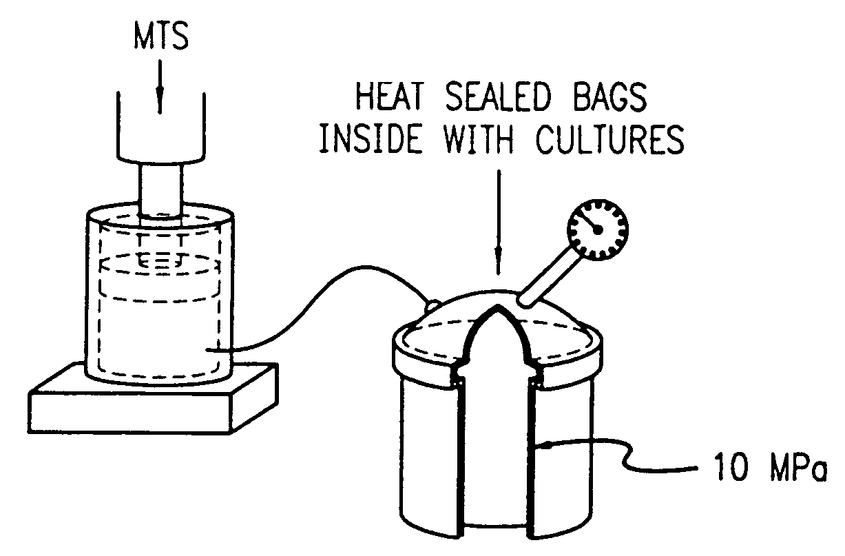
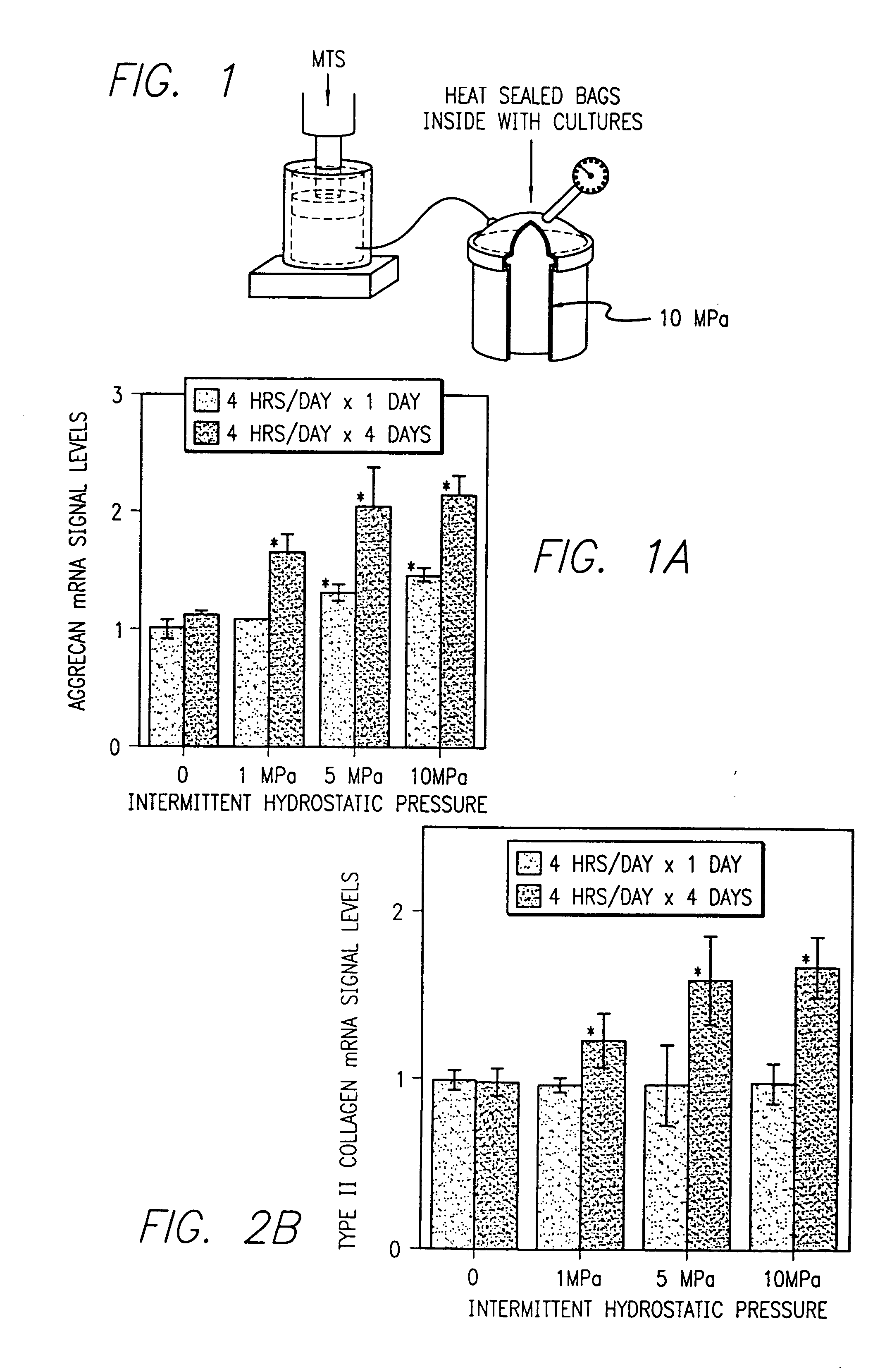
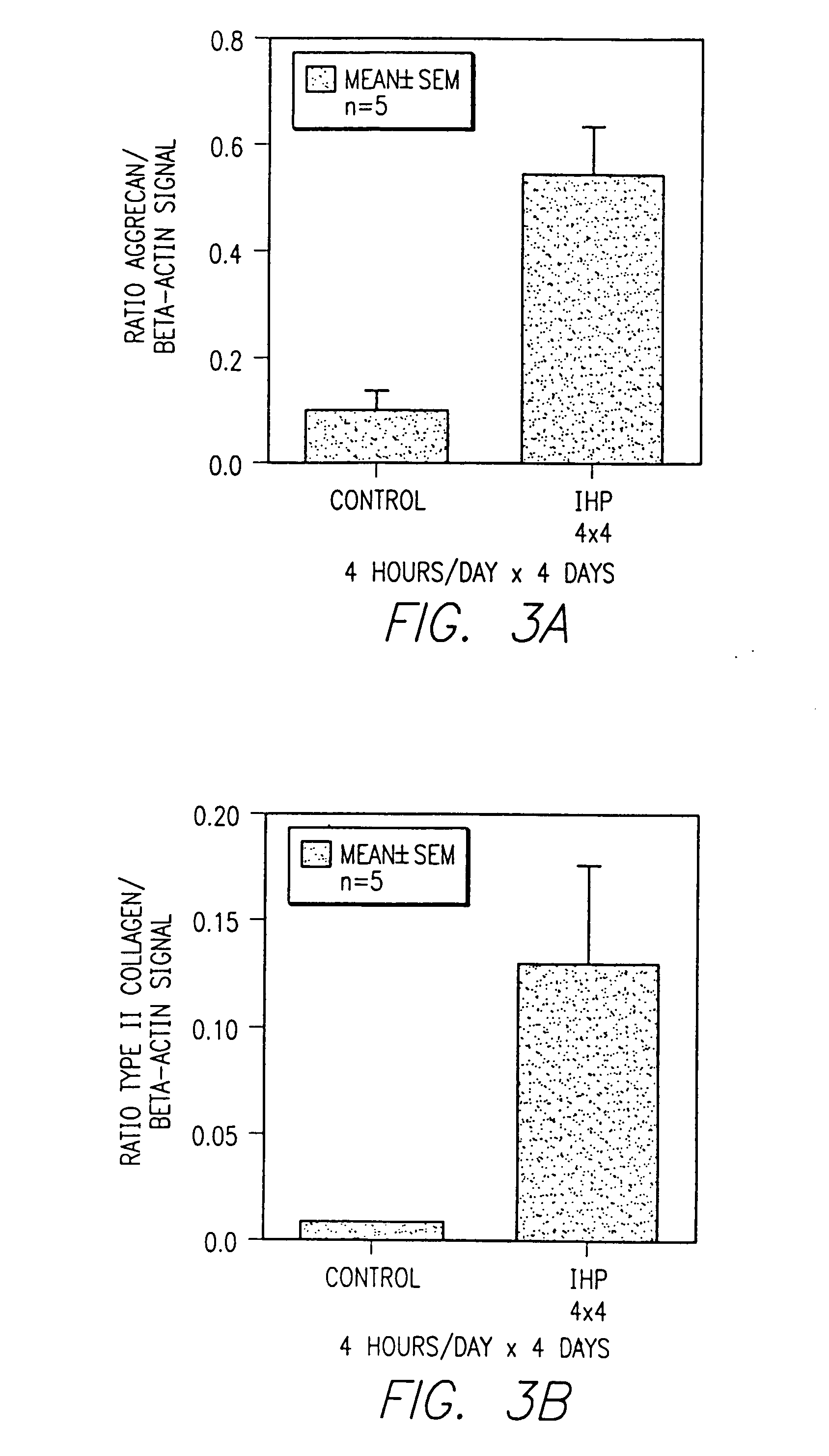
[0178]Hydrostatic pressure was cyclically applied at a loading dose of 10 MPA and at a frequency of 1 Hz. The intermittent hydrostatic pressure was applied continuously with cells removed at periods of 2, 4, 8, 12 and 24 hours, or through an interval loading protocol with the cells removed after a 4 day period during which intermittent hydrostatic pressure was applied for and limited to 4 hours per day followed by 20 hours of recover. This was repeated daily for four days or more. Each experimental time point was tested in triplicate and each experiment was carried out for a minimum of three independent trials.

[0179]The pressure was generated with a commercially available stainless steel pressure vessel interfaced to a servo-hydraulic loading instrument seen in FIG. 1. The design provided for the complete evacuation of ...


PUM

PropertyMeasurementUnit
frequencyaaaaaaaaaa
hydrostatic pressureaaaaaaaaaa

Abstract

A method for in vivo, ex vivo and in vitro regeneration of cartilage and collagen. In vivo, ex vivo and in vitro regeneration and de novo formation of articular cartilage and collagen by intermittently applied hydrostatic pressure. The application of external interval loading consisting of repeated periods of applied hydrostatic pressure followed and interrupted by periods of recovery. The application of the intermittent hydrostatic pressure at physiological levels 5-10 MPA for an interval of 4 hours followed by a recovery period up to about 20 hours, said pressure applied to the cartilage cells in vitro, explants of cartilage ex vivo and in vivo to cartilage that remains intact within te joint space of diarthrotic joints. The interval loading results in the selective inhibition of matrix degrading enzymes, pro-inflammatory cytokines and chemokines that attract inflammatory cells into the joint cavity and in selective decrease of gene expression of growth factors that are inhibitory to type II collagen expression.

Description

[0001]This application is based on and claims priority of the Provisional Application Ser. No. 60 / 157,337 filed on Oct. 1, 1999.[0002]This invention was made with U.S. Government support under Veteran's Administration Rehabilitation Research and Development Merit Review Grant No. A857-RC. The U.S. Government has certain rights in this invention.BACKGROUND OF THE INVENTION[0003]1. Field of the Invention[0004]This invention concerns a method for in vivo, ex vivo and in vitro repair, regeneration, de novo formation and remodeling of diseased and normal mesenchymal or mesenchymally derived cells, cartilage, collagen and bone. In particular, this invention concerns in vivo, ex vivo and in vitro regeneration of articular cartilage and collagen and bone remodeling by intermittently applied hydrostatic pressure. The method involves the application of external interval loading consisting of repeated periods of applied hydrostatic pressure followed and interrupted by periods of recovery. The ...

Claims


Application Information

Patent Timeline
Patent Type & Authority Applications(United States)
IPC IPC(8): C12N5/08C12N5/06C12Q1/68A61F2/00A61F2/08A61F2/28A61F2/30A61L27/00C12N5/077
CPCA61F2/08A61F2/28A61F2/30756A61F2/3094A61F2240/001C12N2521/00A61K35/12C12N5/0655C12N2500/02A61K35/32A61F2310/00365A61P19/00A61F2/30
Inventor SMITH, R. LANECARTER, DENNIS R.SCHURMAN, DAVID J.
Owner THE BOARD OF TRUSTEES OF THE LELAND STANFORD JUNIOR UNIV
Why Eureka
Try Eureka