Clinical assessment and training system

Active Publication Date: 2009-08-20
MEDICAL BRACING SYS INC
View PDF22 Cites 46 Cited by
  • Summary
  • Abstract
  • Description
  • Claims
  • Application Information

AI Technical Summary

Problems solved by technology

Both procedures have potentially serious complications if not done correctly.
The most feared complication, usually due to operator experience or patient anatomy is a ‘missed intubation’ or inability to insert the endotracheal tube into the airway.
Even if successful, potential complications that may occur during intubation include injury to the teeth, lips and oral cavity; edema; bleeding; tracheal and esophageal perforation; pneumothorax (collapsed lung); and aspiration.
Further life-threatening complications of airway problems may occur and these signs and symptoms include, but are not limited to, sore throat, pain or swelling of the face and neck, chest pain, subcutaneous emphysema, and difficulty swallowing.
Needle biopsy or aspiration also has risks.
A recent study showed that in one case a needle biopsy of a liver tumor resulted in major bleeding.
Other complications depend upon the body part on which the biopsy takes place: Lung biopsies are frequently complicated by pneumothorax (collapsed lung).
Although it is impossible to predict in whom this will occur, collapsed lungs are more frequent and more serious in patients in whom the biopsy is difficult to perform.
Breast biopsies may also be complicated by pneumothorax.
Deaths have been reported from needle aspiration biopsies, but such outcomes are extremely rare.
This procedure results in a cephalad rotation of the symphysis pubis and a flattening of the sacral promontory.
Thus, simulators that enhance the training of practitioners for procedures involving direct and indirect contact are desperately needed.

Method used


Image

  • Clinical assessment and training system
  • Clinical assessment and training system
  • Clinical assessment and training system

Examples


Embodiment Construction

[0031]As described below and illustrated in FIGS. 1-10B, systems for medical training comprise an anatomical simulator having internal and external sensors. At least one internal sensor is positioned at an internal location of the simulator and at least one external sensor is positioned at an external location of the simulator. A feedback display system in communication with the sensors simultaneously provides external sensor reading from external contact and internal sensor readings from internal contact. The systems can be used for training a user for various procedures or examinations in medical, veterinary, and other fields.

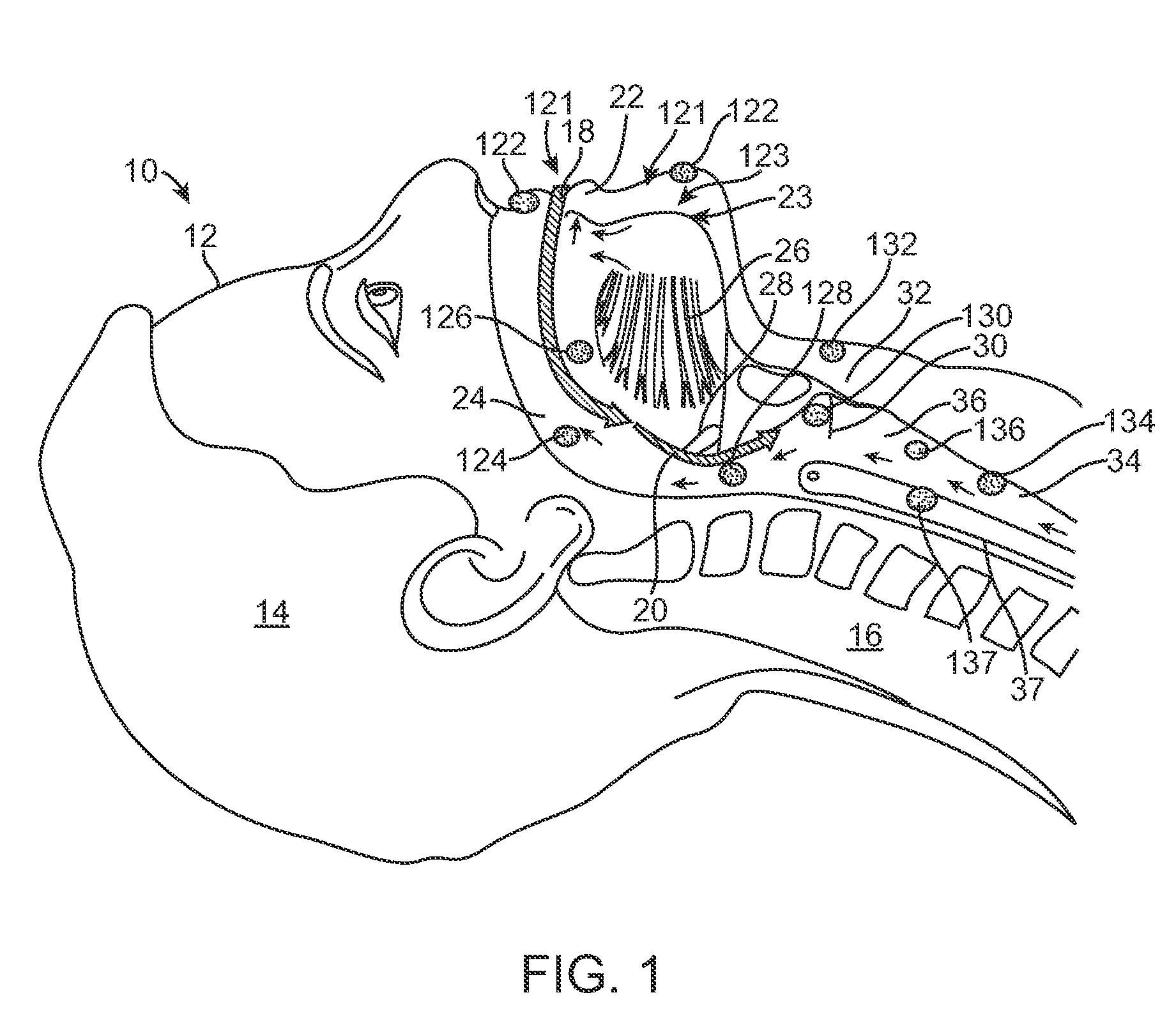
[0032]FIG. 1 illustrates one embodiment of an anatomical simulator system 10 for medical training for endotracheal intubation in accordance with the present invention. System 10 comprises an anatomical simulator 12. Anatomical simulator 12 includes a head portion 14, a neck portion 16 and an internal cavity defined by the various anatomical structures of the ...


PUM


Abstract

A system for medical training includes an anatomical simulator modeled after at least a portion of a body, the simulator including at least one external surface, and at least one cavity in fluidic communication with the external surface via a cavity. The system further includes at least one internal sensor positioned at an internal location of the anatomical simulator, the internal sensor positioned to receive an internal input based on forces applied from within the cavity, at least one external sensor positioned at an external location of the anatomical simulator and to receive an external input based on forces applied to the external surface, and a feedback display system in communication with the sensors to simultaneously record external sensor readings from the at least one external sensor and internal sensor readings from the at least one internal sensor and at least one time measurement device.

Description

RELATED APPLICATIONS[0001]This application claims the benefit of, and priority to U.S. Provisional Patent Application 61 / 029,202 filed Feb. 15, 2008, the entirety of which is incorporated herein by reference.FIELD OF THE INVENTION[0002]The present invention relates generally to a system and method for practicing and teaching medicine. More specifically, the invention relates to an anatomical simulator system having sensors and a means for providing feedback to a user in medical training.DESCRIPTION OF THE RELATED ART[0003]Anatomical simulators have been developed for training and assessment of medical students, nursing students, medics and practitioners. These simulators have enabled health care professionals of all backgrounds to practice clinical procedures in a safe environment, away from the patient. The majority of simulators that have been developed thus far focus mainly on emergency care, anesthesia and laparoscopic surgery. In addition, simulators have been developed for exa...

Claims


Application Information

Patent Timeline
IPC IPC(8): G09B23/30G09B23/28
CPCG09B23/30
Inventor PUGH, CARLA M.
Owner MEDICAL BRACING SYS INC
Why Eureka
Try Eureka