Apparatus and Methods for the Measurement of Cardiac Output

a technology of cardiac output and apparatus, applied in the direction of catheters, packaged goods, packaged foodstuffs, etc., can solve the problems of more complex and riskier intubation process

Active Publication Date: 2009-09-10
MICROPEN TECH CORP +1
View PDF3 Cites 97 Cited by
  • Summary
  • Abstract
  • Description
  • Claims
  • Application Information

AI Technical Summary

Benefits of technology

[0010]The apparatus includes a collection of at least three sense electrodes, and may be three, four, five or more than five sense electrodes. For example, the combination of the five sense electrodes provides three orthogonal pairs of sense electrodes. The tube of the apparatus may be an endotracheal tube. Also, the current electrode is disposed on the distal portion of the tube between the termini of the electrode runners (near the middle of the tube in a proximal-distal direction) and the inflatable cuff. In certain embodiments, the current electrode is at least one centimeter in length (e.g., one, two or more centimeters) as measured in the proximal-distal dimension of the tube. In some embodiments the current electrode extends over about 90°, 120°, or about 180° of the circumference of the tube. Optionally, the ground electrode is also placed on the tube.
[0011]In another aspect, the invention provides a method of fabricating an apparatus by providing an apparatus having a first portion and a second portion that is capable of being inflated, at least partially inflating the second portion, imaging the inflated second portion so as to obtain imaging data, directing a positive displacement dispensing system to apply to a region of the inflated second portion a polymeric underlayer based upon the imaging data, applying to at least a portion of the polymeric underlayer a conductive material based upon the imaging data, and applying to a portion of the conductive material a polymeric overlayer based upon the imaging data. In certain embodiments, no polymeric overlayer is applied to a plurality of regions of the conductive material, thereby forming a plurality of electrode patches. The imaging step comprises capturing images, such as dynamic video images. The imaging step includes in certain embodiments the capturing of a plurality of images that are used to identify one or more contours of the second portion.
[0012]In certain embodiments, one or more of the polymeric underlayer, the conductive material, or the polymeric overlayer are applied by a positive displacement dispensing system. The positive displacement dispensing system includes a pen tip that is kept substantially perpendicular to the surface of the second portion during application of the polymeric underlayer, the conductive material, or the polymeric overlayer.

Problems solved by technology

The presence of electrodes on the inflatable cuff and electrode leads on the external surface of the endotracheal tube results in a more complex and riskier intubation process.
Further, the electrodes are attached to the inflatable cuff when inflated, resulting in irregularities (e.g., sharp edges of the electrode, broken electrode leads) when the cuff is deflated prior to insertion.

Method used


Image

  • Apparatus and Methods for the Measurement of Cardiac Output
  • Apparatus and Methods for the Measurement of Cardiac Output
  • Apparatus and Methods for the Measurement of Cardiac Output

Examples


Embodiment Construction

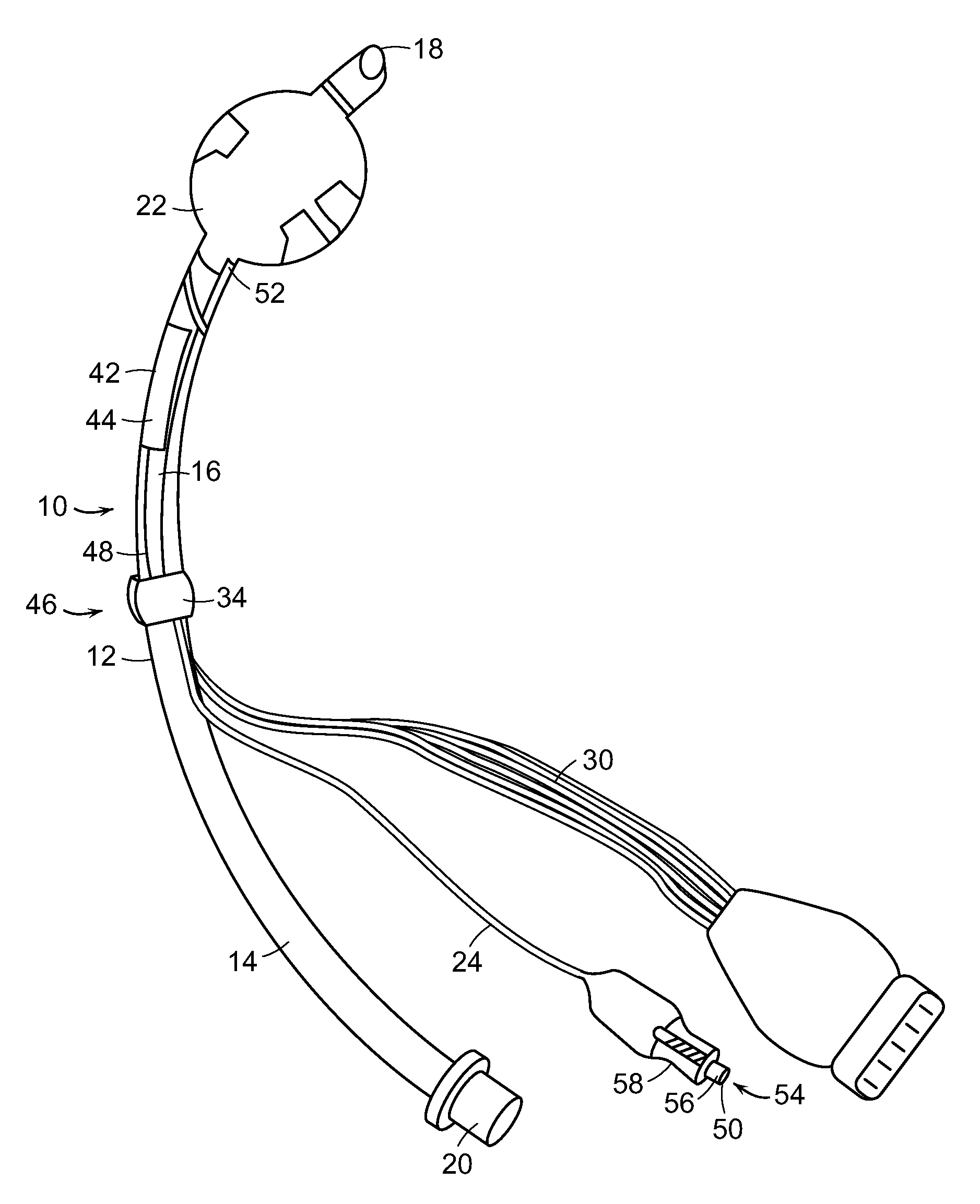
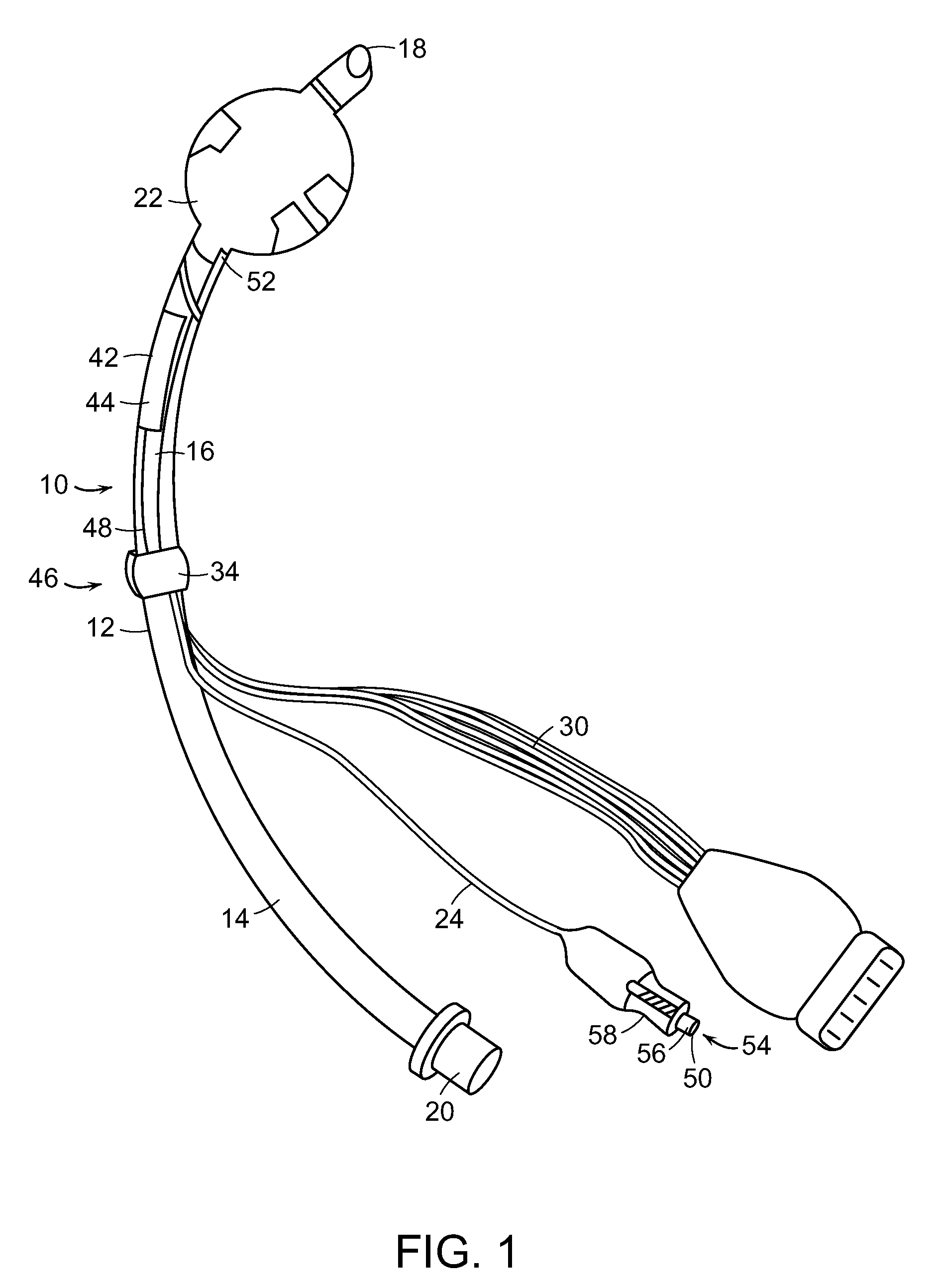
[0025]In some embodiments described herein, the present invention relates generally to an apparatus useful as an endotracheal tube (also known as an intratracheal tube or ET tube). The endotracheal tube is useful in measuring physiological characteristics of a mammalian subject, particularly human subjects suffering from acute or chronic injury or illness. For example, the endotracheal tube is used to measure cardiac output in a mammalian subject. The endotracheal tube is inserted into the trachea, generally via the mouth, but sometimes through the nares of the nose or even through a tracheostomy.

[0026]The apparatus 10 for measuring a mammalian subject's cardiac output shown in FIG. 1 contains tube 12 having proximal portion 14 and distal portion 16. The tube 12 is generally formed of a medically approved synthetic polymeric material such as silicone rubber, polyvinyl chloride or polypropylene. See, U.S. Pat. Nos. 3,599,642 and 4,593,690, the contents of which are incorporated herei...


PUM


Abstract

The current invention provides an endotracheal tube fabricated with an array of electrodes disposed on an inflatable cuff on the tube. The array of electrodes includes multiple sense electrodes and a current electrode. The array of electrodes on the inflatable cuff is applied using a positive displacement dispensing system, such as a MicroPen®. A ground electrode is disposed on the tube approximately midway between the inflatable cuff and the midpoint of the endotracheal tube. The endotracheal tube is partially inserted into a mammalian subject's airway such that when the inflatable cuff is inflated, thereby fixing the tube in position, the array of electrodes is brought into close contact with the tracheal mucosa in relative proximity to the aorta. The endotracheal tube is useful in the measurement of cardiac parameters such as cardiac output.

Description

RELATED APPLICATIONS[0001]This application claims the benefit of priority to U.S. Provisional Application No. 60 / 998,682, filed Oct. 12, 2007, the teachings of which are incorporated herein by reference in their entirety.FIELD OF THE INVENTION[0002]The invention provides an apparatus for measuring cardiac output in a mammalian subject. The apparatus includes a tube and an inflatable cuff and is configured so that electrodes on the inflatable cuff located in close proximity to the subject's aorta measure voltage changes following stimulation of the tissue with a current delivered by an electrode. The electrodes are printed on the tube and the cuff with a positive displacement dispensing system to improve the durability of the apparatus.BACKGROUND OF THE INVENTION[0003]Cardiac output is a calculation of the volume of blood being pumped by the heart, for example a ventricle, per minute. Cardiac output is equivalent to the heart rate multiplied by the stroke volume. Understanding of a s...

Claims


Application Information

Patent Timeline
Patent Type & Authority Applications(United States)
IPC IPC(8): A61M16/04A61B5/029B05D5/12A61B5/296
CPCA61B5/029A61B5/0421A61B5/0535A61B5/0538A61B5/6853A61B2562/0209H05K2201/0949A61B2562/125H05K1/0284H05K1/117H05K1/141H05K1/147H05K3/4069A61B2562/046A61B2562/0215A61B5/285A61B5/0295
Inventor LOWERY, GUY RUSSELLGRANDE, WILLIAM J.
Owner MICROPEN TECH CORP
Why Eureka
Try Eureka