Method and Apparatus for Ultrasound Synthetic Imagining

a synthetic imaging and ultrasound technology, applied in ultrasonic/sonic/infrasonic diagnostics, instruments, applications, etc., can solve the problems of loss or poor spatial coherence of raw data, and achieve the effect of accurately combining data received, recovering spatial coherence, and losing spatial coherence of wavefield

Active Publication Date: 2009-09-17
SUPER SONIC IMAGINE
View PDF14 Cites 56 Cited by
  • Summary
  • Abstract
  • Description
  • Claims
  • Application Information

AI Technical Summary

Benefits of technology

[0019]Thanks to these dispositions, the spatial coherence of the raw data is recovered at step b) prior to beamforming, thus enabling to accurately combine the data received from the transmission of the various ultrasonic waves. The need to recover spatial coherence is due the fact that, when illuminating the imaged region with spatially widely spread wavefield, the echoes comi

Problems solved by technology

The need to recover spatial coherence is due the fact that, when illuminating the imaged region with spatially widely spread wavefield, the echoes coming back from the medium can be seen as a

Method used


Image

  • Method and Apparatus for Ultrasound Synthetic Imagining
  • Method and Apparatus for Ultrasound Synthetic Imagining
  • Method and Apparatus for Ultrasound Synthetic Imagining

Examples


Embodiment Construction

[0046]In the Figures, the same references denote identical or similar elements.

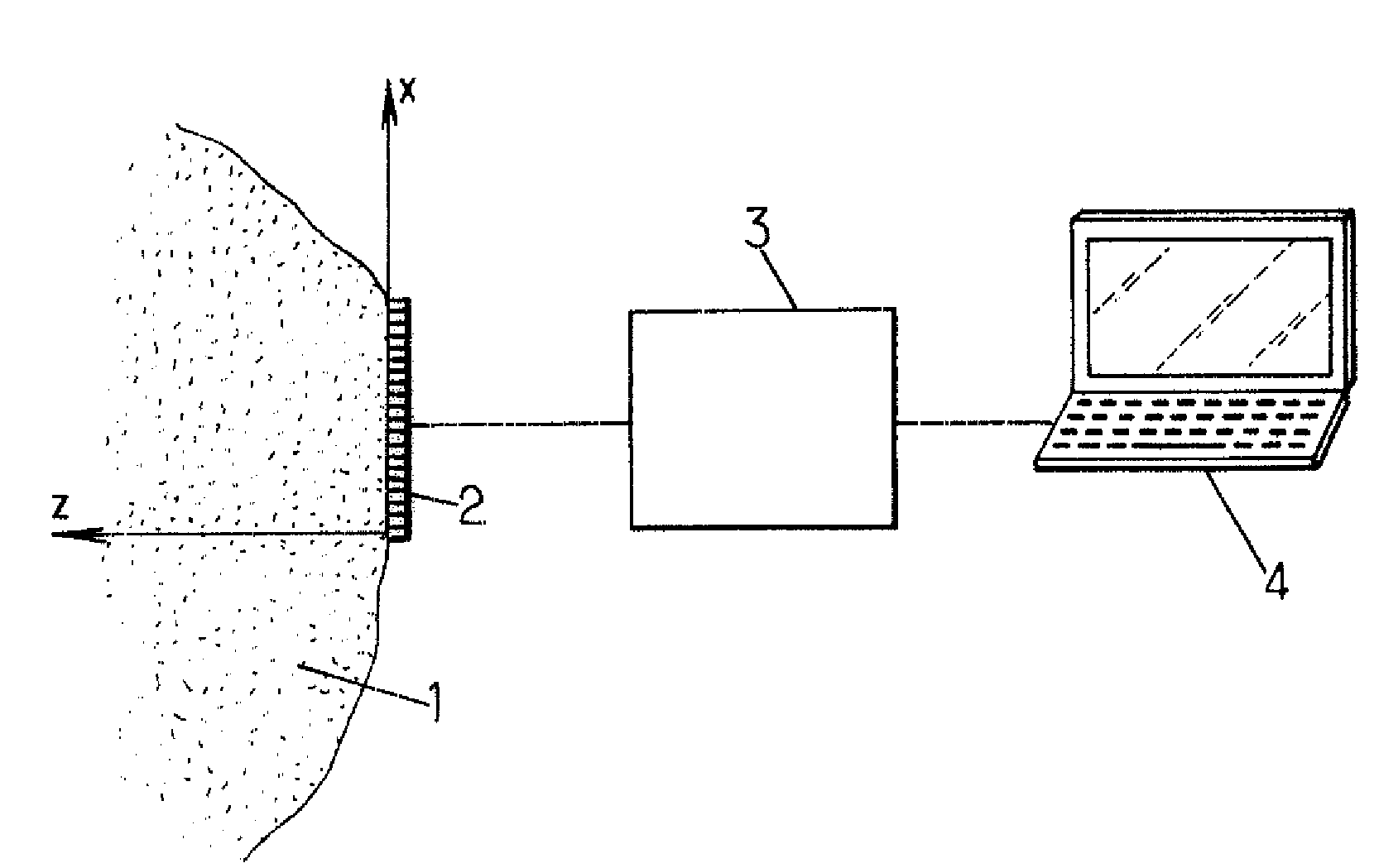
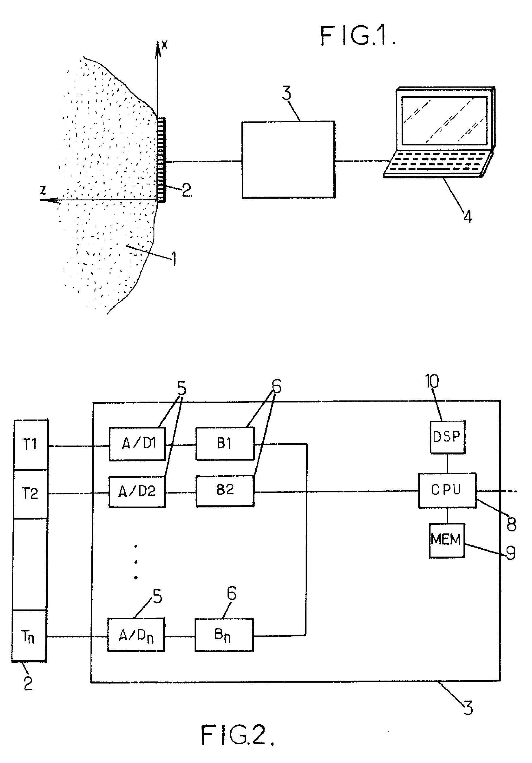
[0047]The apparatus shown on FIG. 1 is adapted for synthetic ultrasound imaging of a region 1, for instance living tissues and in particular human tissues of a patient. The apparatus may include for instance:[0048]an ultrasound transducer array 2, for instance a linear array typically including a few tens of transducers (for instance 100 to 300) juxtaposed along an axis X as already known in usual echographic probes (the array 2 is then adapted to perform a bidimensional (2D) imaging of the region 1, but the array 2 could also be a bidimensional array adapted to perform a 3D imaging of the region 1);[0049]an electronic bay 3 controlling the transducer array and acquiring signals therefrom;[0050]a microcomputer 4 for controlling the electronic bay 3 and viewing ultrasound images obtained from the electronic bay (in a variant, a single electronic device could fulfill all the functionalities of the electroni...


PUM


Abstract

Method for ultrasound imaging comprising at least the following steps:
    • a) a transmission step in which a plurality of ultrasonic waves are transmitted into an imaged region and a set of raw data is acquired by an array of transducers in response to each ultrasonic wave, the ultrasonic waves having different spatial frequency content;
    • b) a coherence enhancing step in which, for each of a plurality of virtual transmit focal zones in the imaged region, at least one set of coherent data is synthesized from the sets of raw data;
    • c) a beamforming step in which, for each of a plurality of locations included in each of the virtual transmit focal zones, an image pixel is computed by beamforming, using the set of coherent data.

Description

FIELD OF THE INVENTION[0001]The invention relates to methods and apparatus for ultrasound synthetic imaging, in particular for medical imaging.BACKGROUND OF THE INVENTION[0002]Ultrasound Standard Imaging[0003]Standard ultrasound imaging consists of an insonification of the medium with a cylindrical wave that focuses on a given point. Using the backscattered echoes of this single insonification, a complete line of the image is computed using a dynamic receive beamforming process To build a complete image, this procedure is repeated by sending a set of focused waves that scan along a lateral line at given depth (named the focal plane). For each focused wave, a dynamic beamforming is performed and the complete image is obtained line by line. The dynamic beamforming guarantees a uniform focusing in the receive mode, whereas, in the transmit mode the focus is fixed at a given depth. The final image is optimal in the focal plane and in a limited region of the medium corresponding to the f...

Claims


Application Information

Patent Timeline
IPC IPC(8): A61B8/00
CPCG01S7/52049G01S15/8997G10K11/346G01S15/8977G01S15/8995G01S15/8915G01S7/52046A61B8/00G06T1/00G01S15/8927
Inventor BERCOFF, JEREMYCOHEN, CLAUDE BACRIETANTER, MICKAELFINK, MATHIASMONTALDO, GABRIEL
Owner SUPER SONIC IMAGINE
Why Eureka
Try Eureka