
Radiation image pick-up device, radiation image pick-up method and program

a technology of radiation image and pick-up method, which is applied in the field of radioation detection system, can solve the problems of inevitably selecting the retry of photographing, requiring a lot of time to search the patient film, and requiring a large amount of time, so as to prevent the increase of the dose of radiation exposure, reduce the frequency of redoing image pick-up, and reduce the search time for searching the desired tomogram

Inactive Publication Date: 2005-04-14
CANON KK
View PDF14 Cites 9 Cited by
  • Summary
  • Abstract
  • Description
  • Claims
  • Application Information

AI Technical Summary

Benefits of technology

The present invention provides a radiation image pick-up device, method, and program for obtaining tomography images in a single-time photographing. The device includes a radiation generation means and a radiation detection means with a matrix of radiation detection elements. The device is moved to capture the object being detected, and the electrical signals generated by the detection elements are read every frame over a plurality of frames. The method involves setting the object between the radiation generation means and the detection means, and moving them to create an intersection point between a straight line connecting a point at which an optional detection element is located and a point at which the radiation generation means is located. The electrical signals generated by the detection elements are read every frame over a plurality of frames. The program includes a computer control for executing the method. The invention solves the problem of increased dosage of exposure to radiation and prevents the need for multiple redoing of image pick-up. It also solves the problem of film storage and space in a hospital and reduces the search time for finding a desired tomogram.

Problems solved by technology

Moreover, when a desired tomography image is not obtained, there is a case in which retry of photographing is inevitably selected again.
Furthermore, the exposure time to a film generally requires several seconds in the case of the tomography photographing method.
Therefore, a tomography image around the heart of the patient frequently becomes unclear in a chest tomography image due to the movement of the heat in an exposure period or the movement of breathing of the patient and it is difficult to obtain a preferable tomogram.
Moreover, in the case of a method using a film not applied only to the tomography photographing method, many problems are left on management and operation in a hospital by including a problem on a film storage space and a problem that a lot of time is required to search a necessary patient film out of a great number of films in a hospital.

Method used


Image

  • Radiation image pick-up device, radiation image pick-up method and program
  • Radiation image pick-up device, radiation image pick-up method and program
  • Radiation image pick-up device, radiation image pick-up method and program

Examples


Embodiment Construction

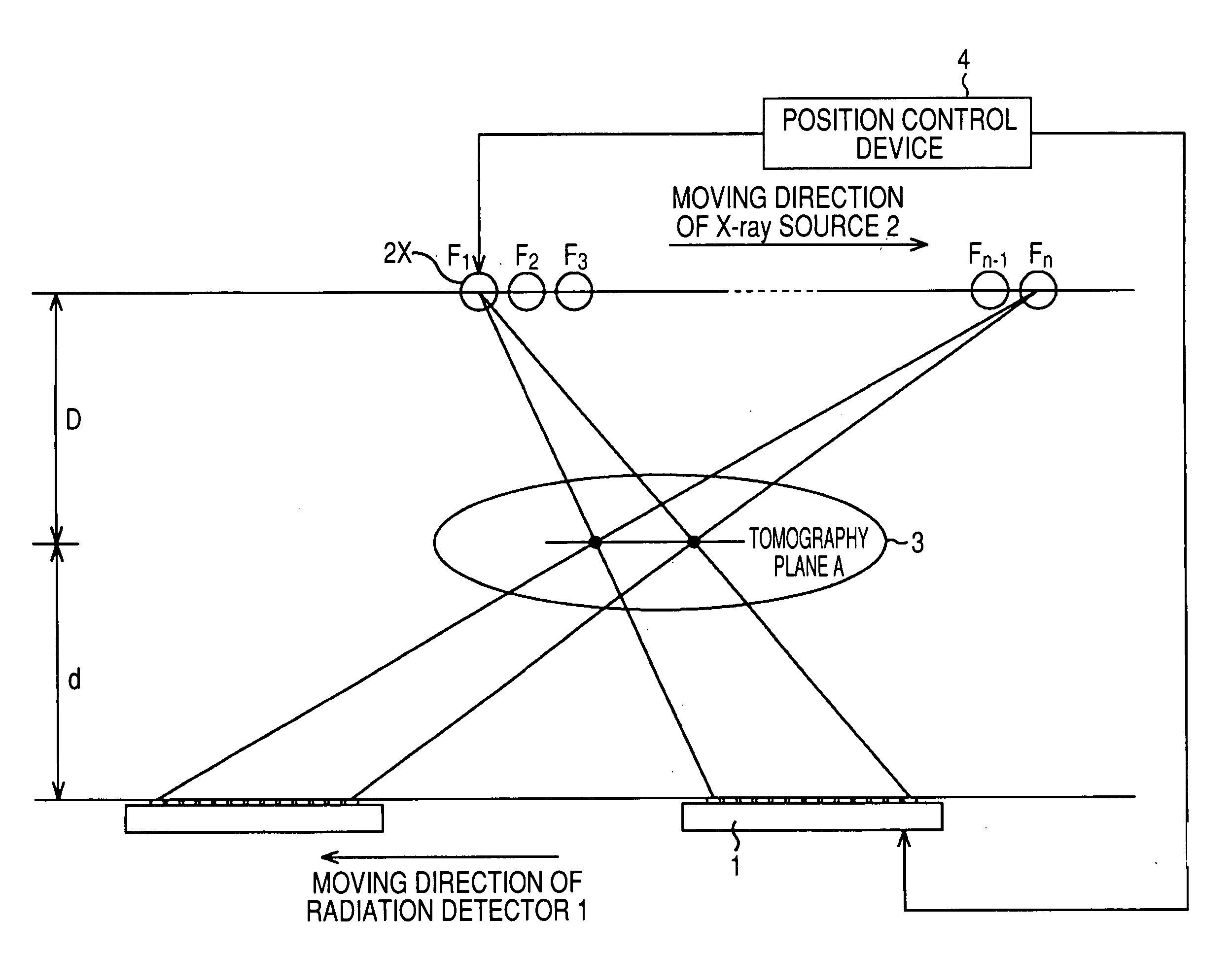
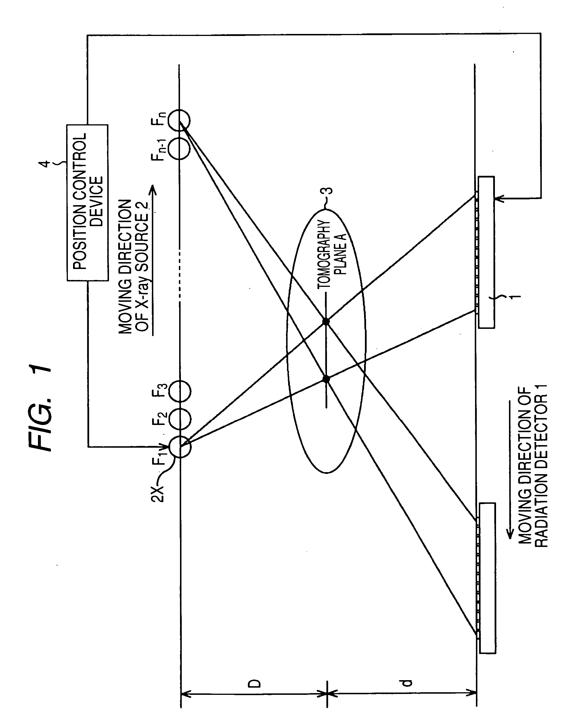
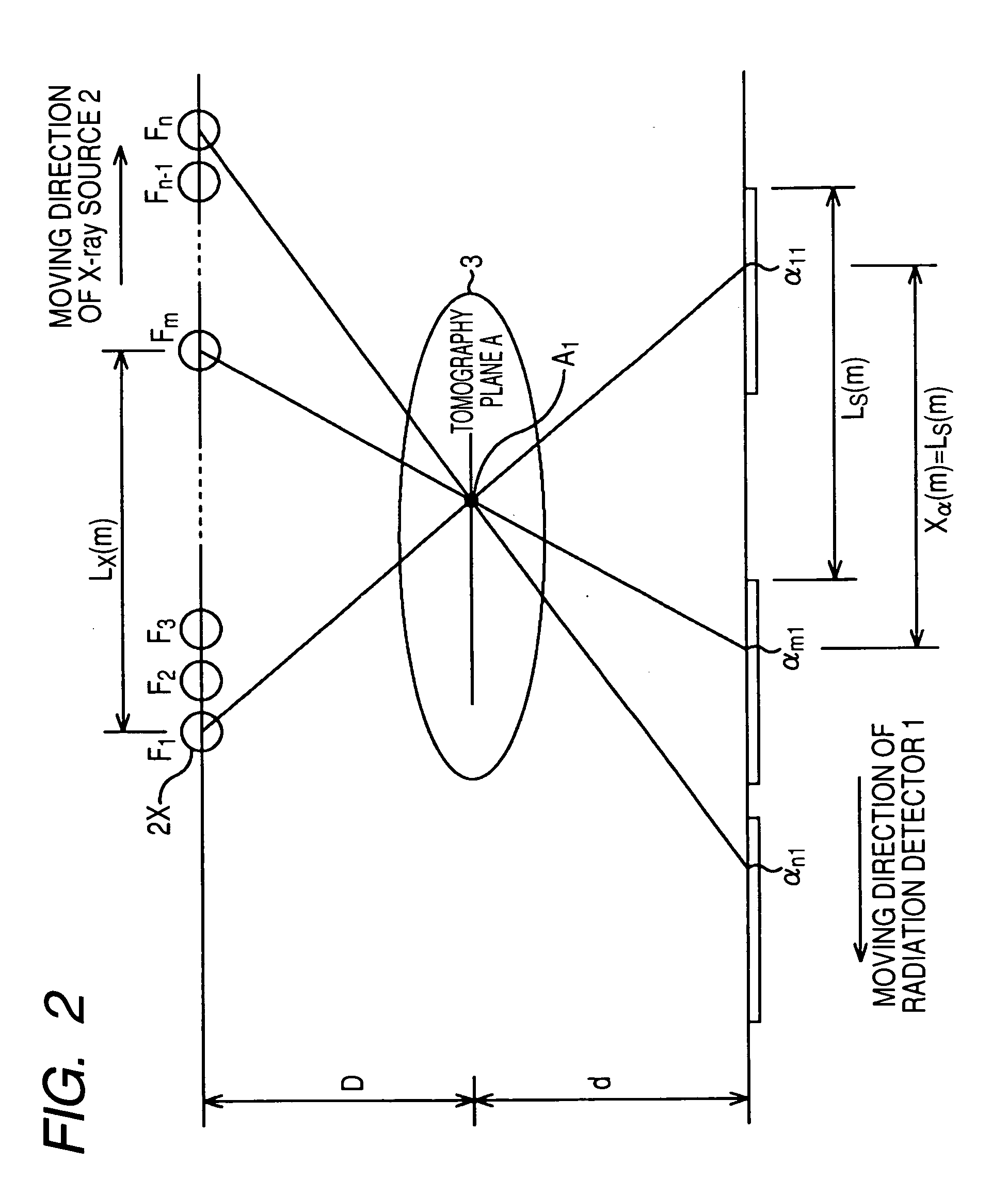
[0037] Embodiments of the present invention are specifically described below by referring to the accompanying drawings. FIG. 1 is a schematic view showing a configuration of an X-ray image pick-up device (radiation image pick-up device) of an embodiment of the present invention.

[0038] In the case of this embodiment, an X-ray source 2 is set above an object to be detected (patient) 3 and a radiation detector 1 is set under the object to be detected 3. The radiation detector 1 is provided with a plurality of X-ray detection elements (pixels) for converting X rays passing through the object to be detected 3 irradiated from the X-ray source 1 into electrical signals. FIG. 1 shows 13 X-ray detection elements arranged in one direction.

[0039] Moreover, this embodiment is provided with a position controller 4 for controlling positions of the radiation detector 1 and X-ray source 2. In the case of this embodiment, the position controller 4 moves the X-ray source 2 to an optional tomography...


PUM

PropertyMeasurementUnit
thicknessaaaaaaaaaa
electricalaaaaaaaaaa
pressureaaaaaaaaaa

Abstract

To provide a radiation image pick-up device capable of obtaining a plurality of tomography images through single-time image pick-up, a radiation image pick-up method and a program. When a position controller moves an X-ray source to an optional tomography plane in an object to be detected in parallel and the X-ray source is present at geometric focuses set at equal interval, X rays are pulsatively generated by the X-ray source and detection of X rays by a radiation detector is performed by the total of n times. Moreover, a position controller moves the radiation detector in accordance with the movement of the X-ray source.

Description

BACKGROUND OF THE INVENTION [0001] 1. Field of the Invention [0002] The present invention relates to a radiation detection system used for a diagnosis in a hospital, a radiation image pick-up device preferably used for an industrial non-destructive inspection, a radiation image pick-up method and a program. [0003] 2. Related Background Art [0004] At present, a film system for irradiating a patient with X-rays and exposing its transmitted X-ray image to a film is a main stream of an X-ray still image photographing system in medical treatment. A film is widely spread because it has a function for displaying and recording information, a large area and a high gradation characteristic and is lightweight and easily handled. [0005] There is a tomography photographing method as one of photographing methods using a film. FIG. 15 is a schematic view showing a conventional tomography photographing method using a film. A point F denotes the position of the focus of an X-ray generation source. T...

Claims


Application Information

Patent Timeline
Patent Type & Authority Applications(United States)
IPC IPC(8): G21K5/02A61B6/00A61B6/02G01N23/04G01T1/20G01T1/24H01L27/14H01L27/146H04N5/225H04N5/32
CPCA61B6/4441A61B6/4476G01N2223/612A61B6/507G01N2223/419G01N23/046
Inventor ENDO, TADAO
Owner CANON KK
Learn More