Tissue biopsy needle apparatus

a biopsy needle and tissue technology, applied in the field of tissue biopsy needle apparatus, can solve the problems of inability to puncture the target site, failure of manual puncture attempts with the distal end of the puncture needle,

Inactive Publication Date: 2009-10-22
OLYMPUS MEDICAL SYST CORP
View PDF9 Cites 25 Cited by
  • Summary
  • Abstract
  • Description
  • Claims
  • Application Information

AI Technical Summary

Problems solved by technology

However, attempts to puncture a target site manually with the distal end of the puncture needle may fail if the target site is hard.
That is, during an attempt to puncture the hard target site with the puncture needle, the puncture needle pushed in manually can pass by the target site making it impossible as a result to puncture the target site.
The puncture needle apparatus configured with an urging spring member to eject the puncture needle at high speed can cause malfunctions due to misoperations such as puncturing body tissue at an unintended site due to an operation mistake, puncturing a treatment instrument passage channel of an ultrasonic endoscope with the puncture needle ejected by an operation mistake during passage of the puncture needle into the treatment instrument passage channel of the ultrasonic endoscope, and ejecting the puncture needle by mistake during cleaning of the puncture needle.
However, although the puncture needle operation aid disclosed in Japanese Patent Laid-Open No. 2001-37765 includes the switch mechanism which lifts the restraint temporarily imposed by the restraining mechanism on the urging force of the urging mechanism which urges the puncture needle and the safety device which disables the switch mechanism, if a surgeon forgets to disable the switch mechanism by means of the safety device with the urging force of the urging mechanism restrained temporarily by the restraining mechanism and touches or operates the switch mechanism by mistake during insertion of the puncture needle into the insertion portion of the ultrasonic endoscope, during passage of the puncture needle through a non-target site, or during cleaning, the puncture needle can puncture the non-target site or the treatment instrument passage channel of the ultrasonic endoscope or a puncturing operation can occur during cleaning.

Method used


Image

  • Tissue biopsy needle apparatus
  • Tissue biopsy needle apparatus
  • Tissue biopsy needle apparatus

Examples


Embodiment Construction

[0037]The present invention will be described below with reference to an illustrated embodiment.

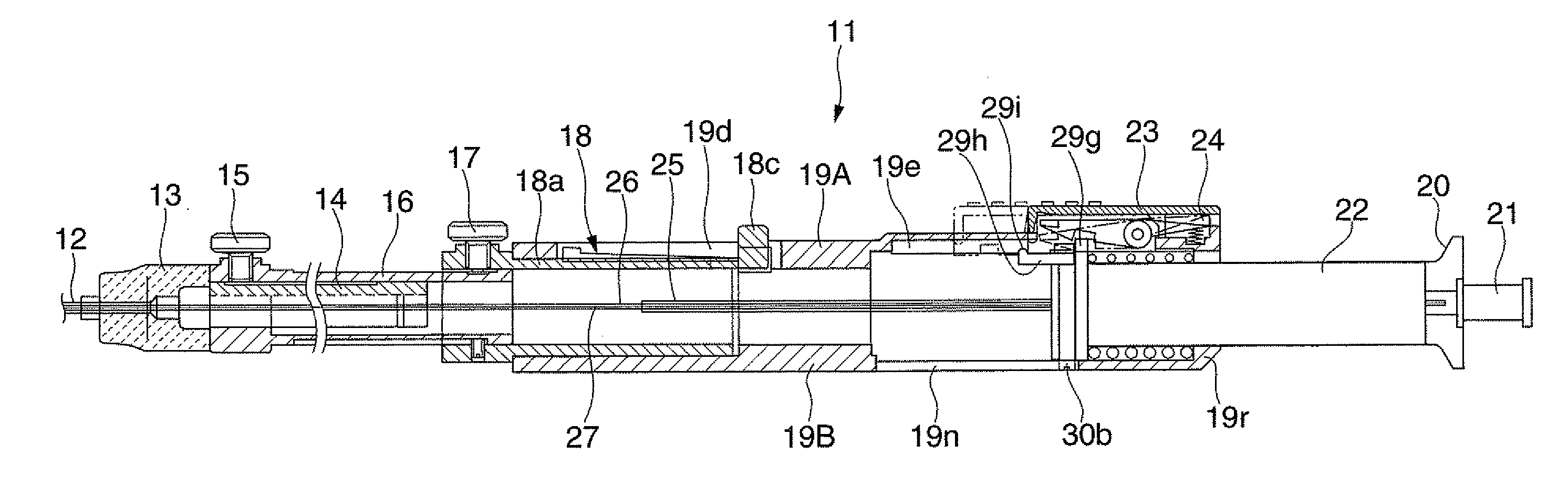
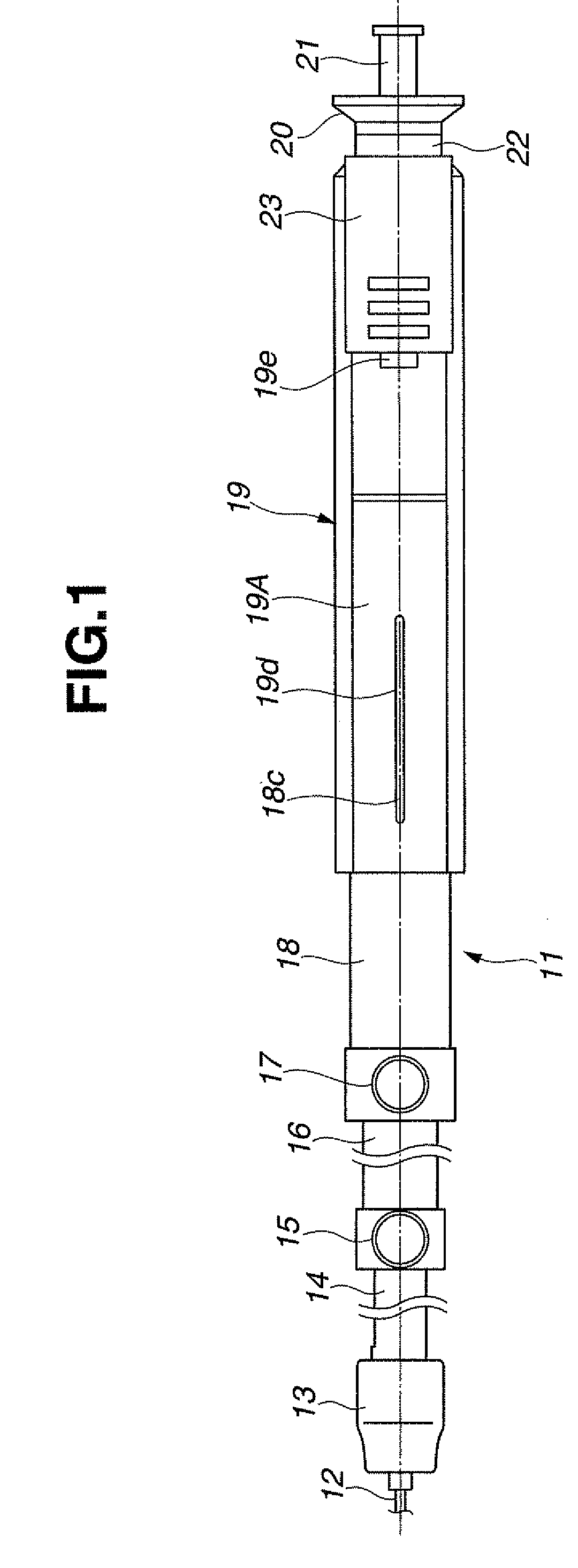
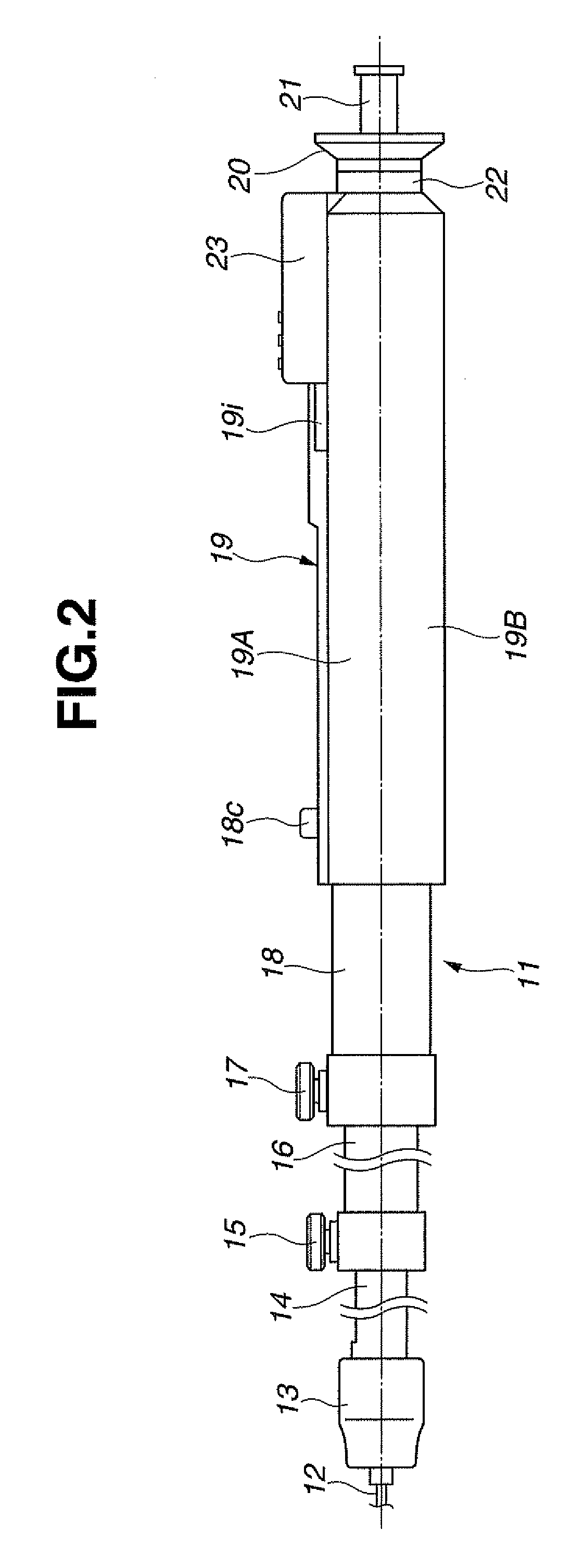
[0038]FIGS. 1 to 19 show a tissue biopsy needle apparatus according to the embodiment of the present invention. Specifically, FIGS. 1 and 2 show an external configuration of an operation portion of the tissue biopsy needle apparatus according to the embodiment of the present invention, where FIG. 1 is a top view and FIG. 2 is a side view. FIG. 3 is a cross-sectional view showing an internal configuration of a handle unit of the tissue biopsy needle apparatus according to the embodiment of the present invention in a preparatory stage for ejection of a puncture needle. FIG. 4 is a cross-sectional view showing an internal configuration of the handle unit of the tissue biopsy needle apparatus according to the embodiment of the present invention before a needle tube is drawn. FIGS. 5 and 6 show an upper part of a handle body installed in the handle unit of the tissue biopsy needle apparatus ac...


PUM


Abstract

The present invention is intended to provide a tissue biopsy needle apparatus equipped with a protective cover which prevents a needle tube from being ejected unnecessarily when an urging member is compressed in preparation for ejection of the needle tube. For that, the tissue biopsy needle apparatus includes a spring 28 which ejects the needle tube 26; a drawing unit which draws the spring 28 to a compressed position, the drawing unit including a drawing knob 20 and a drawing tube 29; an operation switch 24 which causes the drawing unit to draw and hold the spring 28 at the compressed position and causes the spring 28 to be released from the compressed position, thereby ejecting the needle tube 26; a protective cover 23 which disables the operation switch 24 when the spring 28 is drawn and held at the compressed position; and an elastic tongue 23c and an abutting strip29b which move the protective cover 23 from a working position to a non-working position of the operation switch 24 in accordance with drawing of the spring 28 to the compressed position by the drawing unit, the elastic tongue 23c being installed on the protective cover and the abutting strip 29b being installed on the drawing tube 29.

Description

TECHNICAL FIELD[0001]The present invention relates to a tissue biopsy needle apparatus, and more particularly, to a tissue biopsy needle apparatus used to puncture a subject site in a body cavity and take a sample of body tissue by being passed into a treatment instrument passage channel of an endoscope inserted into a body cavity.BACKGROUND ART[0002]Recently, a series of medical acts has found a general use which involves acquiring ultrasonic tomographic images of organs in a body cavity using an ultrasonic endoscope inserted in the body cavity, puncturing a body tissue with a puncture needle and taking samples of the body tissue while watching the ultrasonic tomographic images, and conducting a pathologic diagnosis using the tissue samples.[0003]The tissue biopsy needle apparatus configured to take samples of body tissue by watching ultrasonic tomographic images by means of an ultrasonic endoscope is used to take a sample of body tissue by passing a puncture needle into a treatmen...

Claims


Application Information

Patent Timeline
Patent Type & Authority Applications(United States)
IPC IPC(8): A61B10/02
CPCA61B10/0233A61B2019/4805A61B2010/0208A61B2090/0801
Inventor KODAMA, HIROSHI
Owner OLYMPUS MEDICAL SYST CORP
Why Eureka
Try Eureka