Medical Device for Constricting Tissue or a Bodily Orifice, for example a mitral valve

Inactive Publication Date: 2009-11-19
KARDIUM
View PDF99 Cites 490 Cited by
  • Summary
  • Abstract
  • Description
  • Claims
  • Application Information

AI Technical Summary

Benefits of technology

[0015]The present design of a medical device with enhanced capabilities for percutaneous deployment and annulus cinching employs a superior method for constricting tissue or a bodily orifice, such as the mitral valve, tricuspid valve, or aortic valve. The device may employ methods that enable a complete ring to be anchored to tissue and may enable reduction of the circumference of said ring during installation or at a later time. Reference throughout this spe

Problems solved by technology

However, the use of percutaneous technologies also raises some particular challenges.
Medical devices used in percutaneous surgery need to be deployed via narrow tubes called catheter sheaths, which significantly increase the complexity of the device structure.
As well, doctors do not have direct visual contact with the medical tools used once they are placed within the body, and positioning the tools correctly and operating the tools successfully can often be very challenging.
Improper function of any one of these structures or in combination can lead to mitral regurgitation.
The mechanical valve carries the risk of thromboembolism and requires anticoagulation with all of its potential hazards, whereas the biological prosthesis suffers from limited durability.
Another hazard with replacement is the risk of endocarditis.
Such a procedure is expensive, requires considerable time, and is associated with high morbidity and mortality.
Due to the risks associated with this procedure, many of the sickest patients are denied the potential benefits of surgical correction of mitral regurgitatio

Method used


Image

  • Medical Device for Constricting Tissue or a Bodily Orifice, for example a mitral valve
  • Medical Device for Constricting Tissue or a Bodily Orifice, for example a mitral valve
  • Medical Device for Constricting Tissue or a Bodily Orifice, for example a mitral valve

Examples


Embodiment Construction

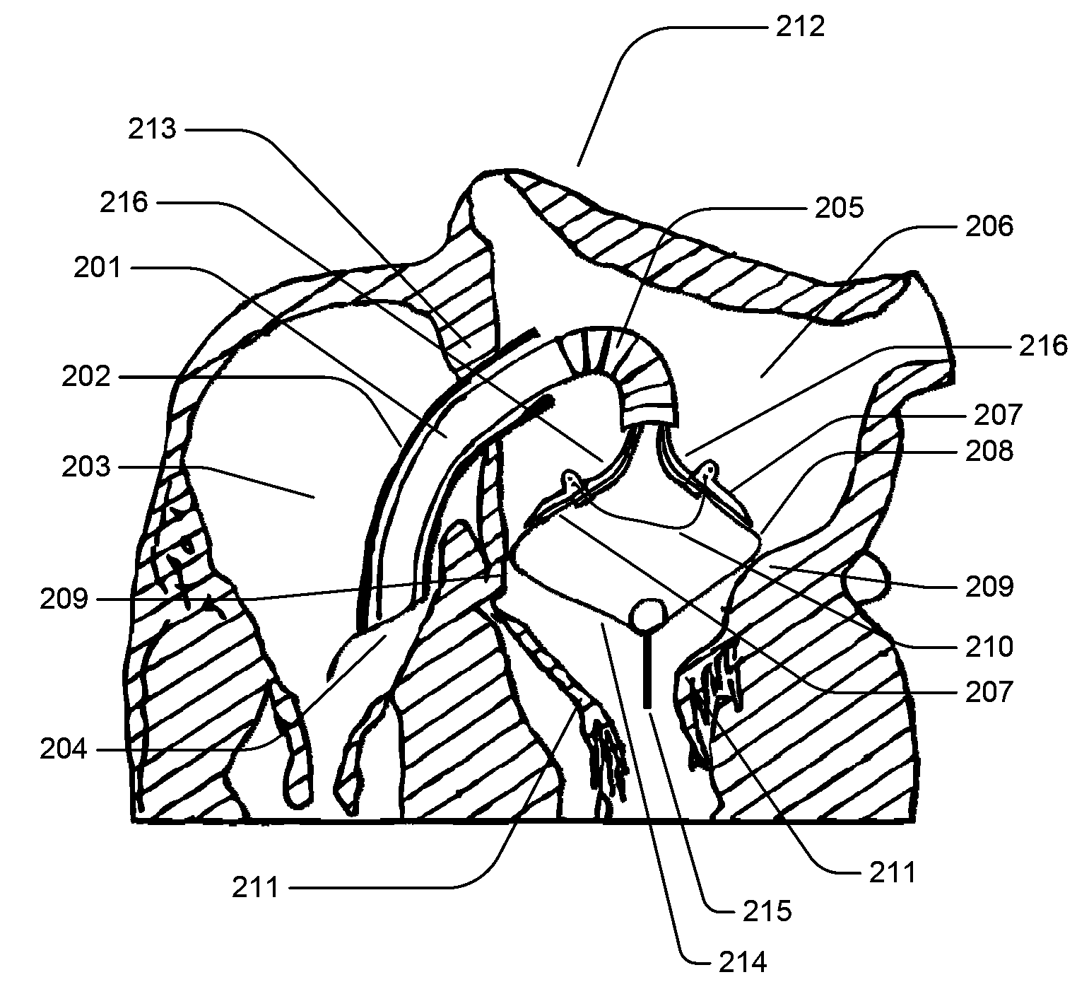
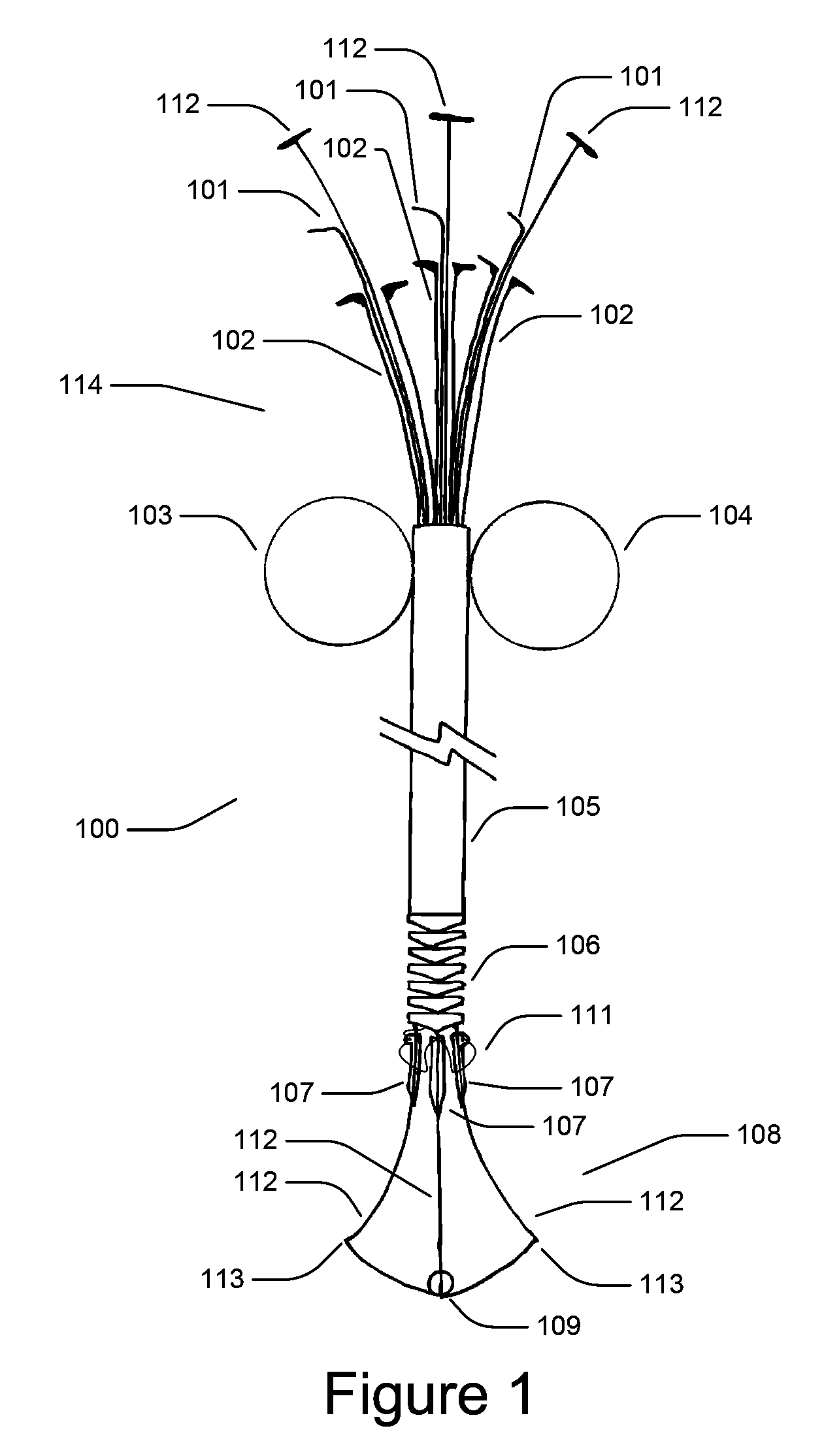
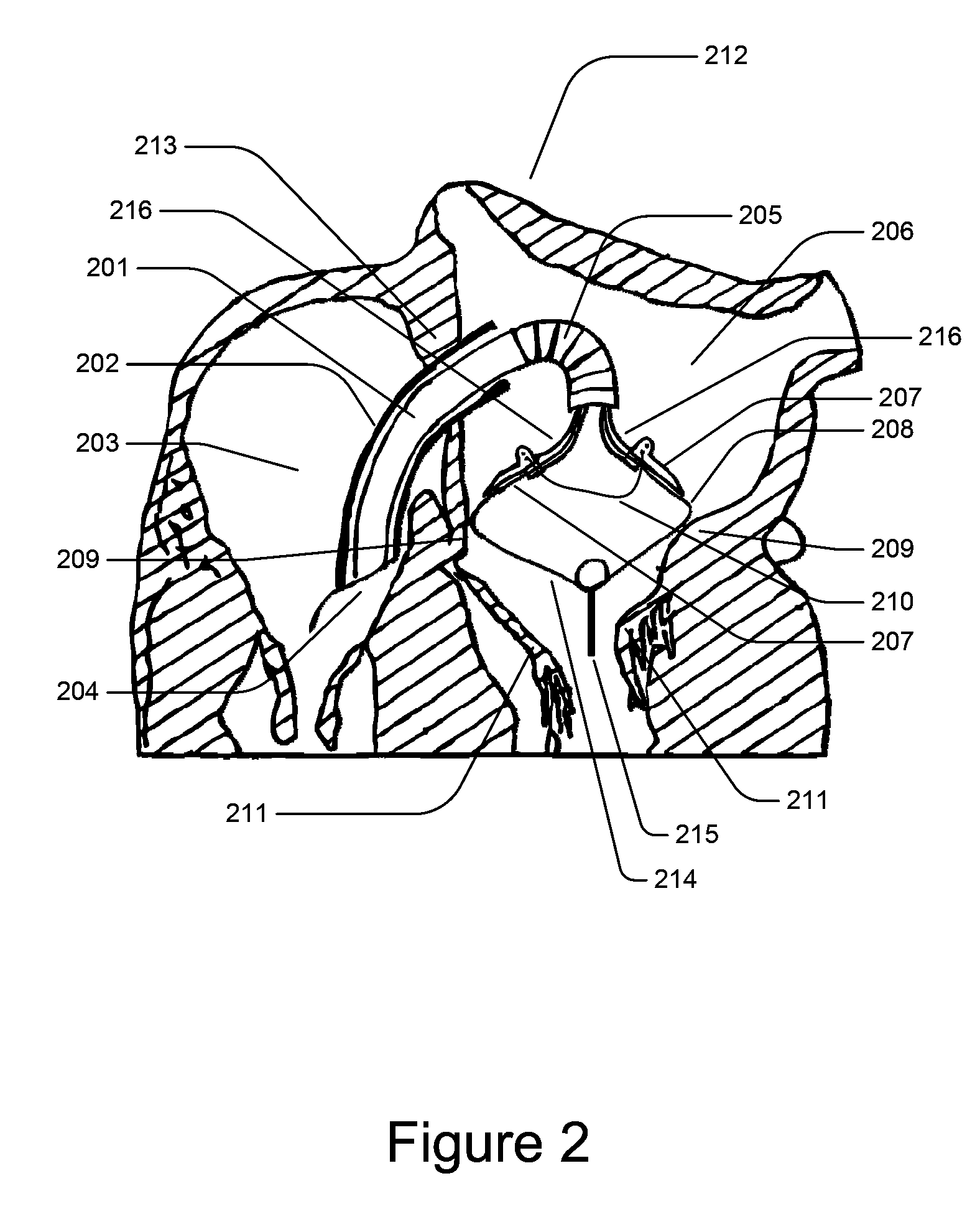
[0040]In the following description, certain specific details are set forth in order to provide a thorough understanding of various embodiments of the invention. However, one skilled in the art will understand that the invention may be practiced without these details. In other instances, well-known structures have not been shown or described in detail to avoid unnecessarily obscuring descriptions of the embodiments of the invention.

[0041]Unless the context requires otherwise, throughout the specification and claims which follow, the word “comprise” and variations thereof, such as, “comprises” and “comprising” are to be construed in an open, inclusive sense, that is as “including, but not limited to.”

[0042]Reference throughout this specification to “one embodiment” or “an embodiment” means that a particular feature, structure or characteristic described in connection with the embodiment is included in at least one embodiment of the present invention. Thus, the appearances of the phras...


PUM


Abstract

A medical apparatus positionable in a cavity of a bodily organ (e.g., a heart) may constrict a bodily orifice (e.g., a mitral valve). The medical apparatus may include tissue anchors that are implanted in the annulus of the orifice. The tissue anchors may be guided into position by an intravascularly or percutaneously deployed anchor guiding frame. Constriction of the orifice may be accomplished by cinching a flexible cable attached to implanted tissue anchors. The medical device may be used to approximate the septal and lateral (clinically referred to as anterior and posterior) annulus of the mitral valve in order to move the posterior leaflet anteriorly and the anterior leaflet posteriorly and thereby improve leaflet coaptation and eliminate mitral regurgitation.

Description

BACKGROUND[0001]1. Field[0002]This disclosure is generally related to percutaneous or minimally invasive surgery, and more particularly to percutaneously deployed medical devices suitable for constricting tissue or a bodily orifice such as a mitral valve[0003]2. Description of the Related Art[0004]Cardiac surgery was initially undertaken only by performing a sternotomy, a type of incision in the center of the chest, which separates the sternum (chest bone) to allow access to the heart. In the previous several decades, more and more cardiac operations are performed using a percutaneous technique, which is a medical procedure where access to inner organs or other tissue is gained via a catheter.[0005]Percutaneous surgeries benefit patients by reducing surgery risk, complications, and recovery time. However, the use of percutaneous technologies also raises some particular challenges. Medical devices used in percutaneous surgery need to be deployed via narrow tubes called catheter sheat...

Claims


Application Information

Patent Timeline
IPC IPC(8): A61B17/12A61F2/24
CPCA61B17/0401A61B17/0469A61B17/0485A61B2017/00331A61B2017/0409A61F2/2454A61B2017/0441A61B2017/0464A61B2017/0496A61B2017/22038A61F2/2445A61B2017/0437
Inventor DAHLGREN, JONGOERTZEN, DOUGGELBART, DANIEL
Owner KARDIUM
Why Eureka
Try Eureka