
Compound Imaging with HIFU Transducer and Use of Pseudo 3D Imaging

a technology of hifu transducer and compound imaging, which is applied in the field of compound imaging with hifu transducer and use of pseudo 3d imaging, can solve the problems of bulky and bandwidth intensive imaging equipment to be used, the unharmed interfacial tissue between the hifu source and the hifu focal point remains, and the imaging equipment is bulky and intensiv

Inactive Publication Date: 2009-12-31
MIRABILIS MEDICA
View PDF91 Cites 23 Cited by
  • Summary
  • Abstract
  • Description
  • Claims
  • Application Information

AI Technical Summary

Benefits of technology

[0008]Generally, the present invention contemplates the use of ultrasound imaging and therap...

Problems solved by technology

However, when the HIFU energy is focused at locations deep in tissue, the intervening tissue between the HIFU source and the HIFU focal point remains unharmed.
These approaches require bulky and bandwidth intensive imaging equipment to be used external to the tissue to be treated.

Method used


Image

  • Compound Imaging with HIFU Transducer and Use of Pseudo 3D Imaging
  • Compound Imaging with HIFU Transducer and Use of Pseudo 3D Imaging
  • Compound Imaging with HIFU Transducer and Use of Pseudo 3D Imaging

Examples


Embodiment Construction

[0023]Certain specific details are set forth in the following description and figures to provide an understanding of various embodiments of the invention. Certain well-known details, associated electronics and devices are not set forth in the following disclosure to avoid unnecessarily obscuring the various embodiments of the invention. Further, those of ordinary skill in the relevant art will understand that they can practice other embodiments of the invention without one or more of the details described below. Finally, while various processes are described with reference to steps and sequences in the following disclosure, the description is for providing a clear implementation of particular embodiments of the invention, and the steps and sequences of steps should not be taken as required to practice this invention.

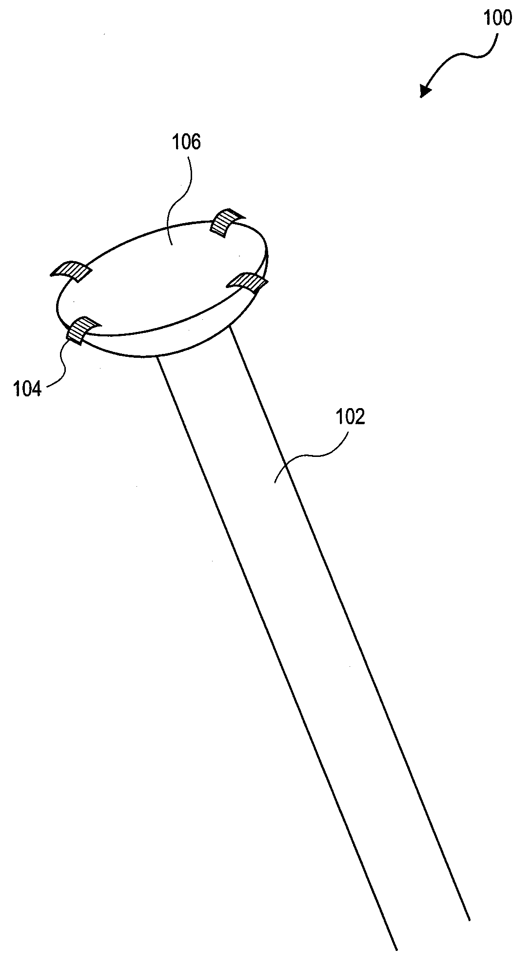
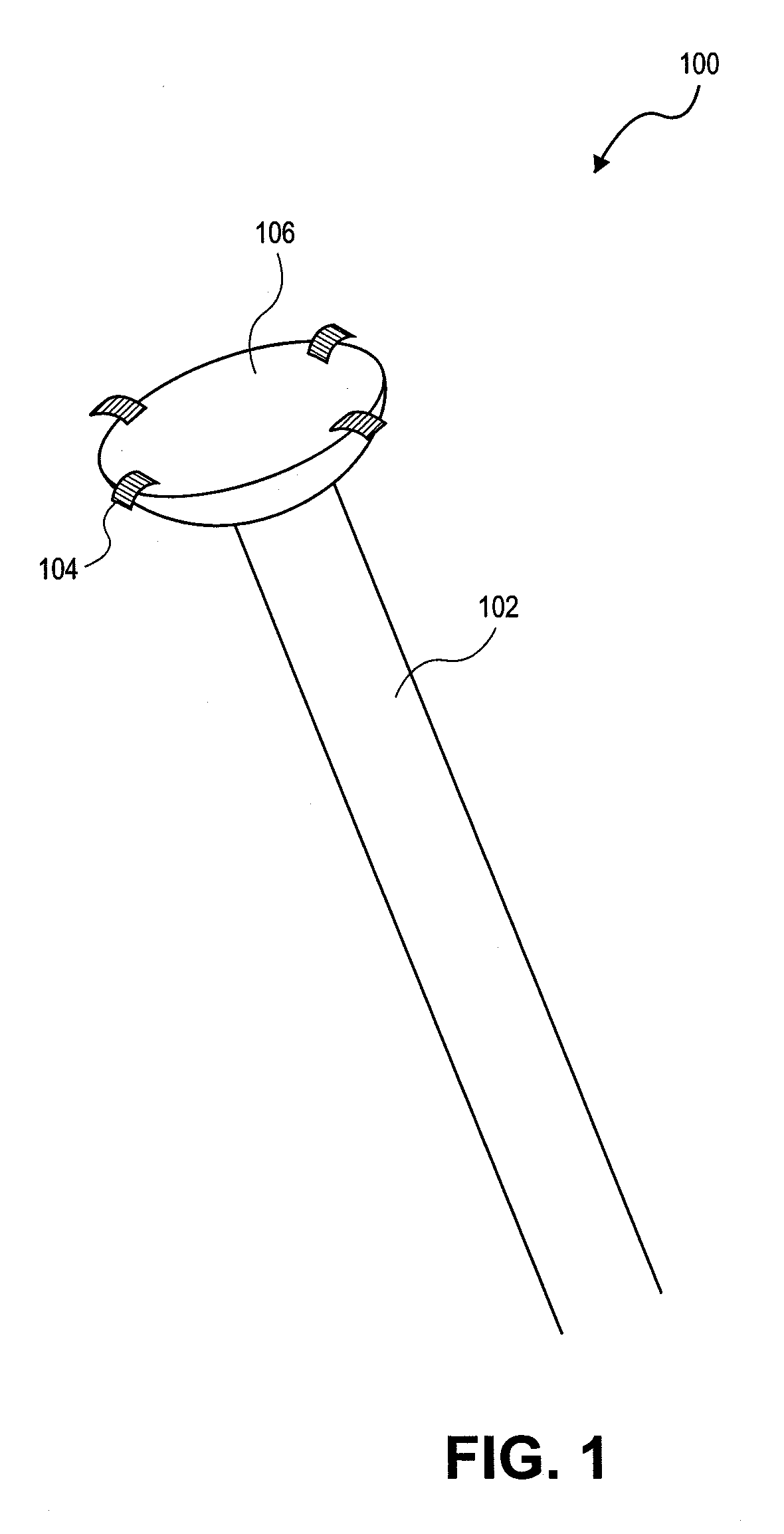
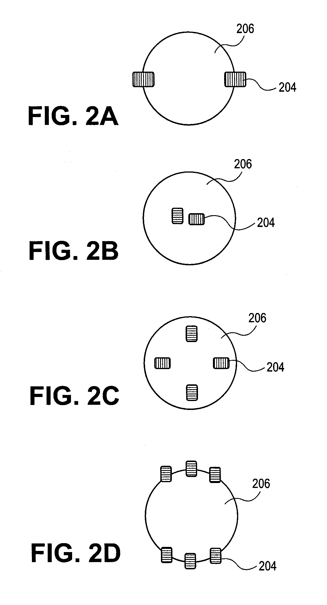
[0024]FIG. 1 illustrates a medical imaging and therapy applicator 100 comprising applicator body 102, ultrasound imaging transducers 104, and therapy transducer 106. Any...


PUM


Abstract

A medical imaging and therapy applicator is provided that may include any of a number of features. One feature of the medical imaging and therapy applicator is that it can image target tissue of a patient with ultrasound imaging transducers. Another feature of the medical imaging and therapy applicator is that it can display the imaging information and provide therapeutic energy to the target tissue. Methods associated with use of the medical imaging and therapy applicator are also covered.

Description

INCORPORATION BY REFERENCE[0001]All publications and patent applications mentioned in this specification are herein incorporated by reference to the same extent as if each individual publication or patent application was specifically and individually indicated to be incorporated by reference.FIELD OF THE INVENTION[0002]The present application is direct to methods and apparatus that provide ultrasound imaging and therapeutic treatment of internal pathological conditions using high intensity focused ultrasound energy.BACKGROUND OF THE INVENTION[0003]Ultrasound is used in medical settings as a diagnostic aid for imaging internal structures. Advantages of ultrasound over other imaging forms include low cost, portability, and safety.[0004]A variety of therapeutic uses of ultrasound have been described. Some applications take advantage of the ability of high intensity focused ultrasound (HIFU) waves to generate heat and thus destroy tissue such as tumors or blood vessels. Currently, HIFU ...

Claims


Application Information

Patent Timeline
IPC IPC(8): A61N7/00A61B8/00
CPCA61B8/14A61B8/4483A61N7/022A61B2017/4216A61B2019/5276A61B2017/2253A61B2090/378
Inventor DARLINGTON, GREGORYLAU, MICHAELCONNOLLY, MICHAEL
Owner MIRABILIS MEDICA
Learn More