Method of image manipulation to fade between two images

a technology of image manipulation and image, applied in the field of medical imaging, can solve the problems of undesirable glint, unpredictably better, etc., and achieve the effect of easy fading and better results

Inactive Publication Date: 2010-02-11
STI MEDICAL SYST
View PDF12 Cites 21 Cited by
  • Summary
  • Abstract
  • Description
  • Claims
  • Application Information

AI Technical Summary

Benefits of technology

[0016]This invention newly recognizes that it is desirable to provide the clinician with information from both the image with glint and the glint-free image, and to develop a method for easily fading between the two, to desired degrees. Thus, the present invention of a method of controlling the fading between an image with glint and a glint-free image is quite advantageous to cancer detection because it maintains the relationship between the image with glint and the glint-free image, meaning that the clinician is enabled to detect important features that may be masked by glint, and (by varying fading) it permits the three-dimensional shape and surface texture information in the image with glint to also be discerned. The ability to maintain the glint in an image to a user-selected extent to aid in the diagnosis of cancer provides unexpectedly and unpredictably better results over the prior art (which teaches that glint is undesirable). Another way to describe the invention is a method by which one can

Problems solved by technology

The ability to maintain the glint in an image to a user-selected extent to aid in the diagnosis of cancer provi

Method used


Image

  • Method of image manipulation to fade between two images
  • Method of image manipulation to fade between two images
  • Method of image manipulation to fade between two images

Examples


Embodiment Construction

1. System Framework

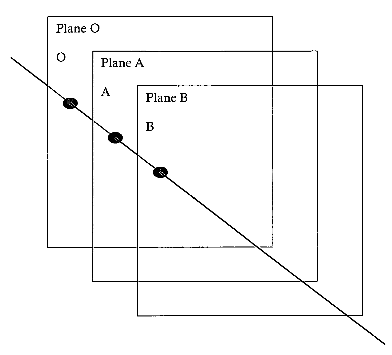
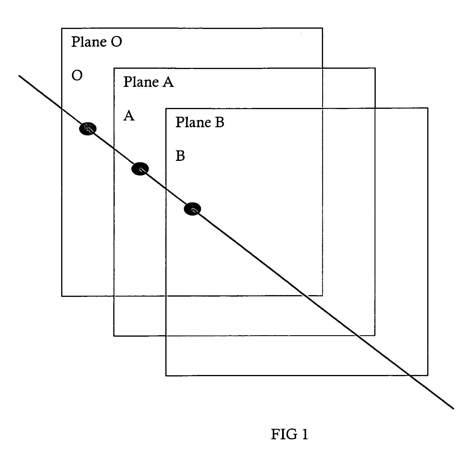
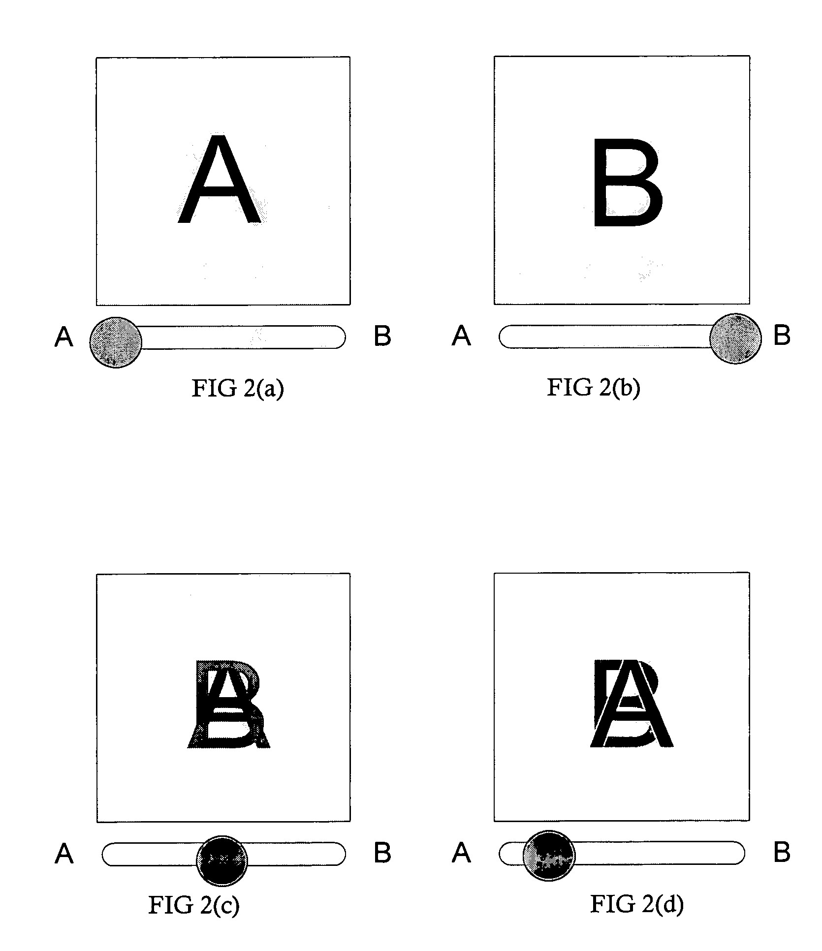
[0021]The presently preferred embodiment of the invention discloses a process for fading between (adjusting the comparative opacity or transparency) two digital images of tissue or an organ (such as the cervix) obtained during an examination with a digital imager (such as a colposcope) in order to provide the user with a means to choose to combine an actual image (image with glint) with a glint-free image, to a user-controllable extent, to aid in the diagnosis of cancer.

[0022]First, an image with glint (such as an unpolarized, parallel-polarized, or singly-polarized image) and a glint-free image (such as a cross-polarized image) are obtained (collected) using a digital imager. Cross-polarized (XP) is when a first polarization orientation is perpendicular to a second polarization orientation. Parallel-polarized (PP) is where a first polarization orientation is parallel to the second polarization orientation. PP can also mean singly-polarized where there is only one...


PUM


Abstract

A process to fade between two frames of a dual frame digital TIFF image of an organ taken during an examination with a colposcope to use in computer-aided-diagnosis (CAD) systems.

Description

TECHNICAL FIELD [0001]This invention generally relates to medical imaging and, more specifically to, a method of fading between an image with glint and an image without glint for diagnostic purposes. The method can be used to achieve high-quality standardized digital imagery to use in archive-quality medical records and Computer-Aided-Diagnosis (CAD) systems.BACKGROUND ART [0002]Although this invention is being disclosed in connection with cervical cancer, it is applicable to many other areas of medicine. Uterine cervical cancer is the second most common cancer in women worldwide, with nearly 500,000 new cases and over 270,000 deaths annually (LARC, “Globocan 2002 database, “International agency for research in cancer, 2002, incorporated herein by reference). Colposcopy is a diagnostic method used to detect cancer precursors and cancer of the uterine cervix (B. S. Apgar, Brotzman, G. L. and Spitzer, M., Colposcopy: Principles and Practice, W. B. Saunders Company: Philadelphia, 2002,...

Claims


Application Information

Patent Timeline
IPC IPC(8): G09G5/00
CPCG06T7/0024G06T13/80G09G2380/08G09G2340/10G06T15/503G06T7/30
Inventor WHITESELL, ANDREW BEAUMONTOFIESH, GREG RAYMOND
Owner STI MEDICAL SYST
Why Eureka
Try Eureka