System and Method for Coronary Digital Subtraction Angiography

a digital subtraction angiography and digital subtraction technology, applied in the field of fluoroscopic image sequences, can solve the problems of blood-filled structures that cannot be distinguished from surrounding tissue using conventional radiology, information that is difficult to decipher, and it is difficult to accurately distinguish between these structures in the fluoroscopic image sequence using the traditional intensity based approach

Active Publication Date: 2010-02-11
SIEMENS HEALTHCARE GMBH
View PDF15 Cites 45 Cited by
  • Summary
  • Abstract
  • Description
  • Claims
  • Application Information

AI Technical Summary

Problems solved by technology

Since blood has the same radiodensity as the surrounding tissues, these blood-filled structures cannot be differentiated from the surrounding tissue using conventional radiology.
Such fluoroscopic image sequences contain useful information that can be difficult to decipher due to the collapsing of 3-dimensional information into the 2-dimensional images.
However, such traditional techniques can yield erroneous results in medical image sequences, such as fluoroscopic image sequences, which are generated using the phenomenon of transparency.
Since various internal structures have different levels of transparency in the fluoroscopic images, these structures can overlap, and it may be difficult to accurately distinguish between these structures in the fluoroscopic image sequences using the traditional intensity based approaches.

Method used


Image

  • System and Method for Coronary Digital Subtraction Angiography
  • System and Method for Coronary Digital Subtraction Angiography
  • System and Method for Coronary Digital Subtraction Angiography

Examples


Embodiment Construction

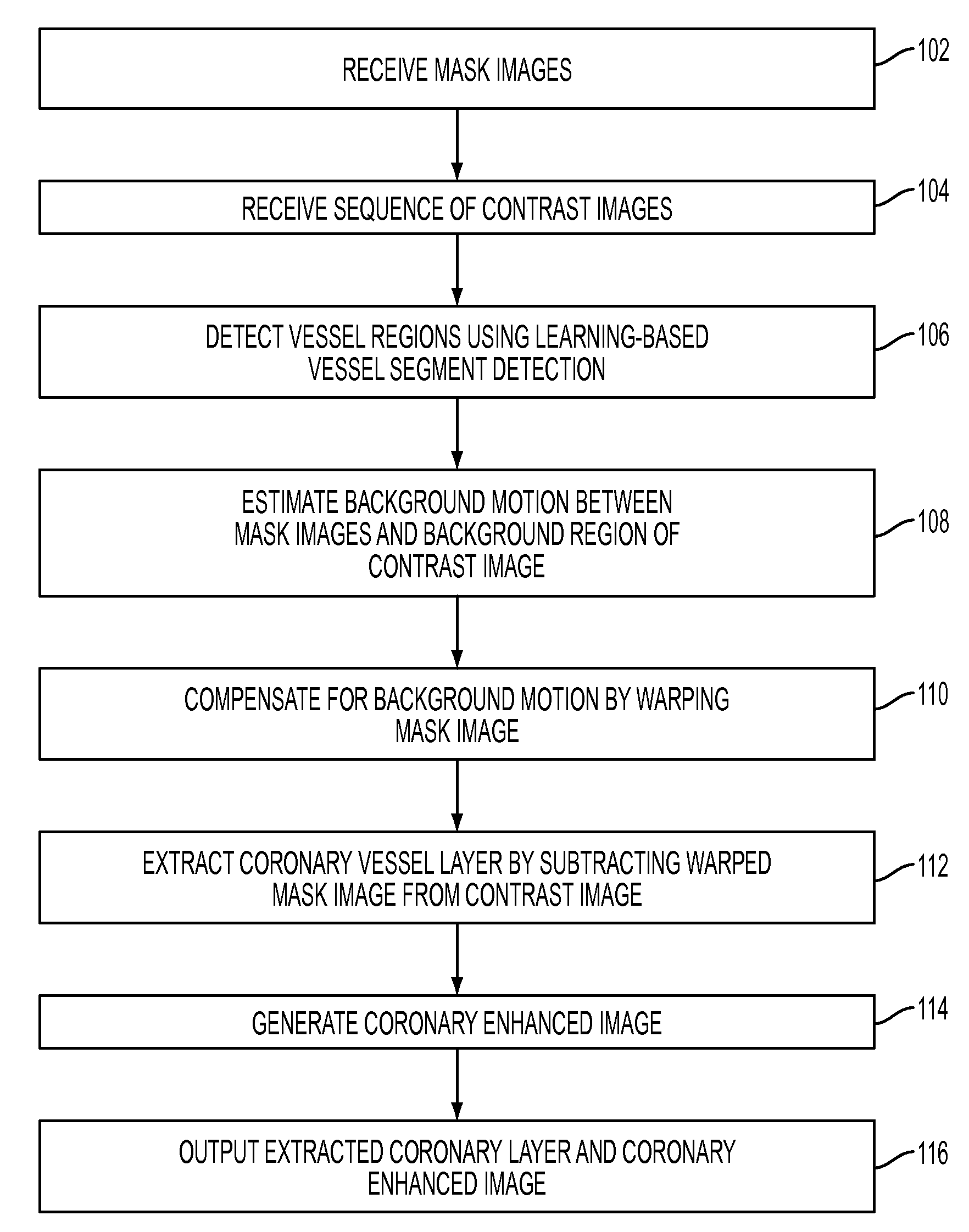
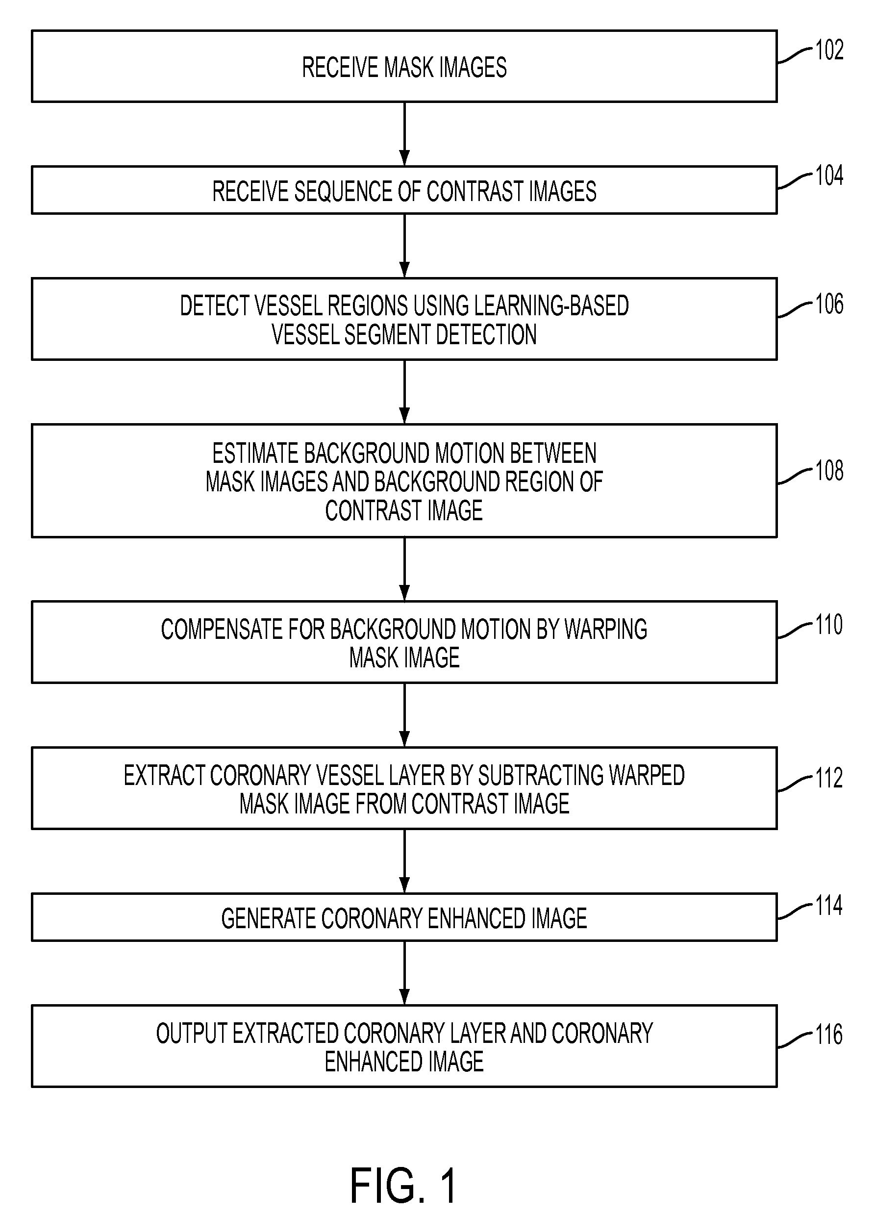
[0015]The present invention is directed to a method for detecting coronary vessels from fluoroscopic images. Embodiments of the present invention are described herein to give a visual understanding of the coronary vessel extraction method. A digital image is often composed of digital representations of one or more objects (or shapes). The digital representation of an object is often described herein in terms of identifying and manipulating the objects. Such manipulations are virtual manipulations accomplished in the memory or other circuitry / hardware of a computer system. Accordingly, is to be understood that embodiments of the present invention may be performed within a computer system using data stored within the computer system.

[0016]Digital subtraction angiography (DSA) is a technique for visualizing blood vessels in the human body. In DSA, a sequence of fluoroscopic images, referred to as contrast images, is acquired to show the passage of contrast medium that is injected into ...


PUM


Abstract

A method and system for extracting coronary vessels fluoroscopic image sequences using coronary digital subtraction angiography (DSA) are disclosed. A set of mask images of a coronary region is received, and a sequence of contrast images for the coronary region is received. For each contrast image, vessel regions are detected in the contrast image using learning-based vessel segment detection and a background region of the contrast image is determined based on the detected vessel regions. Background motion is estimated between one of the mask images and the background region of the contrast image by estimating a motion field between the mask image and the background image and performing covariance-based filtering over the estimated motion field. The mask image is then warped based on the estimated background motion to generate an estimated background layer. The estimated background layer is subtracted from the contrast image to extract a coronary vessel layer for the contrast image.

Description

[0001]This application claims the benefit of U.S. Provisional Application No. 61 / 086,505, filed Aug. 6, 2008, the disclosure of which is herein incorporated by reference.BACKGROUND OF THE INVENTION[0002]The present invention relates to fluoroscopic image sequences, and more particularly to detecting coronary vessel layers from fluoroscopic image sequences.[0003]Angiography is a medical imaging technique in which X-ray images are used to visualize internal blood filled structures, such as arteries, veins, and the heart chambers. Since blood has the same radiodensity as the surrounding tissues, these blood-filled structures cannot be differentiated from the surrounding tissue using conventional radiology. Thus, in angiography, a contrast agent is added to the blood, usually via a catheter, to make the blood vessels visible via X-ray. In many angiography procedures, X-ray images are taken over a period of time, which results in a sequence of fluoroscopic images, which show the motion o...

Claims


Application Information

Patent Timeline
Patent Type & Authority Applications(United States)
IPC IPC(8): G06K9/00G06V10/28
CPCG06K9/38G06K2009/00932G06T7/0014G06T2207/30101G06T2207/20081G06T2207/20144G06T2207/20076G06T7/194G06V40/14G06V10/28
Inventor ZHU, YINGPRUMMER, SIMONECHEN, TERRENCEOSTERMEIER, MARTINCOMANICIU, DORIN
Owner SIEMENS HEALTHCARE GMBH
Why Eureka
Try Eureka