List Mode-Based Respiratory and Cardiac Gating in Positron Emission Tomography

a positron emission tomography and respiratory and cardiac gating technology, applied in tomography, instruments, applications, etc., can solve the problems of reducing image resolution, cardiac pet/ct faces further difficulties, and not all generated gamma photons can be detected

Inactive Publication Date: 2010-03-18
UNIVERSLTAT MUNSTER +1
View PDF2 Cites 26 Cited by
  • Summary
  • Abstract
  • Description
  • Claims
  • Application Information

AI Technical Summary

Problems solved by technology

One problem in PET is the fact that not all generated gamma photons can be detected as many photons travelling through tissue undergo absorption processes.
However, cardiovascular PET / CT faces further difficulties based on the difference in scanning time between CT (usually a few seconds) and PET (a few minutes per bed position).
Thus CT images only represent one respiratory phase, while PET images comprise a superposition of all phases during the PET scan, leading to image blurring, therefore effectively reducing the image resolution.
Furthermore, CT-based attenuation correction may introduce image artifacts in such cases where the CT respiratory phase does not match the averaged PET respiratory phase.
The result may be a spatial mismatch between CT and PET and erroneous tracer quantification (Lang N, Dawood M, Büther F, Schober O, Schäfers M, Schäfers K. Organ Movement-Reduction in PET / CT using Dual-Gated List mode Acquisition.
However, it is not clear if a few breathing cycles really match the situation during the PET scan.
Moreover, this does not solve the problem of motion blurring in the obtained images.
A disadvantage of all these methods is the usage of additional hardware during the PET / CT scan, introducing additional potential errors of measurement.
Unfortunately, these methods are extraordinarily time-consuming (as each time frame has to be reconstructed before extracting motion information) and therefore much too intricate for clinical studies.

Method used


Image

  • List Mode-Based Respiratory and Cardiac Gating in Positron Emission Tomography
  • List Mode-Based Respiratory and Cardiac Gating in Positron Emission Tomography
  • List Mode-Based Respiratory and Cardiac Gating in Positron Emission Tomography

Examples


Embodiment Construction

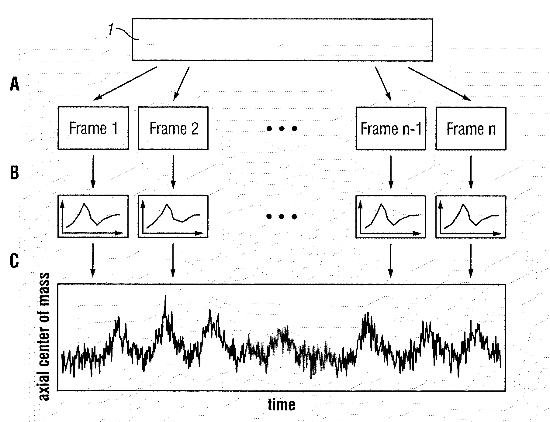
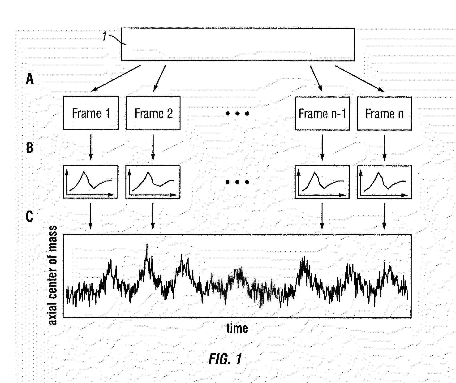
[0028]The present invention is based on extracting internal organ motion from PET coincidence data. The embodiments of the present invention described hereinafter require using a PET scanner with list mode capability.

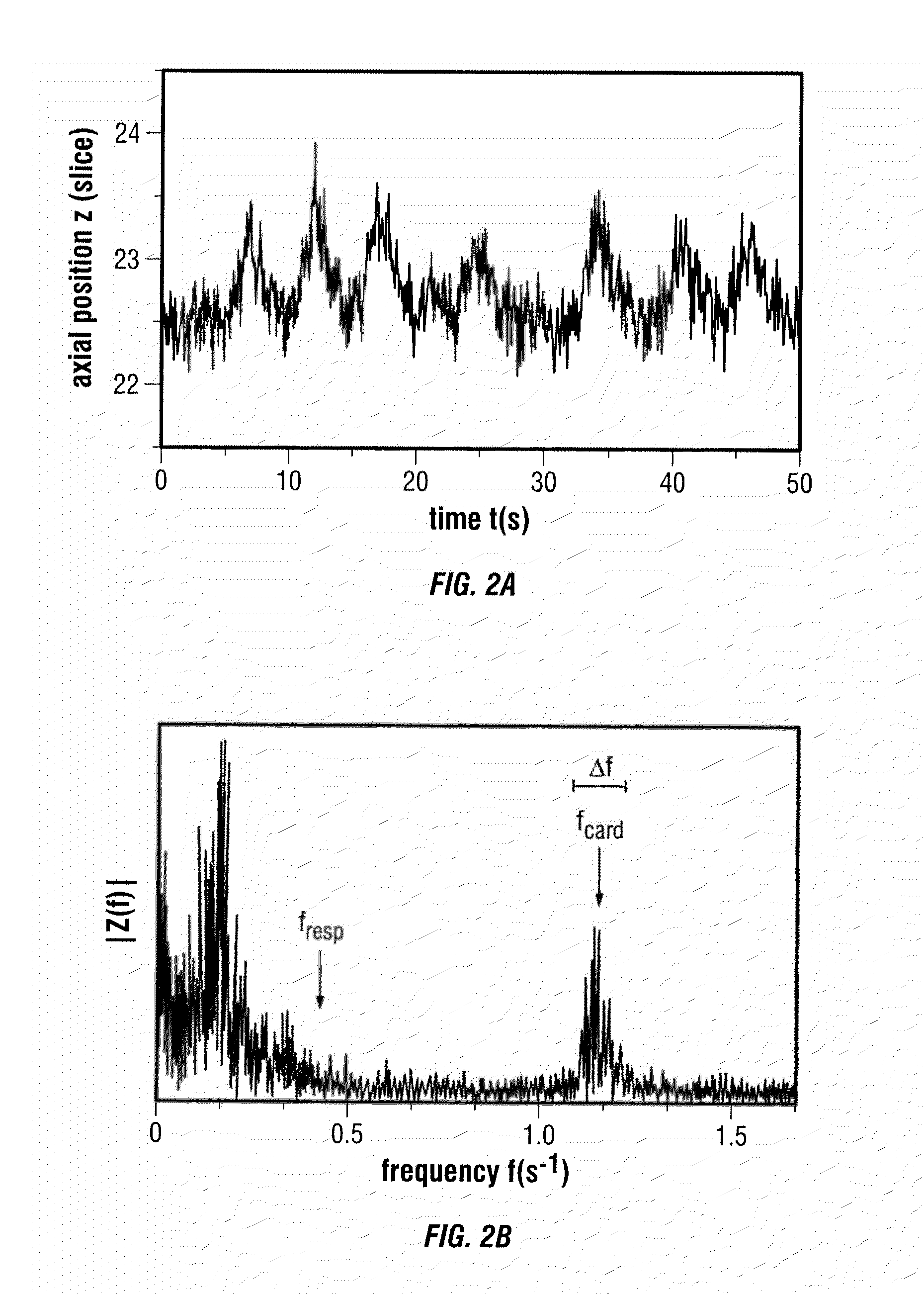
[0029]According to a preferred embodiment of the present invention, the case where heart contraction is connected to an axial motion shift and a heart beat peak is visible in the spectrum is described. The axial center of mass is then plotted as a function of time. Using a Fourier transform, the data is transformed into the frequency domain making it possible to identify and in turn isolate the respiratory and cardiac part of the spectrum respectively. Using an inverse Fourier transform, respiratory and cardiac curves can be computed with which a gating sequence can then be established.

[0030]According to another embodiment of the present invention, the case where heart contraction is not connected to an axial motion shift and no heart beat peak is visible in the spectru...


PUM


Abstract

According to a preferred embodiment, the invention provides a method for extracting internal organ motion from positron emission tomography (PET) coincidence data, the method comprising the following steps: generating a data stream of PET coincidence data using the list mode capability of a PET scanner; dividing the data stream into time frames of a given length; computing a histogram A(i, t) of an axial coincidence distribution for a set of time frames; computing the axial center of mass z(t) for each of the time frames in the set of time frames based on the histogram A(i, t); transforming z(t) into the frequency domain; determining either the frequency contribution caused by respiratory motion, given by fresp, or the frequency contribution caused by heart contractions, given by fcard and Δf, identified in the frequency spectrum |Z(f)|; and carrying out further processing of Z(f) leading to curves zresp(t) and zcard(t) with which a gating sequence is established.

Description

CROSS REFERENCE TO RELATED APPLICATIONS[0001]The present application claims the benefit of prior U.S. Provisional Application No. 61 / 111,360, filed Nov. 5, 2008.BACKGROUND OF THE INVENTION[0002]1. Technical Field[0003]The invention relates to a method for extracting internal organ motion directly from PET coincidence data. More particularly, the invention relates to a method for extracting internal organ motion directly from PET coincidence data in the case of myocardial viability FDG scans.[0004]2. Description of the Prior Art[0005]The combination of Positron emission tomography (PET) and computed tomography (CT) has proven to be a valuable tool in medical imaging, in particular because each of these modalities gathers different information that when combined improve medical diagnosis.[0006]PET is based on positron-emitting isotopes like 18F which are attached to specific molecules (called tracers; e.g., FDG) and which are introduced into the human body. Emitted positrons annihilat...

Claims


Application Information

Patent Timeline
Patent Type & Authority Applications(United States)
IPC IPC(8): G06K9/00
CPCA61B6/032A61B6/541A61B6/5217A61B6/037G16H50/30
Inventor BUTHER, FLORIANSCHAFERS, KLAUSJONES, WILLIAM F.BENDRIEM, BERNARDNEWIGER, HARTWIG
Owner UNIVERSLTAT MUNSTER
Why Eureka
Try Eureka