Imaging Agents Useful for Identifying AD Pathology

a technology of amyloid binding and imaging agent, which is applied in the field of amyloid binding compounds, can solve the problems of increasing the risk of developing alzheimer's disease, requiring invasive procedures such as spinal taps, and the method has not proven reliable in all patients, so as to increase the binding of the compound and improve the effect of treatment or prophylaxis

Active Publication Date: 2010-04-22
SIEMENS MEDICAL SOLUTIONS USA INC
View PDF6 Cites 18 Cited by
  • Summary
  • Abstract
  • Description
  • Claims
  • Application Information

AI Technical Summary

Problems solved by technology

Alzheimer's disease (AD) is the most common form of neurodegeneration and has become a major healthcare issue with the aging population in the United States.
As people advance past the age of 65, the risk for developing Alzheimer's disease increases.
Unfortunately, at the stage of presentation of symptoms in the clinic, the patients have developed significant amyloid deposition in the neurological tissue.
Immunoassay methods have been developed for detecting the presence of neurochemical markers in AD patients and to detect an AD related amyloid protein in cerebral spinal fluid, however such methods have not proven reliable in all patients and require invasive procedures such as a spinal tap.
Monoclonal antibodies and radiolabeled peptides have been used as probes for imaging of Aβ, however these macromolecular structures provide inherently poor brain penetration (see, for example, Majocha, et al., J. Nucl. Med., 33: 2184 (1992)).
Additionally, the peptide probes may lack specificity for AD as they tend to react with diffuse plaques.
Despite the utility of cholinesterase inhibitors, the current array of AD therapeutics merely serves to delay full-onset AD by approximately two to three years, after which they are therapeutically ineffective in inhibiting cognitive decline.

Method used


Image

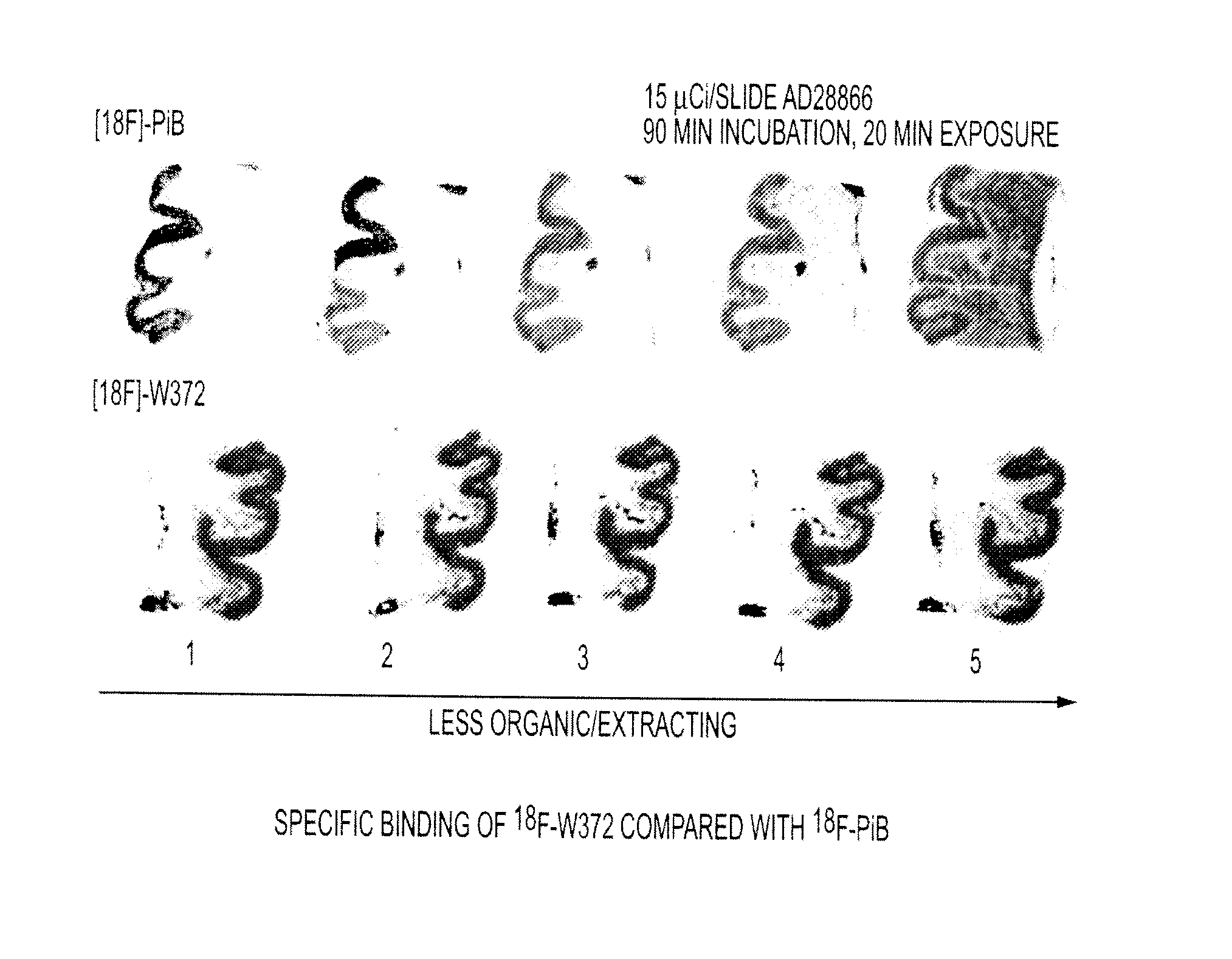
  • Imaging Agents Useful for Identifying AD Pathology
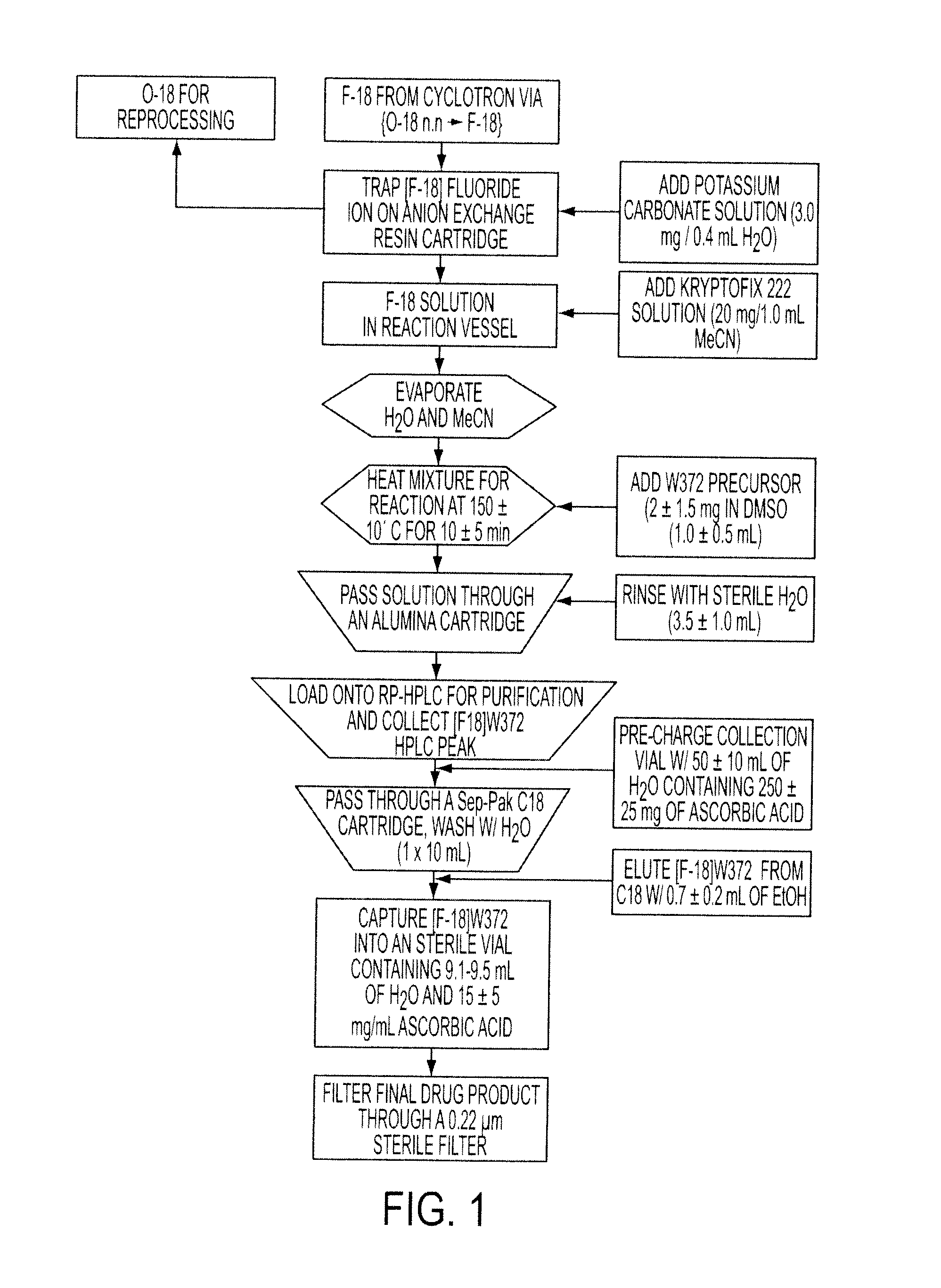
  • Imaging Agents Useful for Identifying AD Pathology
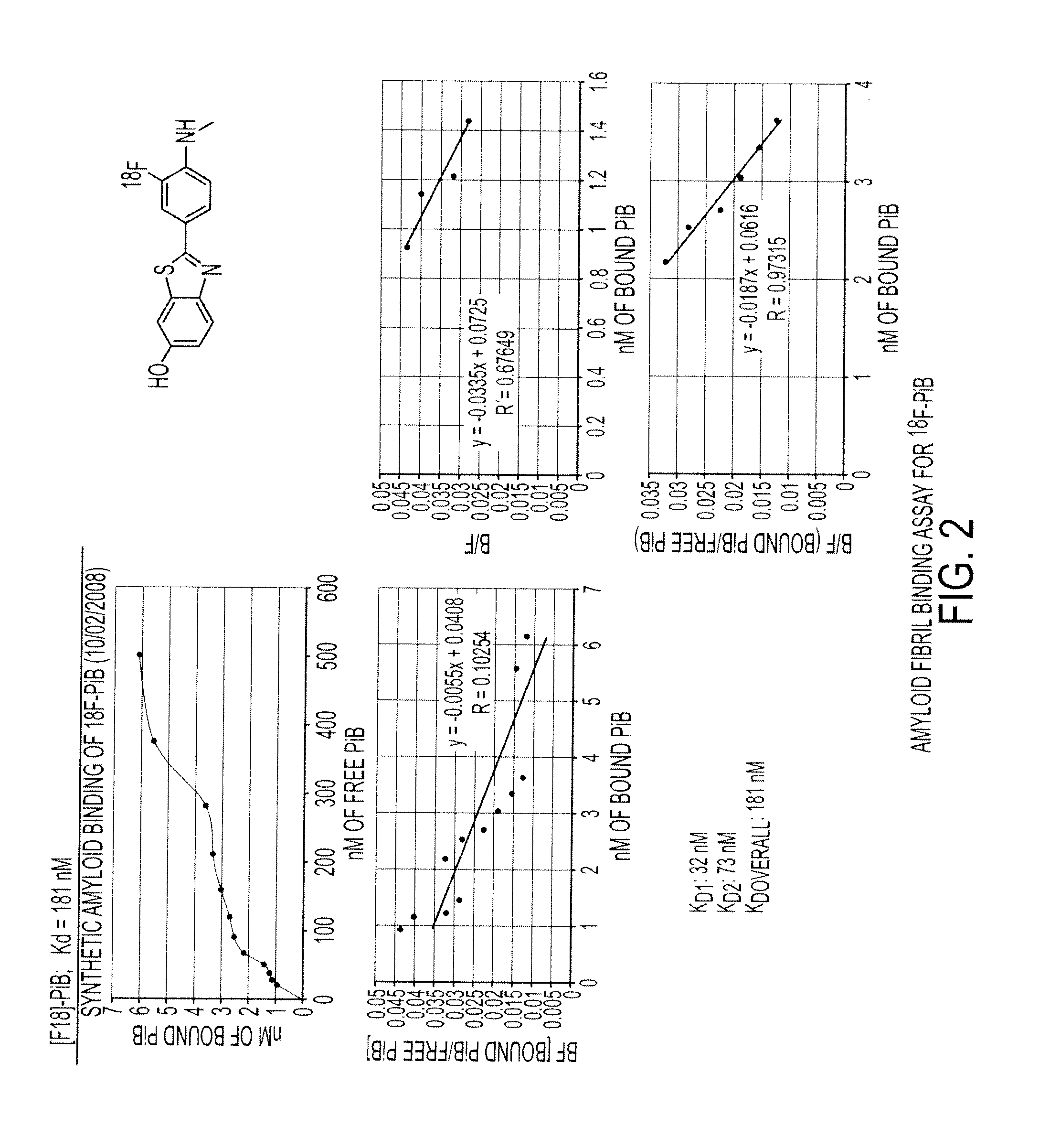
  • Imaging Agents Useful for Identifying AD Pathology

Examples


examples

Synthesis of UG-4-69

[0305]

Preparation of 2-(4-(dimethylamino)phenyl)thiazole-5-carbaldehyde (3)

[0306]To a 10 mL round bottomed flask equipped with a magnetic stir bar and reflux condenser containing dioxane (3 mL) was placed 1 (0.1 g, 0.61 mmol) and 2 (0.14 g, 0.73 mmol). To this solution sat. NaHCO3 (3 mL) and Pd(Ph3P)4 (0.07 g, 0.06 mmol) were added and the reaction was heated to 100° C. for 4 hrs. The reaction was cooled to RT, filtered through celite, then poured into water (20 mL) and extracted into EtOAc (3×15 mL). The combined organic extracts were washed with water (15 mL), brine (15 mL), dried over MgSO4 and concentrated in vacuo. The residue was purified over silica gel using DCM:Hexanes (4:1) as an eluent to afford 3 (0.1 g, 71%) as a white solid.

[0307]LC / MS: Expected for C12H12N2OS: 232.0; found: 233.0 (M+H+).

Preparation of (E)-4-(5-(4-methoxystyryl)thiazol-2-yl)-N,N-dimethylaniline (264)

[0308]To a 10 mL round bottomed flask equipped with a magnetic stir bar and reflux c...


PUM

PropertyMeasurementUnit
temperatureaaaaaaaaaa

Abstract

Provided herein are compounds and compositions which comprise the formulae as disclosed herein, wherein the compound is an amyloid binding compound. An amyloid binding compound according to the invention may be administered to a patient in amounts suitable for in vivo imaging of amyloid deposits, and distinguish between neurological tissue with amyloid deposits and normal neurological tissue. Amyloid probes of the invention may be used to detect and quantitate amyloid deposits in diseases including, for example, Down's syndrome, familial Alzheimer's Disease. In another embodiment, the compounds may be used in the treatment or prophylaxis of neurodegenerative disorders. Also provided herein are methods of allowing the compound to distribute into the brain tissue, and imaging the brain tissue, wherein an increase in binding of the compound to the brain tissue compared to a normal control level of binding indicates that the mammal is suffering from or is at risk of developing a neurodegenerative disease.

Description

CLAIM TO PRIORITY[0001]The present application is based on and claims the priority of U.S. provisional application No. 61 / 083,501, filed Jul. 24, 2008, which is incorporated herein by reference.[0002]The foregoing applications, and all documents cited therein or during their prosecution (“appln cited documents”) and all documents cited or referenced in the appln cited documents, and all documents cited or referenced herein (“herein cited documents”), and all documents cited or referenced in herein cited documents, together with any manufacturer's instructions, descriptions, product specifications, and product sheets for any products mentioned herein or in any document incorporated by reference herein, are hereby incorporated herein by reference, and may be employed in the practice of the invention.FIELD OF THE INVENTION[0003]Embodiments of the present invention are directed to amyloid binding compounds, pharmaceutical compositions comprising the amyloid binding compounds, and method...

Claims


Application Information

Patent Timeline
Patent Type & Authority Applications(United States)
IPC IPC(8): A61K51/04C07D277/02C07D495/02C07D209/34C07D221/02C07D455/00C07D417/00C07D487/00C07D401/02C07D471/00C07D209/82C07D307/91A61P25/28G01N33/53
CPCA61K51/0406A61K51/0453A61K51/0455A61K51/0459A61P25/28C07D277/64C07D277/68C07D417/04C07D417/14C07D471/04C07D498/14C07D513/04C07D513/14A61K51/04
Inventor KOLB, HARTMUTH C.WALSH, JOSEPH C.ZHANG, WEIGANGADHARMATH, UMESH B.KASI, DHANALAKSHMICHEN, KAISINHA, ANJANAWANG, ERICCHEN, GANGSCOTT, PETER J.H.PADGETT, HENRY CLIFTONLIANG, QIANWAGAO, ZHIYONGZHAO, TIEMINGXIA, CHUNFANG
Owner SIEMENS MEDICAL SOLUTIONS USA INC
Why Eureka
Try Eureka