Measurement of geometric quantities intrinsic to an anatomical system

a technology of geometric quantities and anatomical systems, applied in the field of measurement of geometric quantities intrinsic to anatomical systems, can solve problems such as marred values

Active Publication Date: 2010-04-29
EOS IMAGING
View PDF7 Cites 30 Cited by
  • Summary
  • Abstract
  • Description
  • Claims
  • Application Information

AI Technical Summary

Benefits of technology

[0035]These geometric quantities in a plane or on an axis attached to the osteo-articular system of the patient may be more relevant to the practitioner than values measured directly in the 3D space. By using an axis, a plane or a coordinate system attached to the osteo-articular system, it is possible to achieve measurements which are more meaningful to the practitioner.
[0036]In addition

Problems solved by technology

However, the value measured is marred by errors caused by the

Method used


Image

  • Measurement of geometric quantities intrinsic to an anatomical system
  • Measurement of geometric quantities intrinsic to an anatomical system
  • Measurement of geometric quantities intrinsic to an anatomical system

Examples


Embodiment Construction

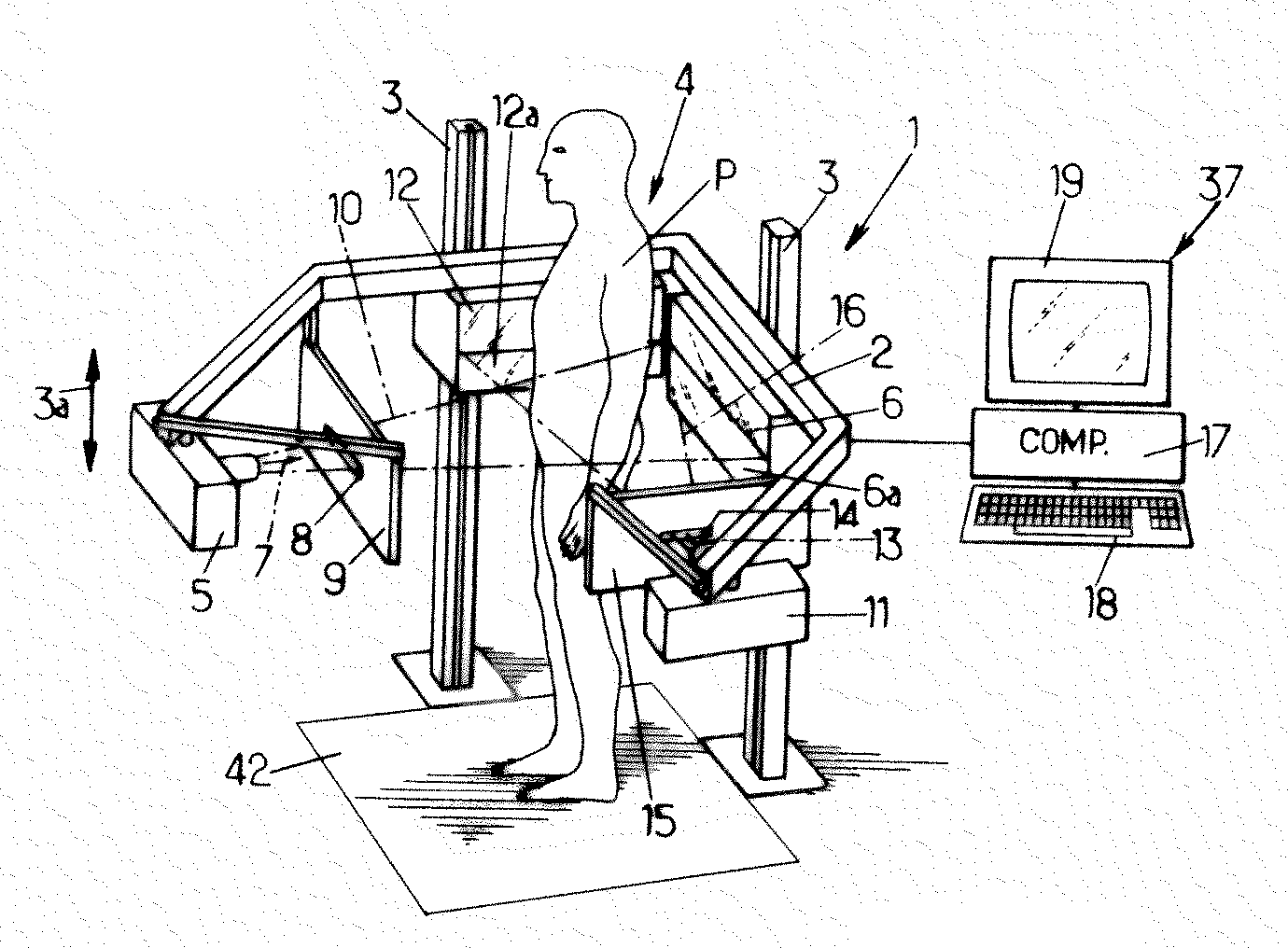
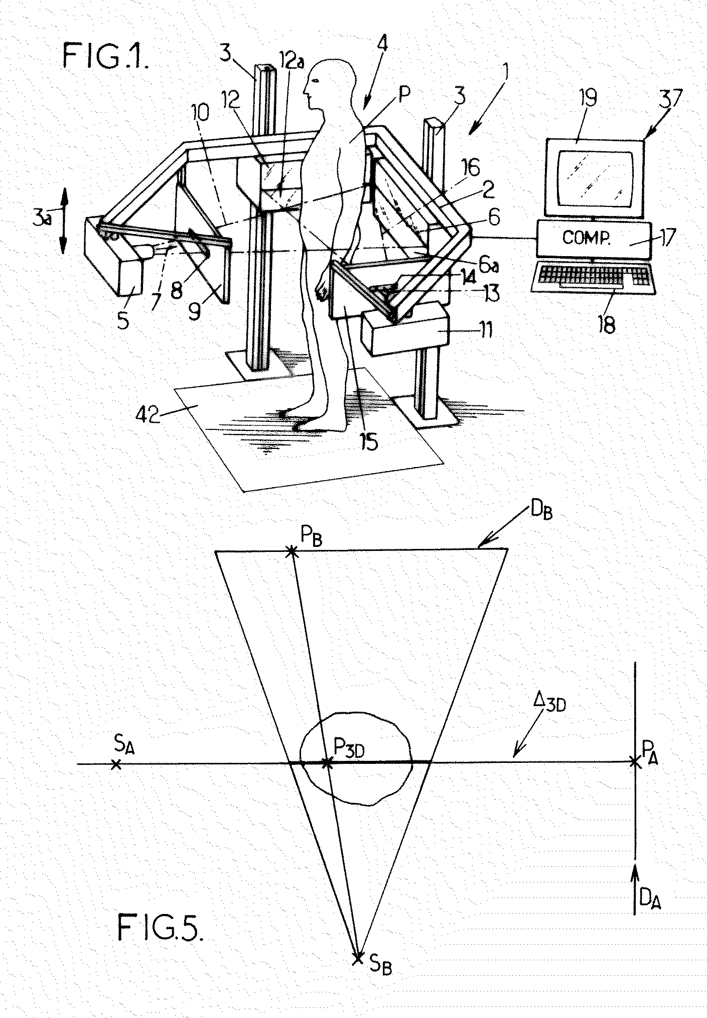
[0055]With reference to FIG. 1, a stereoscopic image acquisition system 1 is shown comprising a support 2 that can be moved along guides 3 in the vertical directions shown by the double arrow 3a.

[0056]The support 2 defines a field of observation 4, in which a patient P may be placed, for example standing upright, for the purposes of observation of the osteo-articular system of this patient.

[0057]On the support 2 are mounted a first X-ray source 5 and a first detector 6 facing the source 5. This first detector 6 comprises at least one horizontal line 6a of detection cells. For example, the detector 6 may comprise a gas detector, for example like that described in document U.S. Pat. No. 5,959,302. Naturally, other types of detectors may be used in the context of the present invention.

[0058]The source 5 is adapted to transmit ionizing rays, in particular X-rays, capable of being detected by the detector 6, in one shooting direction 7. This direction 7 is substantially from front-to-ba...


PUM


Abstract

A method for measuring geometric quantities intrinsic to an anatomical system of a patient, based on two stereoscopic images. Registration data are received on each of the two stereoscopic images. By using geometric calibration information, a three-dimensional geometric primitive is determined defined by at least a portion of the received registration data. Based on the three-dimensional geometric primitive, a value of geometric quantity intrinsic to the anatomical system is computed.

Description

CROSS-REFERENCE TO RELATED APPLICATIONS[0001]The present application claims priority to French Utility Patent Application No. 08 57245 filed Oct. 24, 2008, the entirety of which is incorporated herein by reference.FIELD OF THE INVENTION[0002]The invention relates to the measurement of geometric quantities intrinsic to an anatomical system.BACKGROUND OF THE INVENTION[0003]A practitioner, for example a radiologist, a surgeon, or other practitioner, may use geometric quantities, for example a length of a bone, an angle of a scoliosis, or other element, in order to establish a diagnosis.[0004]It is a known practice to take a radiograph of the osteo-articular system of a patient and to measure geometric quantities on the obtained image.[0005]However, the value measured is marred by errors caused by the process of projection of the rays on the plane of the sensor. This error is capable of varying depending on the position of the patient in the frame of reference of the imaging installatio...

Claims


Application Information

Patent Timeline
IPC IPC(8): G06K9/00
CPCA61B5/1072A61B5/4504A61B6/022A61B6/04A61B6/5235A61B6/4014A61B6/463A61B6/466A61B6/06
Inventor SAINT FELIX, DIDIERDI MONDA, RICHARD
Owner EOS IMAGING
Why Eureka
Try Eureka