Methods for detecting protein in plasma
a protein and plasma technology, applied in the field of methods for detecting protein in plasma, can solve the problem that immunochemical techniques provide merely non-selective results
- Summary
- Abstract
- Description
- Claims
- Application Information
AI Technical Summary
Benefits of technology
Problems solved by technology
Method used
Image

Examples
example 1
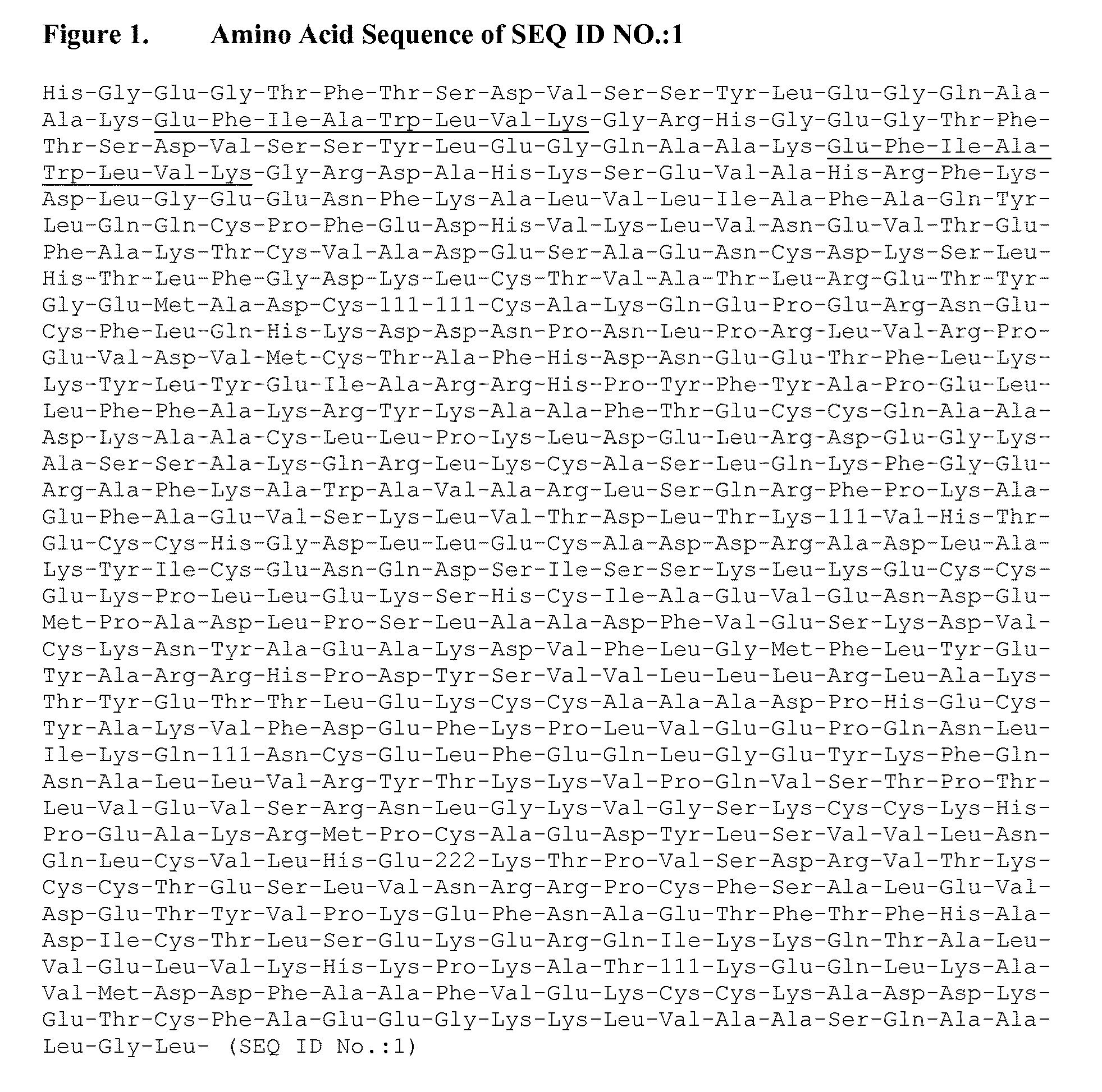
SEQ ID NO.:1 in Human Plasma
[0048]The following parameters were assessed:[0049]Selectivity, sensitivity and linearity[0050]Bias and precision[0051]Stability of SEQ ID NO.:1 in human plasma at room temperature[0052]The effect of freeze-thaw on the stability of SEQ ID NO.:1 in human plasma[0053]Stability of SEQ ID NO.:1 in processed samples[0054]The ability to dilute samples above the higher limit of quantification (HLQ)
Validation Procedure
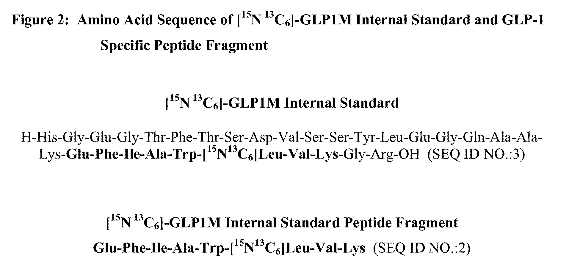
[0055]SEQ ID NO.:1 is a 73,012 Dalton (645 amino acid) genetic fusion protein consisting of two modified 30 amino acid GLP-1 peptides linked in tandem to recombinant human serum albumin. This example describes HPLC-MS / MS methods for the determination of SEQ ID NO.:1 in human plasma. 200 μL human plasma sample from a human administered SEQ ID NO.:1 was combined with an isotopically labeled GLP-1 internal standard, [15N 13C6]-GLP1M (SEQ ID NO.:3) (FIG. 3), and digested using the endoproteinase enzyme Lys-C.
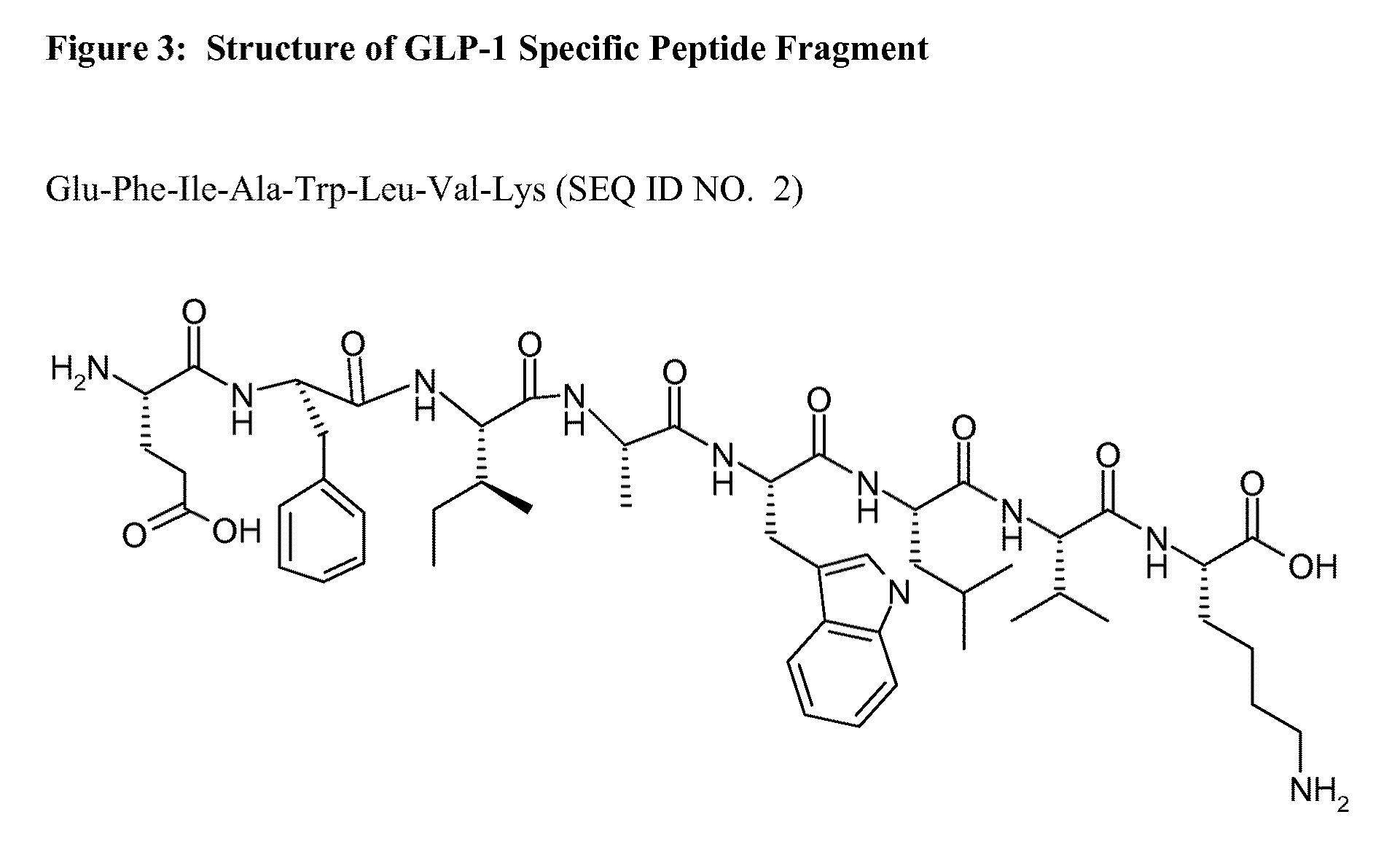
[0056]Two GLP-1 specific peptide fragments (1...
example 2
[0089]An additional assessment of method performance was determined by comparing the results obtained from human clinical study samples with results obtained using sandwich ELISA technique.
[0090]In the validated ELISA procedure, clinical samples were diluted 100-fold with sample buffer prior to analysis. The fusion protein was captured using a rabbit anti-human GLP-1 (7-36) amide and detected using a rabbit anti-HSA conjugated to biotin. Streptavidin HRP conjugate was used as the detection enzyme in conjunction with a chemiluminescent substrate. Sample concentrations were determined by interpolation from the standard curve, which was fitted using a weighted (1 / x). four parameter logistic equation. The validated range of this assay, based on 10 uL of human plasma is 50 to 15000 ng / mL.
[0091]A method correlation plot for data generated using high performance liquid chromatography and mass spectroscopy methods of Example 1 and the ELISA method of this example was generated using sample ...
example 3
[0092]Incurred sample reanalysis of clinical samples was performed in order to confirm both assay robustness and reproducibility. Clinical samples from both healthy and type 2 diabetic patients were reanalyzed on three separate occasions. Incurred sample reproducibility is confirmed when the difference between run CV for the three runs is ≦15%. Results from the incurred sample reanalysis test are presented in Table 7. Incurred sample reproducibility was confirmed in both healthy and type 2 diabetic patients.
TABLE 7Incurred Sample ReproducibilityRun 1Run 2Run 3ng / mLng / mLng / mLHealthy3912.53669.23701.83946.53868.83953.63973.93826.64087.6Mean3944.33788.23914.3% CV0.82.85.0Mean (Between run)3882.3% CV (Between run)2.1T2DM304.9301.6302.6319.8316.6317.3332.1307.6327.3Mean318.9308.6315.7% CV4.32.43.9Mean (Between run)314.4% CV (Between run)1.7
PUM
 Abstract
Description
Claims
Application Information
 - R&D Engineer
- R&D Manager
- IP Professional
- Industry Leading Data Capabilities
- Powerful AI technology
- Patent DNA Extraction
Browse by: Latest US Patents, China's latest patents, Technical Efficacy Thesaurus, Application Domain, Technology Topic.
© 2024 PatSnap. All rights reserved.Legal|Privacy policy|Modern Slavery Act Transparency Statement|Sitemap