X-ray CT IMAGING APPARATUS

a computerized tomography and imaging apparatus technology, applied in tomography, instruments, applications, etc., can solve the problems of apparatus not being able to perform panorama imaging, ct imaging with an offset scan cannot be performed, and the x-ray detector for a wider imaging area is generally more expensive, so as to achieve the effect of changing the magnification factor of the reconstructed imag

Active Publication Date: 2010-09-30
MORITA MFG CO LTD
View PDF19 Cites 27 Cited by
  • Summary
  • Abstract
  • Description
  • Claims
  • Application Information

AI Technical Summary

Benefits of technology

[0016]In an X-ray imaging apparatus of the invention, because an X-ray cone beam irradiating a part of a region of an object to be imaged is incident onto the X-ray detector, the region to be imaged can be enlarged, in contrast to a case wherein the X-ray transmitting the entire region of interest is incident onto the X-ray detector. Further, because the rotary shaft is moved relative to the center of the region to be imaged, the magnifying factor of the reconstructed image can be changed. Thus, CT imaging can be performed in a larger region of interest. For example, if the supporter is revolved while shifted relative to the rotary shaft, CT imaging can be performed in a larger region of interest. Further, the X-ray generator and the object are moved around the object while moving the rotary shaft and the object, CT imag

Problems solved by technology

An X-ray detector for a wider imaging area is generally more expensive.
However, CT imaging wi

Method used


Image

  • X-ray CT IMAGING APPARATUS
  • X-ray CT IMAGING APPARATUS
  • X-ray CT IMAGING APPARATUS

Examples


Embodiment Construction

Explanation of Reference Symbols

[0050]Embodiments the invention are explained below, referring to the appended drawings.

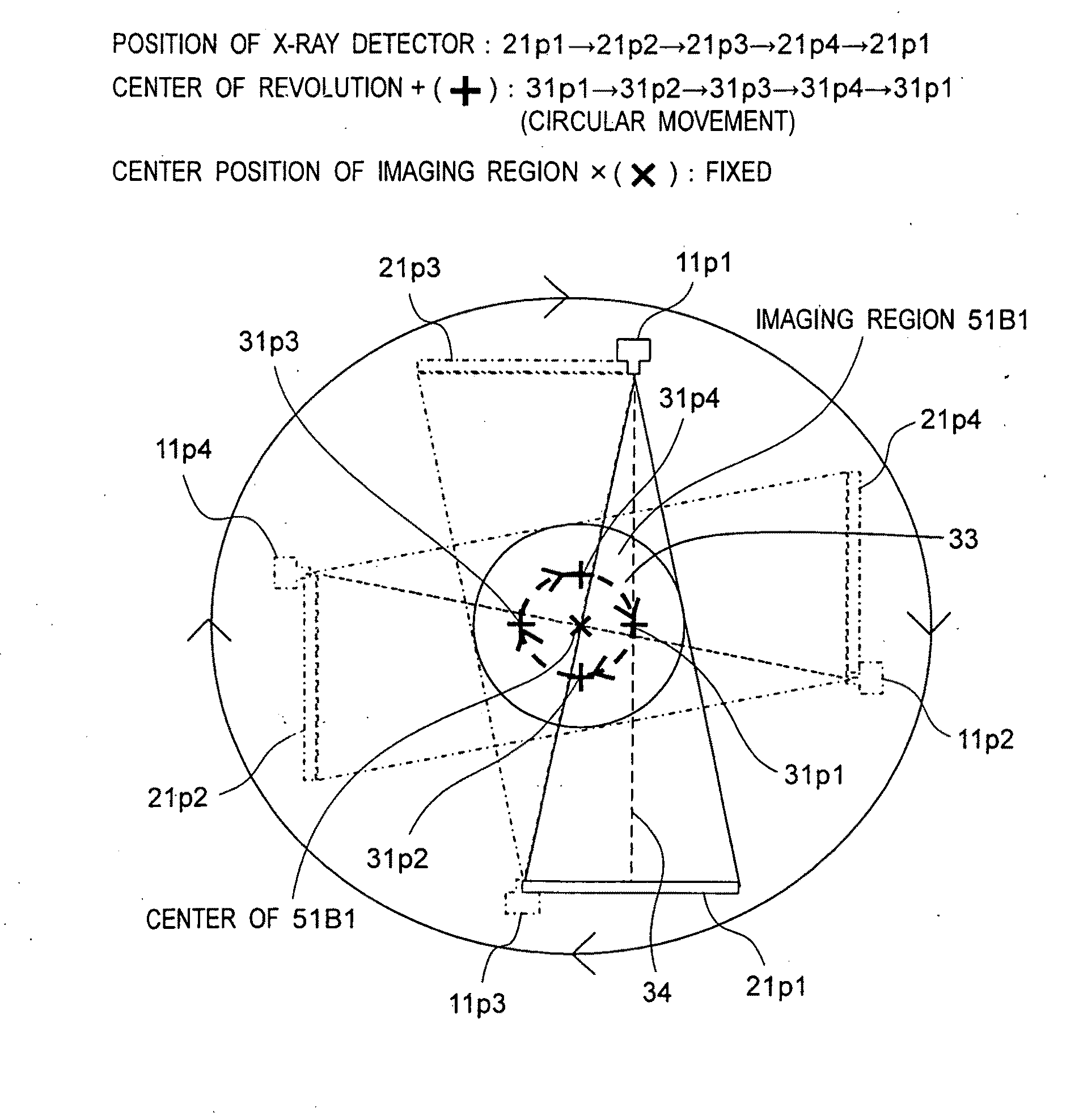
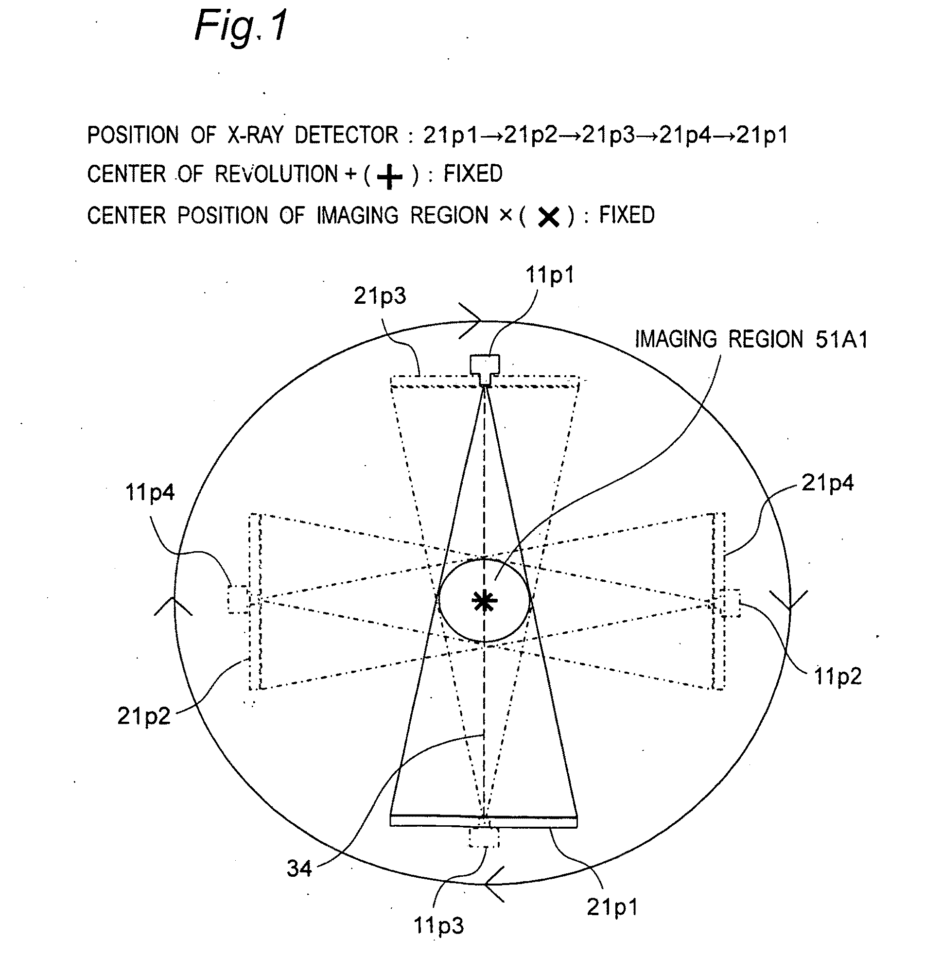
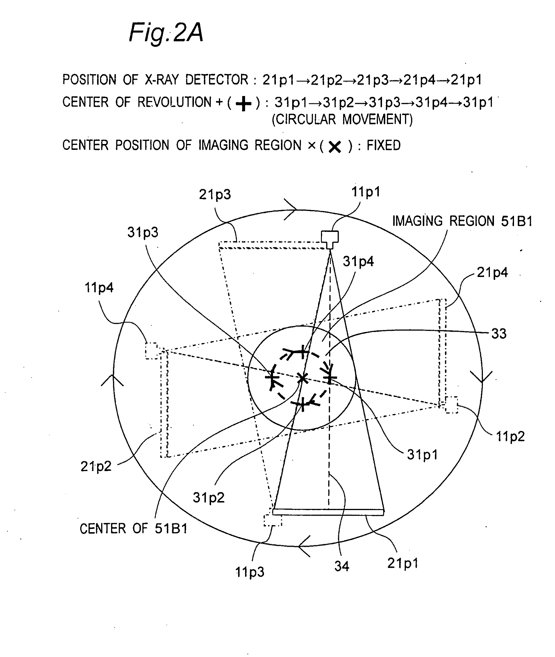
[0051]In CT imaging, an X-ray generator and an X-ray detector are circled around an object relative to the object. The X-ray generator exposes the object to an X-ray cone beam, and the X-ray detector having a two-dimensional detection plane detects X-rays transmitting the object. A larger object is desirable to be imaged in CT imaging. In the invention, in order to image a larger region, the center axis 34 of X-rays (or the symmetrical axis of an X-ray cone beam) does pass the center position (x) of the imaging region of an object and the center axis becomes tangent to an arc 33 having its center at the center position (x). Preferably, X-rays passing through the center of a region of interest of the object enter an edge of the two-dimensional detection plane of the X-ray detector. (A scan for imaging a larger imaging region with an X-ray cone beam by irradiating a ...


PUM


Abstract

In an X-ray CT imaging, an X-ray generator (11) and a two-dimensional X-ray detector (21) are opposed to each other between an object and are rotated by a rotary shaft (32) around the object. At least one of the rotary shaft supporter (61) and an object holder (40) includes a movement mechanism (42, 65) for moving a supporter (30) for the X-ray generator and the X-ray detector relative to the object. In offset scan CT imaging, the rotation of the supporter by the rotary shaft is performed simultaneously with the relative two-dimensional movement of the rotary shaft by the movement mechanism. In the relative two-dimensional movement, the position of the rotary shaft is moved according to the rotary angle of the supporter along a circular orbit around the center of a CT imaging region in a plane crossing the rotary shaft. Thus, it becomes possible to image a larger region of interest of the object.

Description

TECHNICAL FIELD[0001]The invention relates to an X-ray computerized tomography (CT) imaging apparatus, in particular, a head X-ray CT imaging apparatus using an X-ray cone beam, suitable for dentistry, oral surgery, opthalmology, otolaryngology and the like.BACKGROUND ART[0002]In an X-ray computer tomography apparatus using an X-ray cone beam, an X-ray generator and an X-ray detector interposing an object is rotated around the object relative to the object. During the rotation, the X-ray generator generates an X-ray cone beam having a cone or pyramid shape to irradiate the object in various directions, and the X-ray detector having a two-dimensional detection plane measures an intensity distribution of X-rays transmitting the object or a projection of the object. The acquired projection data is back-projected to reconstruct the distribution of X-ray absorption coefficients and to create a two-dimensional tomographic view or a three-dimensional stereoscopic view.[0003]An X-ray detect...

Claims


Application Information

Patent Timeline
IPC IPC(8): A61B6/03
CPCA61B6/032A61B6/587A61B6/4441A61B6/14
Inventor SUZUKI, MASAKAZUYOSHIMURA, TAKAHIROHONJO, MAKOTO
Owner MORITA MFG CO LTD
Why Eureka
Try Eureka