Circular Profile Mapping and Display of Retinal Parameters

a technology of retinal parameters and profile mapping, applied in the field of ophthalmic characterization, can solve the problems of no mechanism for providing consistent local variation detection, no mechanism for detailed tracking of the growth of retinal defects over time, and no mechanism for associating anomalous thickness measurements with the presence of blood vessels or other anatomical features

Active Publication Date: 2010-11-18
TOPCON MEDICAL SYST
View PDF7 Cites 35 Cited by
  • Summary
  • Abstract
  • Description
  • Claims
  • Application Information

AI Technical Summary

Problems solved by technology

There is no mechanism for providing consistent detection of local variations.
(c) There is no mechanism for detailed tracking of the growth of retinal def

Method used


Image

  • Circular Profile Mapping and Display of Retinal Parameters
  • Circular Profile Mapping and Display of Retinal Parameters
  • Circular Profile Mapping and Display of Retinal Parameters

Examples


Embodiment Construction

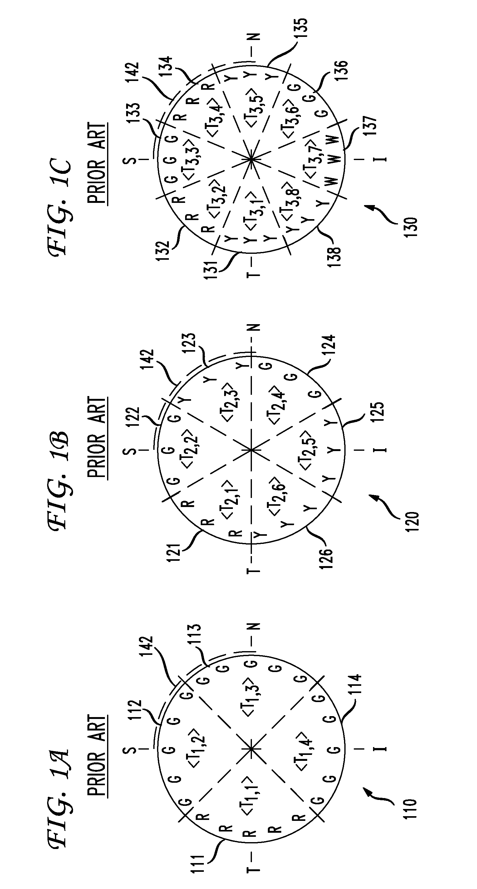
[0022]FIG. 1A-FIG. 1C are schematic representations of conventional retinal thickness plots of data (also referred to as circle scans) measured with a Zeiss Stratus OCT 3 or similar ophthalmological instrument. Measurements are taken along a circle with a center at the nominal center of the optic disc and with a radius fixed at 1.73 mm. The angular orientation of measurement loci along the circle are referenced with respect to TSNIT geometry, where T=Temporal, S=Superior, N=Nasal, and I=Inferior. The measurements along the circle are divided into groups defined by arcs. The number and orientation of the arcs are arbitrary and defined by a user (such as an eye doctor). For measurements along each arc, the average thickness is displayed.

[0023]In FIG. 1A, plot 110 displays measurements divided into four arcs, arc 111-arc 114. Note that the dashed radial lines are used only to aid demarcation of the arcs in the figures. The measurement loci fall only along the arcs, not in the regions ...


PUM


Abstract

Certain diseases of the retina are diagnosed by circular profile analysis of retinal parameters, such as thickness. Retinal thickness around a user-defined circle on the retina is measured by various ophthalmological techniques and ±mapped to a circular profile map. The circular profile map does not use segmentation of measurement data into arbitrary arcs, and thickness is mapped to a quasi-continuous range of display bands. The circular profile map is superimposed on a fundus image, or other two-dimensional image of the retina, allowing association of the circular profile map with the presence of blood vessels and other anatomical features. The simultaneous display of a series of circular profile maps generated from sets of measurement data taken at different times permits the ready visualization of the progression of retinal abnormalities.

Description

CROSS-REFERENCE TO RELATED APPLICATION[0001]This application is related to U.S. patent application Ser. No. ______ (Attorney Docket No. 13857.0008), entitled Characterization of Retinal Parameters by Circular Profile Analysis, which is being filed concurrently herewith and which is herein incorporated by reference in its entirety.BACKGROUND OF THE INVENTION[0002]The present invention relates generally to ophthalmic characterization, and more particularly to circular profile mapping and display of retinal parameters.[0003]Diagnostics for eye disorders typically include a detailed ophthalmic examination of the retina. For initial examination, an eye doctor will view the retina through an ophthalmoscope. For a permanent record, the retina is typically photographed with a fundus camera. A fundus photograph directly records various anatomical features of the retina, such as the optic disc, fovea, blood vessels, and lesions. The imaging capabilities of fundus photography may be enhanced b...

Claims


Application Information

Patent Timeline
IPC IPC(8): A61B3/14A61B3/10
CPCA61B3/0058A61B3/102G06T11/206A61B3/14A61B3/1225A61B3/0025
Inventor HUANG, YIJUNROYAMA, TETSUYOSHIKOTCHKIN, ALEXANDRE
Owner TOPCON MEDICAL SYST
Why Eureka
Try Eureka