Method for Imaging Plaque Using Dual Energy CT

a computed tomography and plaque technology, applied in the field of computed tomography (ct) imaging apparatus, can solve the problems of inability to image advanced (“vulnerable”) lesions, and inability to detect plaque that is vulnerable to becoming symptomatic, and achieve high spatial coincidence and high spatial correspondence

Active Publication Date: 2010-12-16
MAYO FOUND FOR MEDICAL EDUCATION & RES
View PDF14 Cites 35 Cited by
  • Summary
  • Abstract
  • Description
  • Claims
  • Application Information

AI Technical Summary

Problems solved by technology

Many methods are available to detect the presence of plaque, but the ability to detect plaque that is vulnerable to becoming symptomatic is limited.
But up to now, imaging of non-calcified, advanced (“vulnerable”) lesions remains elusive.
The relation of fibro-calcified lesions, as determined by CT using the Agatston score, and cardio-vascular events has been demonstrated in the past, but imaging of advanced, vulnerable lesions with clinical imaging modalities like CT or MRI has remained difficult if not impossible.

Method used


Image

  • Method for Imaging Plaque Using Dual Energy CT
  • Method for Imaging Plaque Using Dual Energy CT
  • Method for Imaging Plaque Using Dual Energy CT

Examples


Embodiment Construction

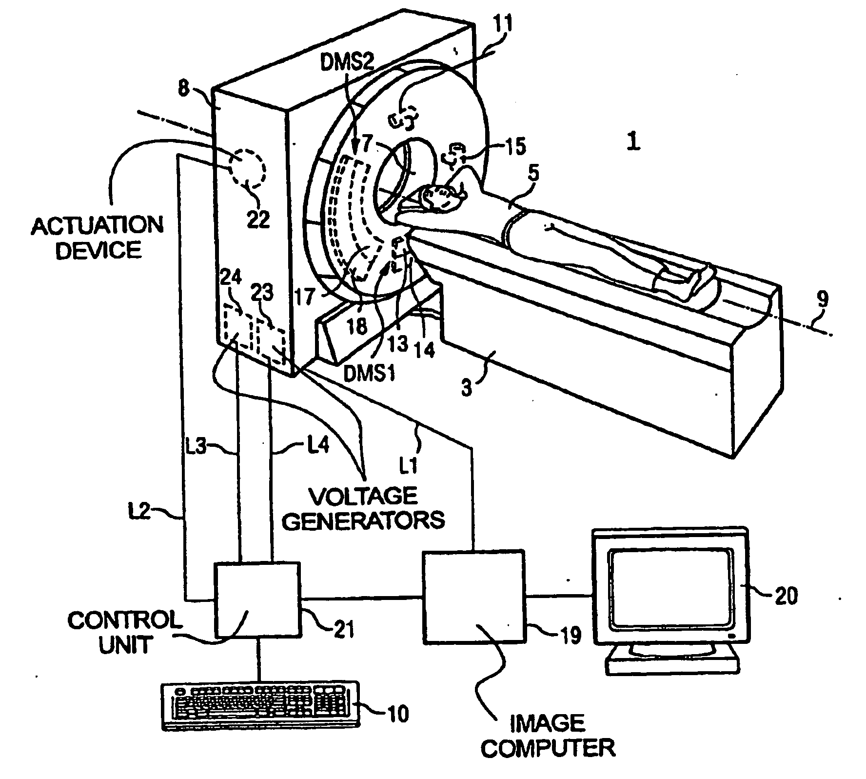
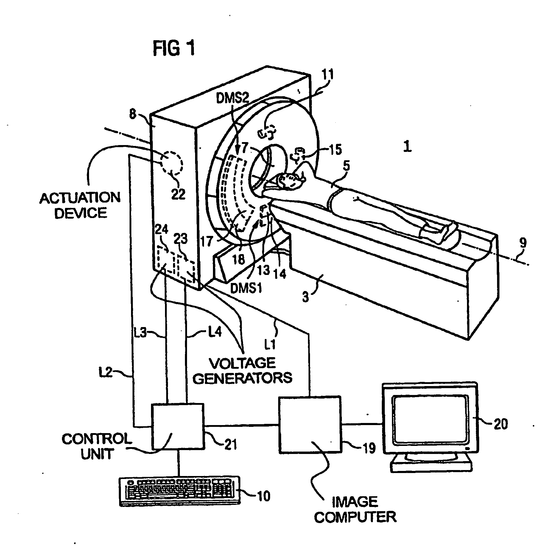
[0015]Referring to FIG. 1, the CT scanner 1 includes a patient table 3 for supporting and positioning an examination subject 5. The region of interest in the patient 5 can be inserted into an opening 7 (diameter 70 cm) in the housing 8 of the tomography apparatus 1 by means of a movable table top. Inside the housing 8, a gantry (not visible) is mounted so as to be rotated with high speed around a rotation axis 9 running through the patient. Moreover, for a spiral, or helical, scan a continuous axial feed is effected with the positioning device 3. A control unit 10 is provided for operation of the tomography apparatus 1 by a doctor or an assistant.

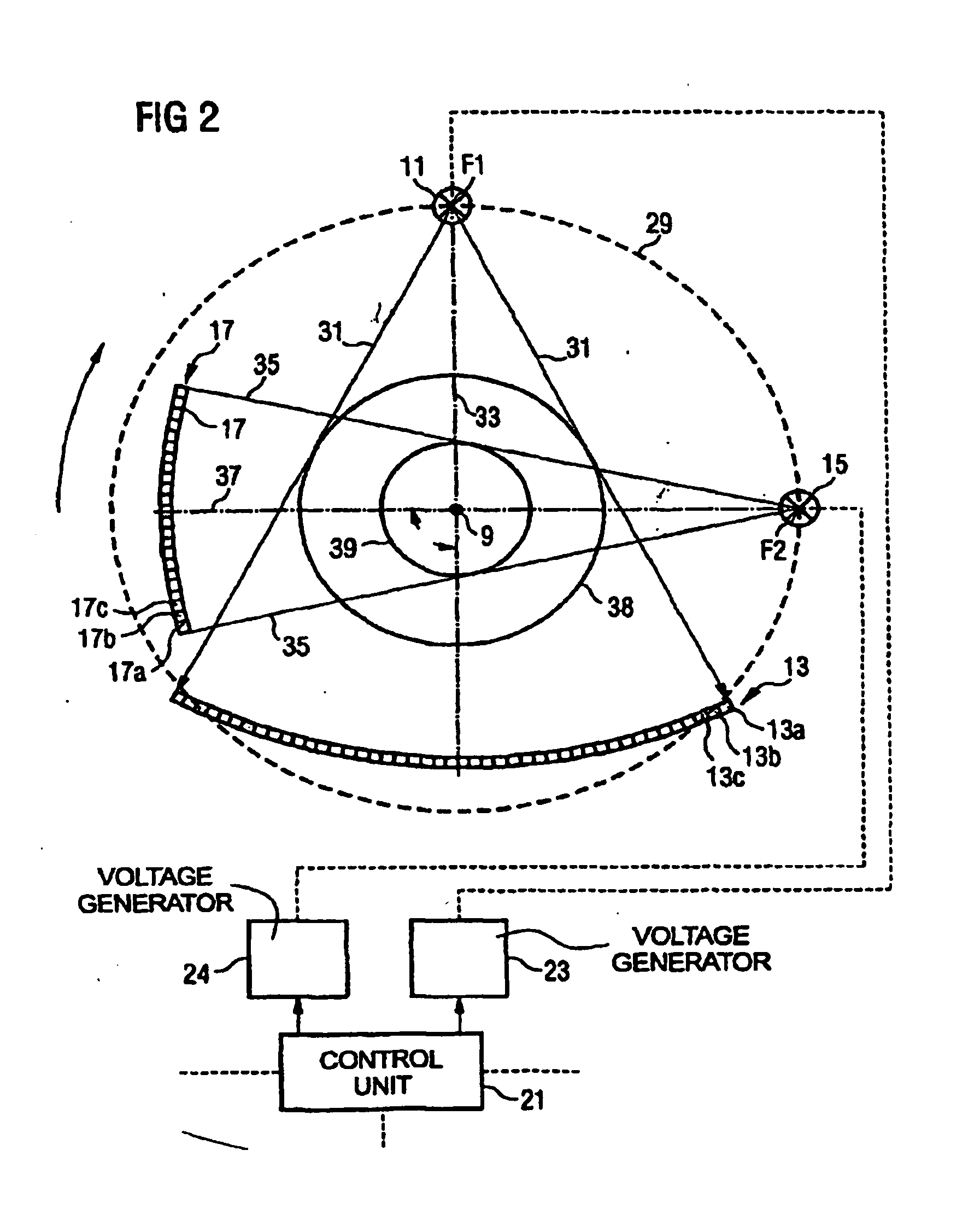
[0016]Two data acquisition systems are mounted on the gantry. A first acquisition system has an x-ray tube as a first radiator 11 and a first data acquisition unit DMS1 formed as a multi row x-ray detector array as a first detector 13. A second acquisition system has a separate x-ray tube as a second radiator 15 and furthermore a second dat...


PUM


Abstract

Two x-ray CT images are acquired of arterial plaque using x-rays at two different energy levels. The reconstructed images are normalized by adjusting pixel brightness until pixels depicting a region containing calcium have substantially the same brightness. The normalized images are subtracted to produce an image that depicts iron in the arterial plaque.

Description

CROSS-REFERENCE TO RELATED APPLICATIONS[0001]This application claims the benefit of U.S. Provisional patent application Ser. No. 60 / 776,645 filed on Feb. 24, 2006 and entitled “Imaging Vulnerable Plaque Using Dual Energy CT”.STATEMENT REGARDING FEDERALLY SPONSORED RESEARCH[0002]This invention was made with government support under Grant No. HL65342 and EB000305 awarded by the National Institute of Health. The United States Government has certain rights in this Invention.BACKGROUND OF THE INVENTION[0003]The present invention relates to computed tomography (CT) imaging apparatus; and more particularly, to the imaging of arterial plaque.[0004]In a current computed tomography system, an x-ray source projects a fan-shaped beam which is collimated to lie within an X-Y plane of a Cartesian coordinate system, termed the “imaging plane.” The x-ray beam passes through the object being imaged, such as a medical patient, and impinges upon an array of radiation detectors. The intensity of the tr...

Claims


Application Information

Patent Timeline
Patent Type & Authority Applications(United States)
IPC IPC(8): G06K9/00
CPCA61B5/02007A61B6/032A61B6/482A61B6/405A61B6/4014
Inventor LANGHEINRICH, ALEXANDER C.RITMAN, ERIK L.
Owner MAYO FOUND FOR MEDICAL EDUCATION & RES
Why Eureka
Try Eureka