Biomarkers of cancer metastasis

a biomarker and cancer technology, applied in the field of cancer diagnosis techniques, can solve the problems of not being able to define the molecular properties of primary tumours that dictate metastaticity

Inactive Publication Date: 2010-12-16
BRITISH COLUMBIA CANCER AGENCY BRANCH
View PDF5 Cites 20 Cited by
  • Summary
  • Abstract
  • Description
  • Claims
  • Application Information

AI Technical Summary

Benefits of technology

[0014]In another aspect, there is provided a method of determining tumour metastatic potential. In one embodiment, the method comprises measuring expression levels in a tumour tissue sample of at least two indicators of metastatic potential which are each independently CAIX, VEGF-C, EFNA5, EPHB2, TGF-β3, PDK3, CAXII, KRT14, HIF-1α, or TNC; and comparing said expression levels to a control to determine metastatic potential.
[0015]In another aspect, there is provided

Problems solved by technology

Furthermore, the molecular properties of primary tumours that dictate

Method used


Image

  • Biomarkers of cancer metastasis
  • Biomarkers of cancer metastasis
  • Biomarkers of cancer metastasis

Examples


example 1

Differential Expression of Hypoxia Inducible Genes in Metastatic Versus Non-Metastatic Primary Tumours

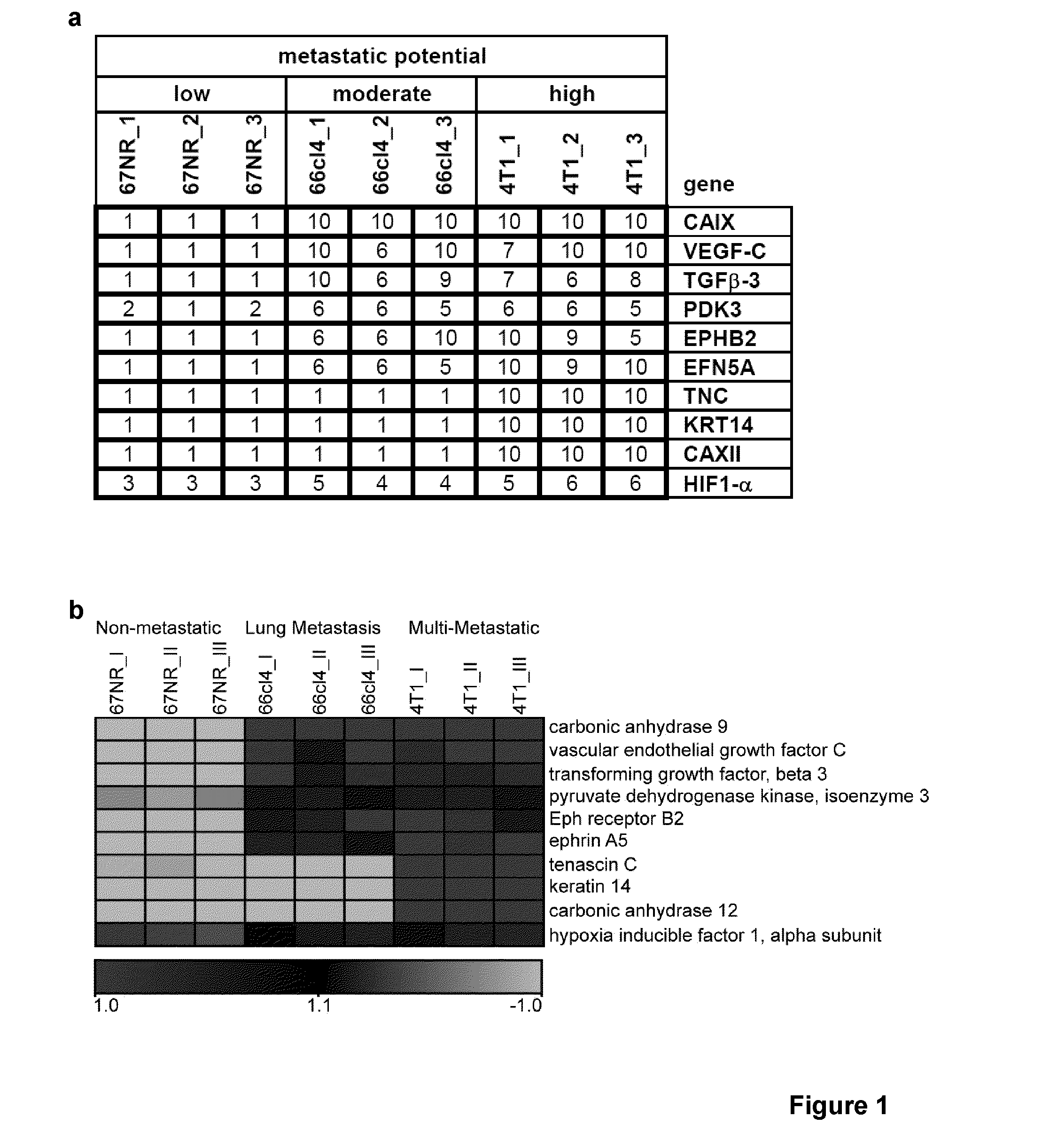
[0141]The complex nature of metastasis requires animal models that can recapitulate the human situation, including spontaneous metastasis from primary tumours and an intact immune system. A well established clinically relevant syngeneic mouse model of spontaneous breast cancer metastasis was used to investigate differential expression of hypoxia inducible genes in metastatic versus non-metastatic primary tumours9,10. The model is highly robust in that several syngeneic tumour cell lines with a spectrum of metastatic phenotypes have been isolated from a spontaneous metastatic mammary tumour in a BALB / cfC3H mouse. When injected into the mammary glands of mice, the tumour cell lines form primary tumours within two weeks10,11, but vary in their metastatic potential and organ specificity. The 67NR cells are non-metastatic, whereas the 66cl4 produce spontaneous metastases to the lungs on...

example 2

Primary Tumour Characterization

[0149]The differential expression of hypoxia inducible genes in the metastatic tumours versus the non-metastatic tumours, led us to investigate the extent of hypoxia, necrosis, apoptosis, proliferation, vascularization and lymphangiogenesis in 4T1, 66cl4 and 67NR tumours. Tumours were grown and labeled in vivo for proliferation (BrdU), hypoxia (pimonidazole) and perfusion (DiOC7). Immunohistochemistry for CD31 (vasculature) and in situ detection of TUNEL (apoptosis) were then performed. Necrosis was assessed by histology. Representative composite, pseudocolored images demonstrated the presence of each marker. Ten individual 67NR, 66cl4 or 4T1 tumours were sectioned and stained for the above parameters.

[0150]FIG. 3, part a, depicts whole tumour averages for microvessel density (MVD) as determined by CD31 staining, proliferation as determined by BrdUrd staining, hypoxia as determined by pimonidazole staining, apoptosis as determined by TUNEL labeling, an...

example 3

Carbonic Anhydrase IX as a Driver of Metastatic Potential

[0154]The differential expression of CAIX in the metastatic versus the non-metastatic primary tumours, led to investigation of whether the differences in hypoxia induced CAIX expression in the metastatic and non-metastatic tumours are innate properties of the cells or whether they are acquired in vivo. The three cell lines were cultured under normoxic or hypoxic conditions for various time periods and the expression of CAIX, as well as other hypoxia inducible proteins, was determined.

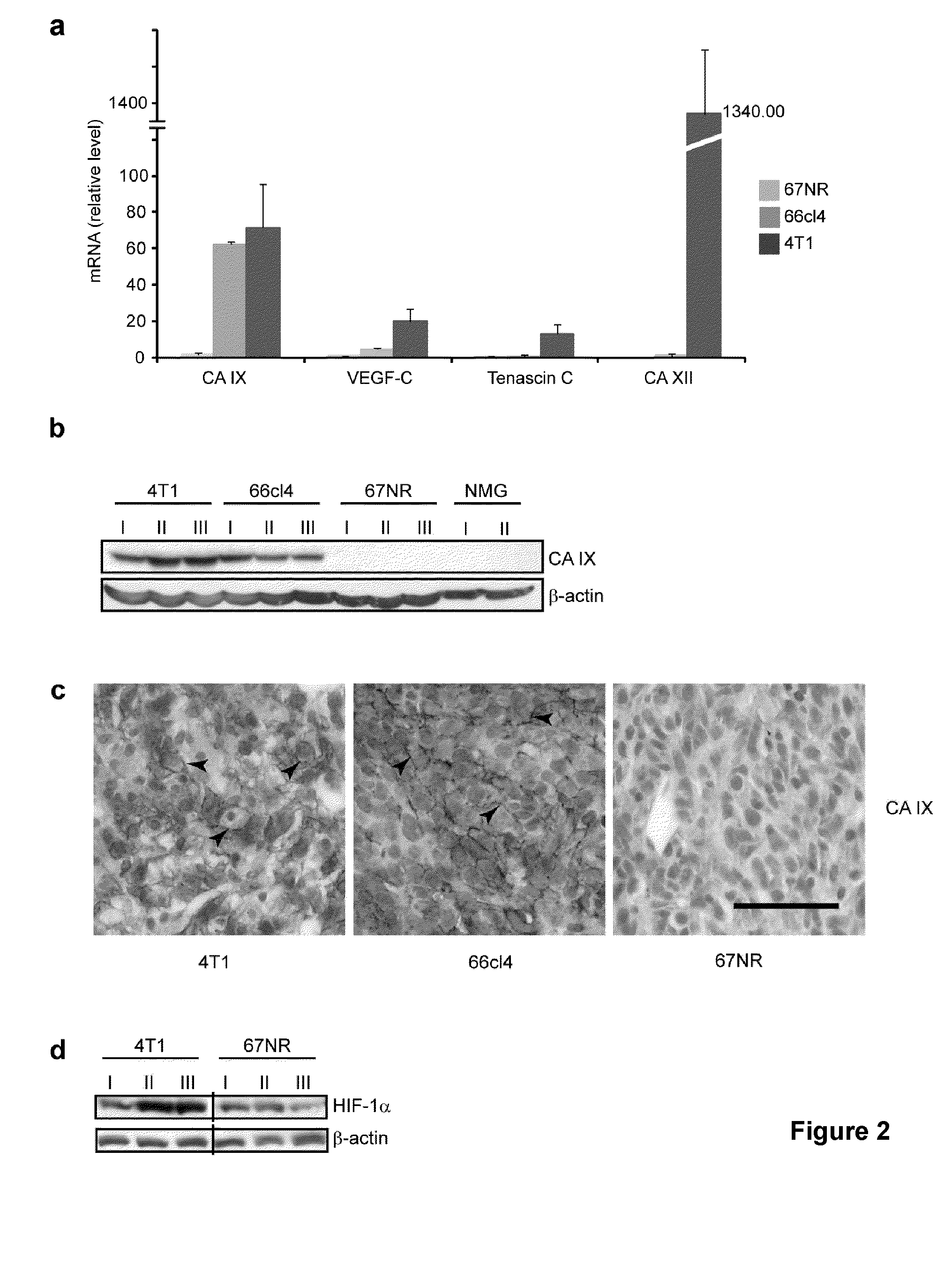
[0155]FIG. 4, part a, shows levels of gene expression as determined by quantitative RT-PCR (qRT-PCR); n=3. Error bars indicate s.e.m. The indicated cell lines were cultured in normoxia or hypoxia for 48 hours. Data shown are representative of three independent experiments. CAIX expression was readily induced after 48 hours in hypoxia in both 66cl4 and 4T1 metastatic cells, but was not induced in the non-metastatic 67NR cells. Similarly, the expres...


PUM

PropertyMeasurementUnit
Magnetic fieldaaaaaaaaaa
Levelaaaaaaaaaa

Abstract

There is provided a panel of biomarkers of tumour metastasis comprising any two of carbonic anhydrase-9 (CAIX), vascular endothelial growth factor C (VEGF-C), ephrin A5 (EFNA5), eph receptor B2 (EPHB2), transforming growth factor beta 3 (TGF-β3), pyruvate dehydrogenase kinase isoenzyme-3 (PDK3), carbonic anhydrase-12 (CAXII), keratin 14 (KRT14), hypoxia inducible factor 1 alpha subunit (HIF-1α), or tenascin C (TNC). CAIX, VEGF-C, EFNA5, EPHB2, TGF-β3 or PDK3 may be indicators of moderate metastatic potential, while CAXII, KRT14, HIF-1α, or TNC may be indicators of high metastatic potential. There is also provided a method of determining risk of tumour metastasis using the aforementioned biomarkers is also provided. The biomarkers may be used in diagnosis, prognosis, treatment selection, or to test putative therapeutics. The biomarkers may be used to assess malignancies or cancers having hypoxic regions, such as breast cancer.

Description

CROSS REFERENCE TO RELATED APPLICATIONS[0001]This application claims the benefit of priority of U.S. Provisional Patent Application No. 61 / 184380 filed Jun. 5, 2009, which is incorporated herein by reference in its entirety.FIELD OF THE INVENTION[0002]The present disclosure relates generally to diagnostic techniques for cancer. More particularly, the present disclosure relates to biomarkers of cancer metastasis.BACKGROUND OF THE INVENTION[0003]Metastasis, the primary cause of cancer mortality is a complex process with multiple steps that include tumour cell invasion, intravasation, extravasation, and establishment of secondary tumours in distant organs1. For breast cancer, genomic analysis of primary tumours and metastases from patients has identified sets of genes whose expression appear to be prognostic of outcome2-4. In addition, this type of analysis has led to sub-classification of human breast cancers into intrinsic subtypes that can also predict outcome and therapeutic respon...

Claims


Application Information

Patent Timeline
IPC IPC(8): C40B30/00C12Q1/68C40B40/08G01N33/68
CPCC12Q1/6886C12Q2600/106C12Q2600/112C12Q2600/118G01N2800/60C12Q2600/178G01N33/57484G01N33/6803C12Q2600/158
Inventor LOU, YUANMEIMCDONALD, PAUL CHRISTOPHEROLOUMI, ARUSHADEDHAR, SHOUKAT
Owner BRITISH COLUMBIA CANCER AGENCY BRANCH
Why Eureka
Try Eureka