
Method and system for anatomy structure segmentation and modeling in an image

a technology of anatomy structure and image, applied in image analysis, image enhancement, instruments, etc., can solve the problems of difficult segmentation of the third ventricle, ventricular region may be left out by these methods, and ventricular region may be left out, etc., to achieve accurate description of features and details.

Inactive Publication Date: 2010-12-23
AGENCY FOR SCI TECH & RES
View PDF6 Cites 35 Cited by
  • Summary
  • Abstract
  • Description
  • Claims
  • Application Information

AI Technical Summary

Benefits of technology

The present invention provides a method and system for segmenting and constructing 3D surface models of structures in an image, specifically brain scan images. The method involves registering a brain model with the image, defining regions of interest, performing region growing to form volumes indicative of ventricles, and segmenting the image using the volumes. The invention also includes constructing a surface model of the segmented anatomy structure and editing it to accurately describe the features and details lost in segmentation. The computer system includes program instructions operable by a computer to perform the steps of the methods.

Problems solved by technology

However, since these methods do not utilize the shape prior-knowledge of the ventricles, “leakage” from the ventricular regions to the non-ventricular regions may arise.
Furthermore, some ventricular regions may be left out by these methods due to the non-homogeneity of the images or the presence of noise and partial volume artifacts in the images.
The accurate segmentation of the third ventricle is especially challenging when using these non model-based methods since the precise boundaries of the third ventricle depend on the shape and topological constraints of themselves and their relationship with surrounding objects.
However, due to the large variation in the shapes and sizes of the ventricles, it is difficult to design a reasonable energy or similarity function to achieve a model deformation adaptable to every variation.
Furthermore, the local minimization problem which causes the false segmentation inevitably exists in these methods.
In general, there are two main difficulties in the segmentation of anatomic structures from images.
Second, some boundaries between the studied structure and its surrounding tissue are too thin to be detected in the image.
As a result, some object regions may “leak” (i.e. connect) to other non-object regions.
Currently, no existing method can detect the transition regions and at the same time, prevent the “leaking” of object regions to non-object regions.

Method used


Image

  • Method and system for anatomy structure segmentation and modeling in an image
  • Method and system for anatomy structure segmentation and modeling in an image
  • Method and system for anatomy structure segmentation and modeling in an image

Examples


example embodiments

ADVANTAGES OF EXAMPLE EMBODIMENTS

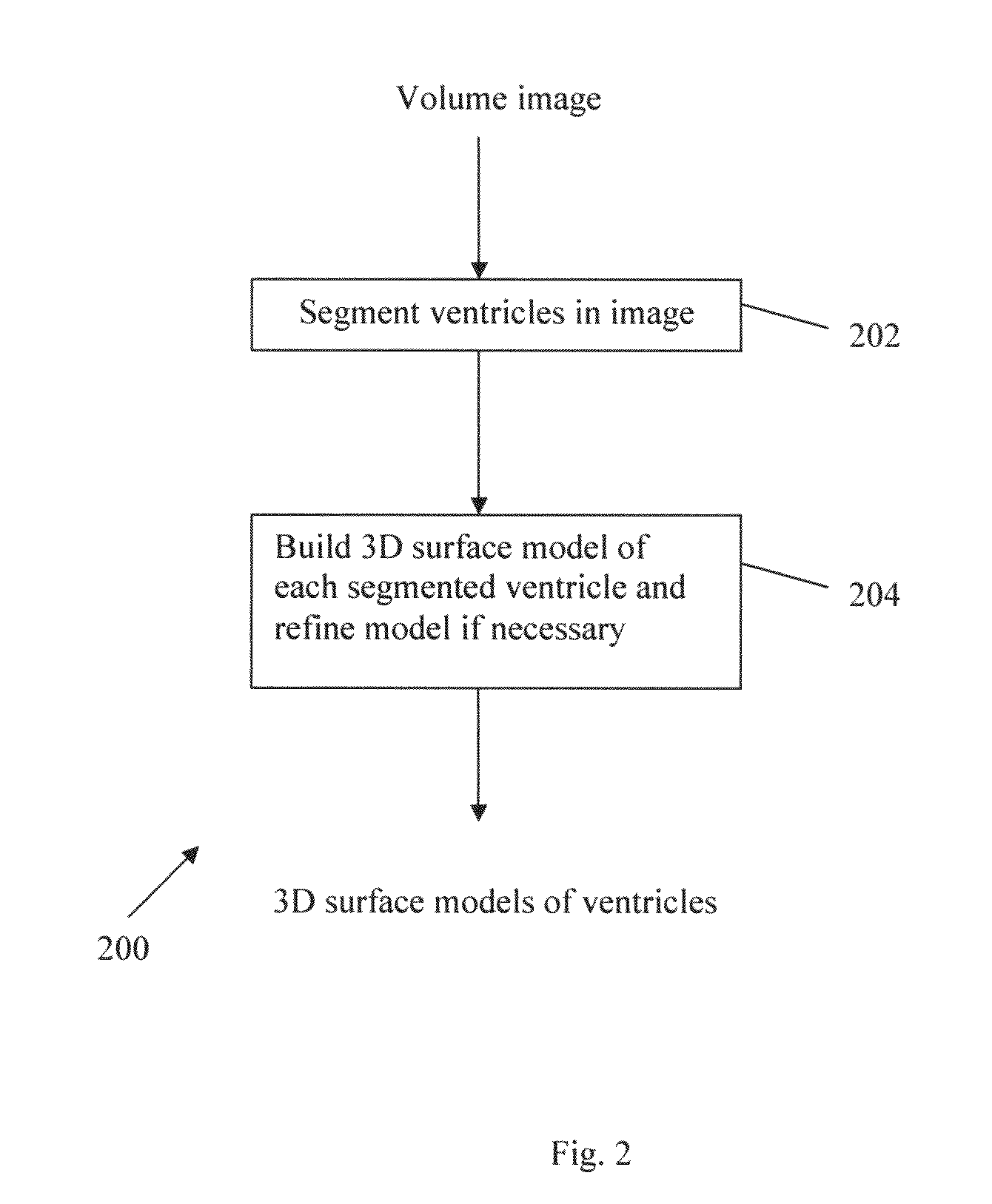
[0099]A volumetric deformable model is used in step 202 as domain knowledge to automatically define a region of interest for the segmentation of the structure to be studied, for example the ventricular structure in the example embodiments. A proper ROI is critical for an accurate segmentation. If the ROI is too small, it may not contain the structure to be studied. On the other hand, if the ROI is too large, it may contain too much unrelated information leading to wrong segmentation. In step 202, the model is first deformed to roughly fit its corresponding structure in the image by a 3D point landmark-based warping approach and the ROI is then defined by expanding (or dilating) the deformed model. The resulting ROI takes the prior shape of the structure to be studied and hence, the amount of unrelated information in the ROI is minimized. Therefore, step 202 is robust to noise and to large shape and size variations.

[0100]Furthermore, a hysteretic thre...


PUM


Abstract

A method is proposed for segmenting one or more ventricles in a three-dimensional brain scan image (e.g. MR or CT). The image is registered against a brain model, which ventricle models of each of the one or more ventricles. Respective regions of interest are defined based on the ventricle models. Object regions are first obtained by applying region growing procedure in the regions of interest, and then trimmed based on anatomical knowledge. A 3D surface model of one or more objects is constructed within a 3D space from the segmented structure. A 3D surface is edited and refined by a user selecting amendment points in the 3D space which are indicative of missing detail features. A region of the 3D surface near the selected points is then warped towards the amendment points smoothly, and the modified patch is combined with the rest of the 3D surface yields the accurate anatomy structure model.

Description

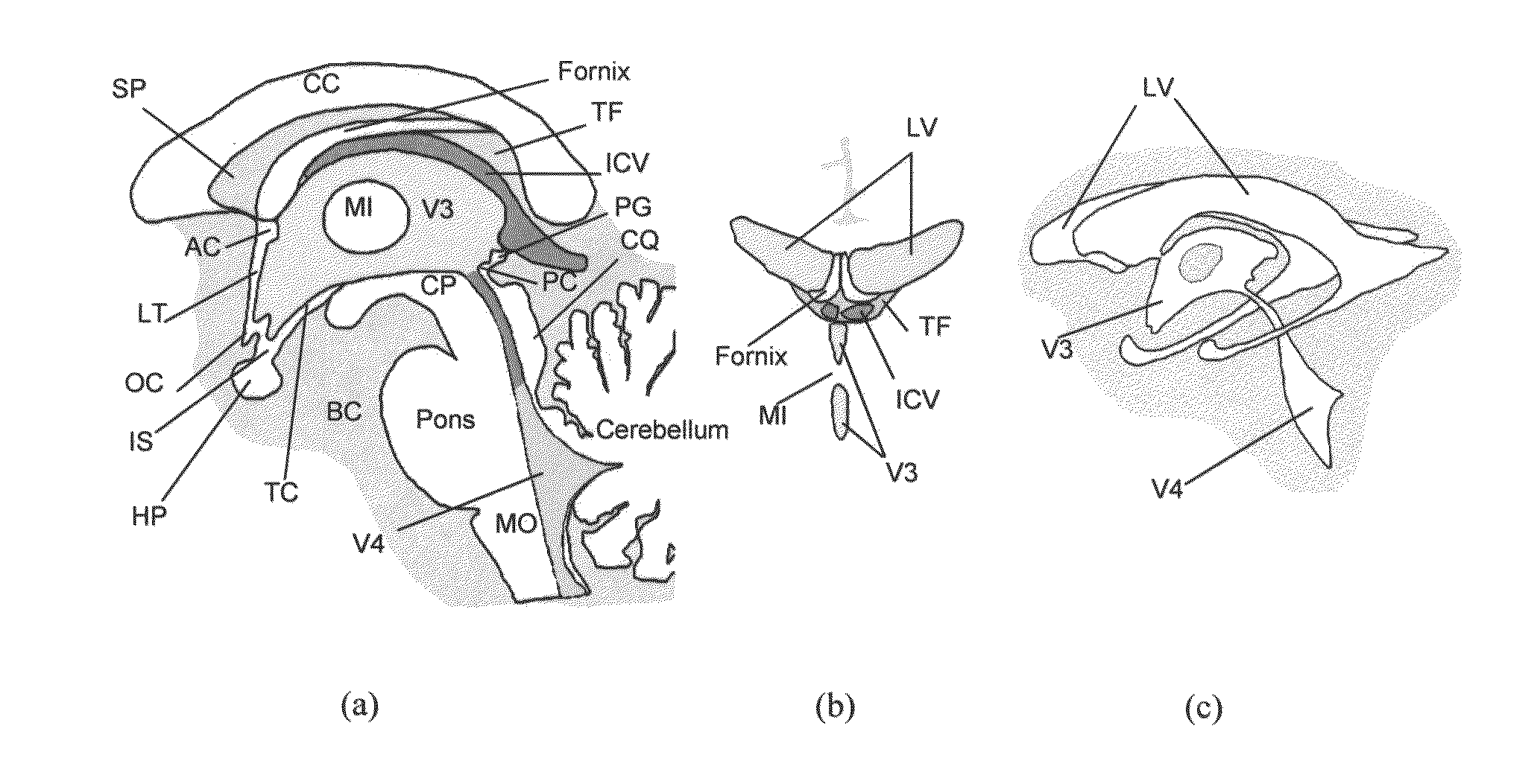
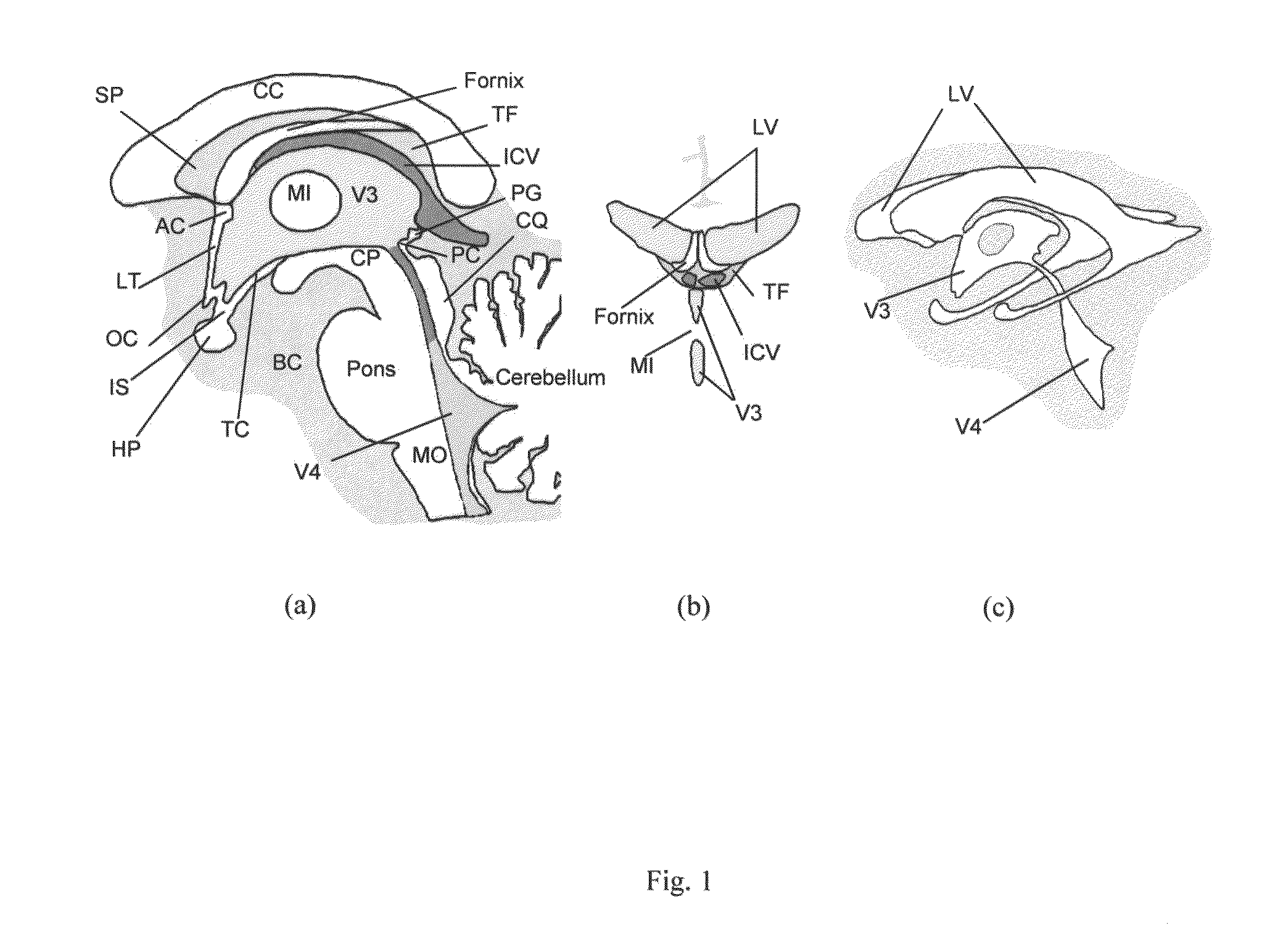
REFERENCE TO RELATED APPLICATIONS[0001]This application is a 371 National Stage application based on PCT / SG2009 / 000075, filed Feb. 27, 2009, which is based on U.S. provisional application 61 / 032,518, filed Feb. 29, 2008.FIELD OF THE INVENTION[0002]The present invention relates to a method and system for segmenting anatomy structures in an image, a method and system for constructing a 3D surface model of a segmented structure. The particular application example is the segmentation and modeling of brain ventricular system in medical images, such as MR images and CT images.BACKGROUND OF THE INVENTION[0003]As shown in FIG. 1, the human cerebral ventricular system consists of four intercommunicating chambers, namely the left lateral ventricle, right lateral ventricle, third ventricle and fourth ventricle. The ventricles are filled with cerebrospinal fluid (CSF), surrounded by white matter (WM) and gray matter (GM). The two lateral ventricles, located within the cerebrum, are relatively l...

Claims


Application Information

Patent Timeline
Patent Type & Authority Applications(United States)
IPC IPC(8): G06K9/00
CPCG06T7/0028G06T7/0081G06T19/00G06T2207/10081G06T2207/10088G06T2210/41G06T2207/20141G06T2207/20148G06T2207/20156G06T2207/30016G06T15/08G06T2207/20128G06T7/33G06T7/11G06T7/187G06T7/136
Inventor LIU, JIMINHUANG, SUNOWINSKI, WIESLAW LUCJAN
Owner AGENCY FOR SCI TECH & RES
Learn More