
Medical apparatus for collecting patient electroencephalogram (EEG) data

a technology for electroencephalograms and medical equipment, applied in the field of medical equipment, can solve the problems of time-consuming data collection process, poor electrode placement, and inability to accurately identify accurate data, so as to improve the identification of accurate data, simple and efficient collection of accurate electroencephalogram data, and eliminate muscle or other physical artifact-related voltages

Inactive Publication Date: 2011-01-20
WAVI
View PDF17 Cites 154 Cited by
  • Summary
  • Abstract
  • Description
  • Claims
  • Application Information

AI Technical Summary

Benefits of technology

[0008]The above-described problems are solved and a technical advance achieved by the present Medical Apparatus For Collecting Patient Electroencephalogram (EEG) Data (termed “EEG Processing Unit” herein), which enables the simple and efficient collection of accurate electroencephalogram data.
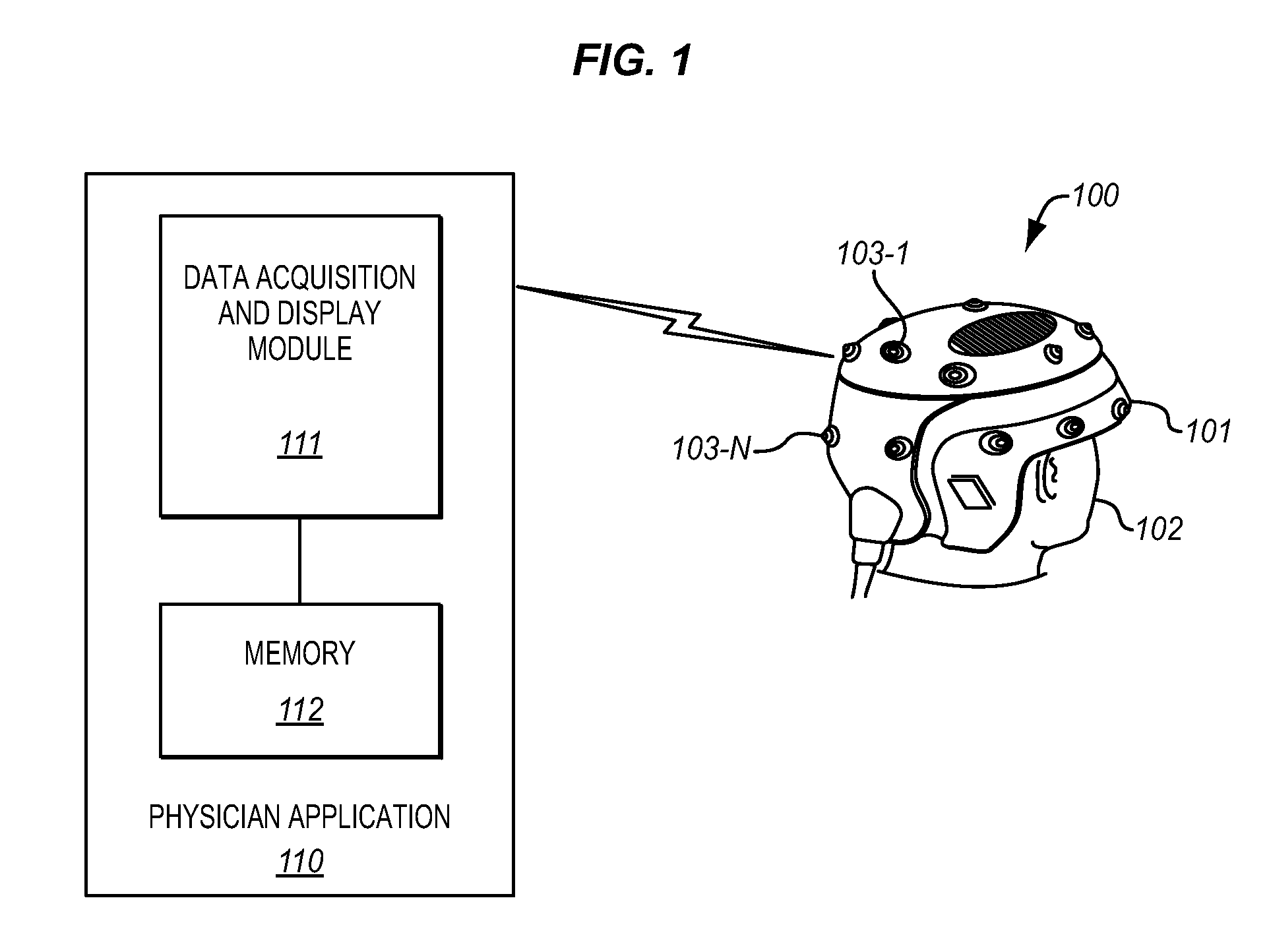
[0010]The voltages generated by the electrodes are amplified and filtered before being transmitted to an analysis platform, which can be a Physician's laptop computer system, either wirelessly or via a set of tethering wires. The EEG Processing Unit includes an automatic artifacting capability which identifies when there is sufficient clean data compiled in the testing session. This process automatically eliminates muscle or other physical artifact-related voltages. Clean data, which represents real brain voltages as opposed to muscle or physical artifact related voltages, thereby are produced. The automatic artifacting capability optionally includes an automatic Patient motion artifacting capability via an accelerometer that produces data indicative of Patient movement, which enhances the identification of accurate data.

Problems solved by technology

There are problems with the existing apparatus and methods used for collecting electroencephalogram data from a patient.
These problems, as described herein, cause the data collection process to be time consuming, potentially inaccurate, and also costly due to the need for semi-skilled medical personnel to place the electrodes on the patient and execute the test process to obtain usable data for the treating Physician.
Marginal voltages and excessive artifacts are indicative of poor electrode placement as well as the long leads that carry the raw measured voltages to the data recorder.
Since the EEG data is complex and can only be properly interpreted to look for Ailments by a trained Physician, the determination of “good” EEG data is difficult to ascertain in the EEG test setting in real time.
Thus, there is presently no system that simplifies the electrode placement, ensures precise and secure electrode attachment, and reduces the effects of artifacts in the electroencephalogram data obtained using EEG tests.

Method used


Image

  • Medical apparatus for collecting patient electroencephalogram (EEG) data
  • Medical apparatus for collecting patient electroencephalogram (EEG) data
  • Medical apparatus for collecting patient electroencephalogram (EEG) data

Examples


Embodiment Construction

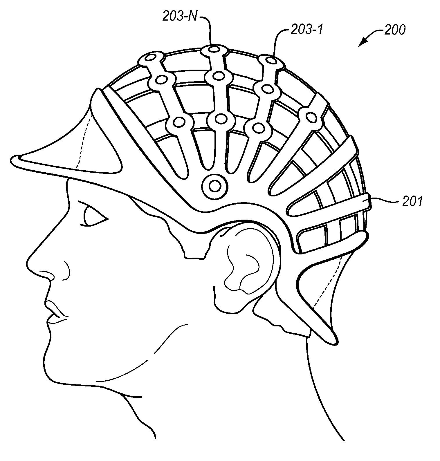
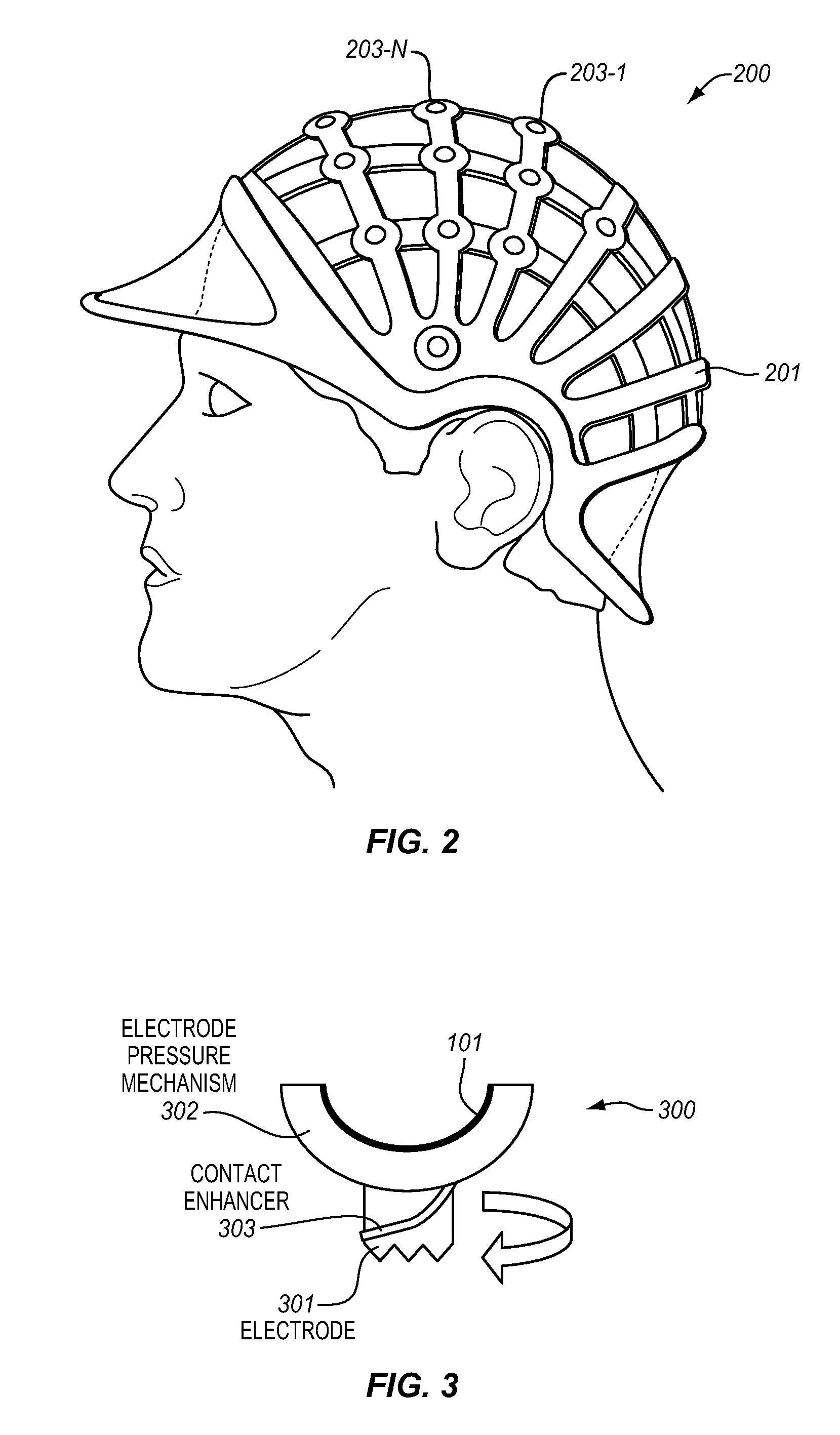
[0018]The EEG Processing Unit comprises a semi-rigid framework which substantially conforms to the head of the Patient. The framework supports a set of electrodes in predetermined loci on the Patient's head to ensure proper electrode placement. The EEG Processing Unit includes automated connectivity determination apparatus can use pressure-sensitive electrode placement to ensure proper contact with the Patient's scalp and also automatically verifies the electrode placement via measurements of the electrode impedance through automated impedance checking. In addition, the EEG Processing Unit can include optional automated electrode movement or rotation apparatus to clean the skin of the Patient to optimize the electrode contact with the Patient's scalp as indicated by the measured impedance.

[0019]The voltages generated by the electrodes are amplified and filtered before being transmitted to an analysis platform, which can be a Physician's laptop computer system, either wirelessly or v...


PUM


Abstract

The EEG Processing Unit comprises a semi-rigid framework which substantially conforms to the Patient's head and supports a set of electrodes in predetermined loci on the Patient's head to ensure proper electrode placement. The EEG Processing Unit includes automated connectivity determination apparatus which can use pressure-sensitive electrode placement ensuring proper contact with Patient's scalp and also automatically verifies electrode placement via measurements of electrode impedance through automated impedance checking. Voltages generated by the electrodes are amplified and filtered before being transmitted to an analysis platform, which can be a Physician's laptop computer system, either wirelessly or via a set of tethering wires. The EEG Processing Unit includes an automatic artifacting capability which identifies when there is sufficient clean data compiled in the testing session. This process automatically eliminates muscle- or other physical-artifact-related voltages. Clean data, which represents real brain voltages as opposed to muscle- or physical-artifact-related voltages, thereby are produced.

Description

CROSS-REFERENCE TO RELATED APPLICATIONS[0001]This application is a continuation-in-part of U.S. patent application Ser. No. 12 / 567,249 filed on Sep. 29, 2009, which application is a continuation-in-part of U.S. patent application Ser. No. 12 / 505,185 filed on Jul. 17, 2009. This application is also related to an application filed concurrently herewith titled “Data Management Apparatus For Comparing Patient Data With Ailment Archetypes To Determine Correlation With Established Ailment Biomarkers” and an application filed concurrently herewith titled “Patient Data Management Apparatus For Comparing Patient EEG Data With Ailment Archetypes To Determine Correlation With Established Ailment Biomarkers.” The foregoing applications are hereby incorporated by reference to the same extent as though fully disclosed herein.FIELD OF THE INVENTION[0002]This invention relates to medical apparatus and, in particular, to an apparatus for collecting patient electroencephalogram (EEG) data.BACKGROUND ...

Claims


Application Information

Patent Timeline
Patent Type & Authority Applications(United States)
IPC IPC(8): A61B5/0476A61B5/11
CPCA61B5/04004G06F19/345A61B5/6843A61B5/6803A61B5/4094G16H50/20A61B5/30A61B5/291
Inventor JOFFE, DAVIDOAKLEY, DAVID
Owner WAVI
Learn More