Methods of analyzing a selected region of interest in medical image data

a medical image and region technology, applied in image analysis, image enhancement, instruments, etc., can solve the problems of increasing the burden on the reporting clinician in terms of roi, and the considerable input of the clinician with the current roi tool availabl

Active Publication Date: 2011-02-03
SIEMENS MEDICAL SOLUTIONS USA INC
View PDF14 Cites 68 Cited by
  • Summary
  • Abstract
  • Description
  • Claims
  • Application Information

AI Technical Summary

Problems solved by technology

However, currently-available ROI tools still require considerable input from the clinician and are not designed specifically to support a PERCIST-style evaluation.
The requirements for quantitative evaluation outlined in the PERCIST criteria clearly place an added burden on the reporting clinician in terms of ROI creation and reporting.

Method used


Image

  • Methods of analyzing a selected region of interest in medical image data
  • Methods of analyzing a selected region of interest in medical image data
  • Methods of analyzing a selected region of interest in medical image data

Examples


Embodiment Construction

[0032]When the following terms are used herein, the accompanying definitions can be applied:

[0033]PET—Positron Emission Tomography

[0034]SUV—Standardized Uptake Value

[0035]SUL—Lean body mass-corrected SUV

[0036]PERCIST—PET Response Criteria in Solid Tumours

[0037]Mediastinum—a non-delineated group of structures in the thorax (chest), surrounded by loose connective tissue. It is the central compartment of the thoracic cavity. It contains the heart, the great vessels of the heart, esophagus, trachea, thymus, and lymph nodes of the central chest

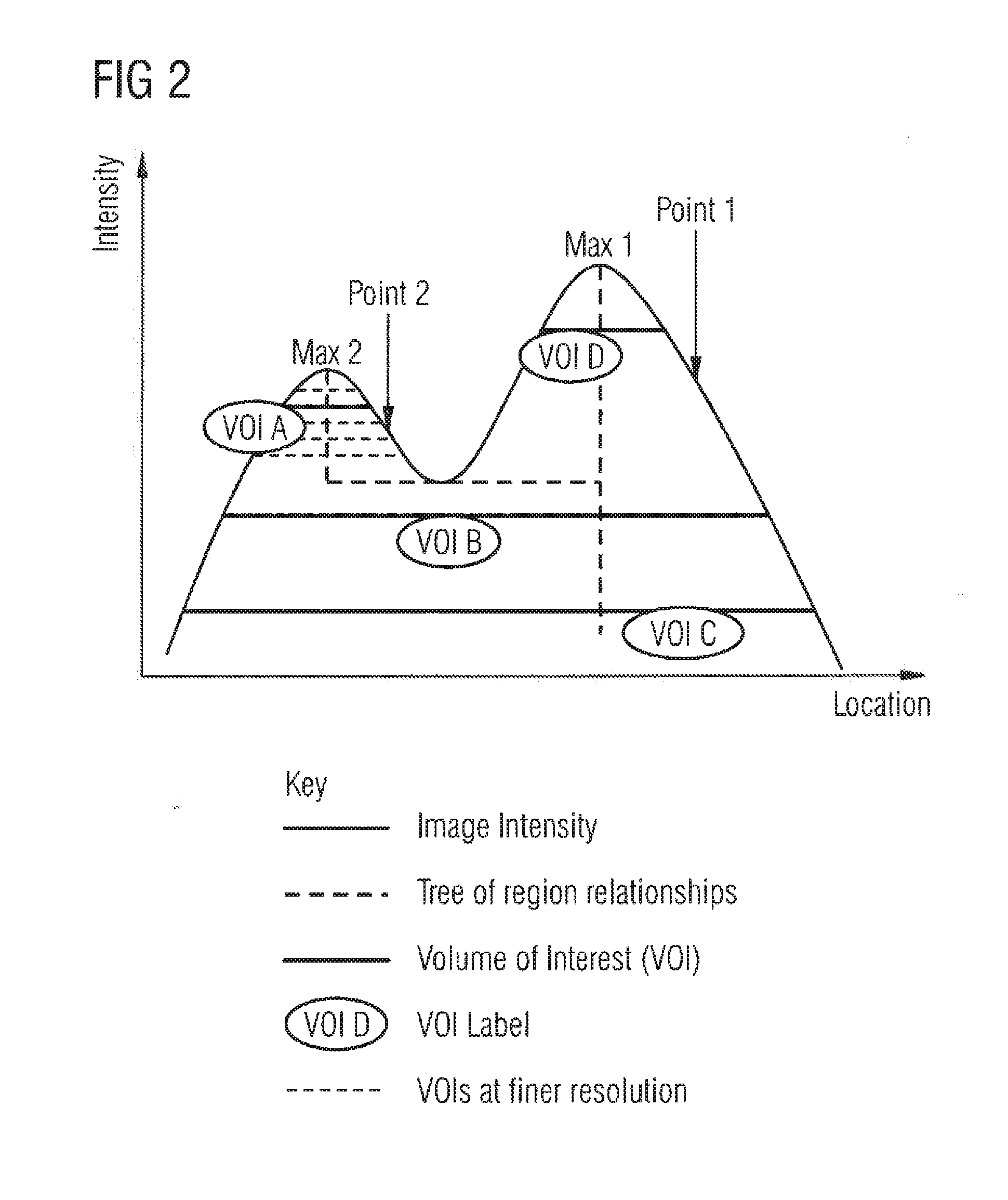
[0038]ROI / VOI—RegionNolume of interest.

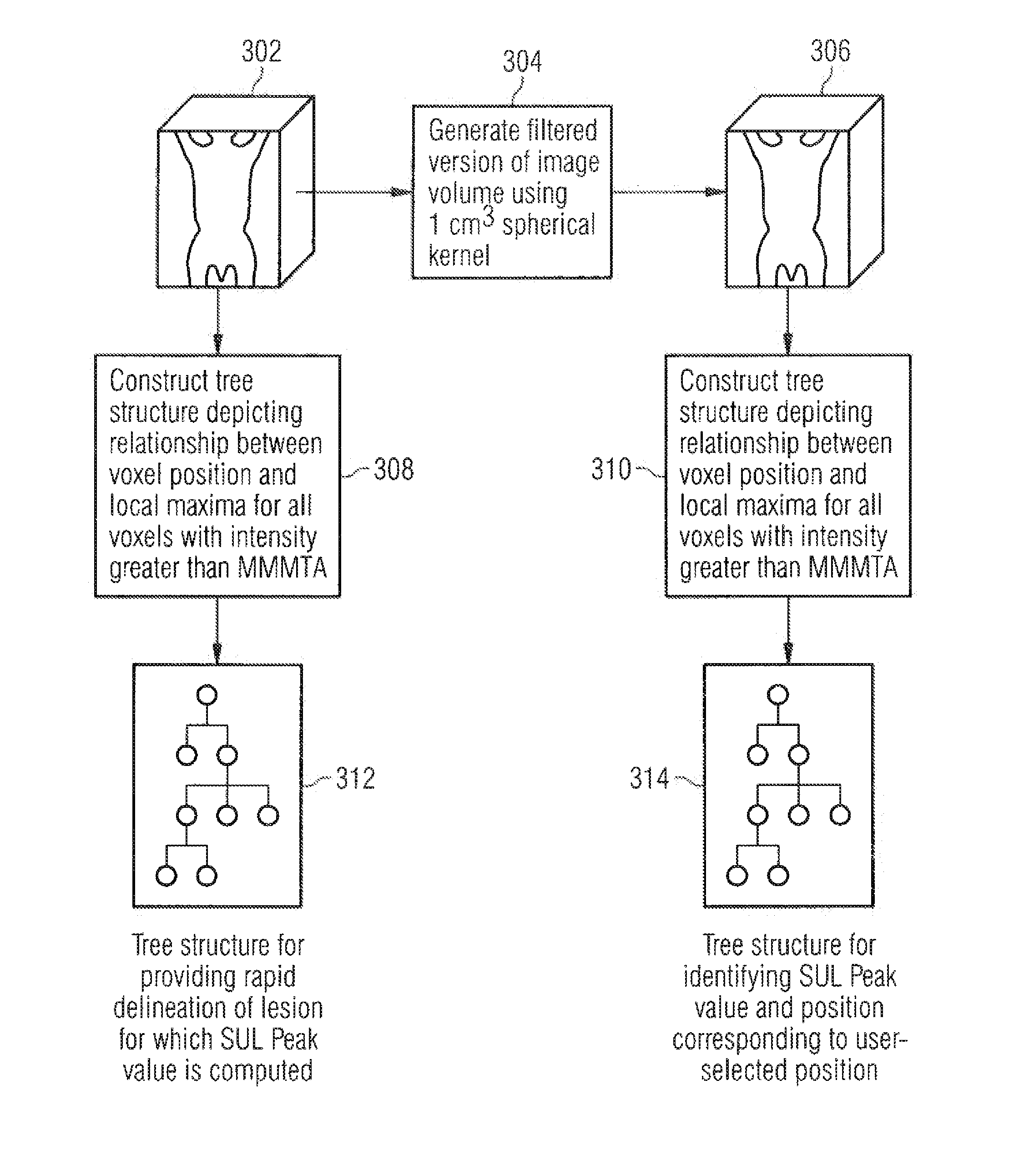
[0039]MMMTA—minimal metabolically measurable tumor activity.

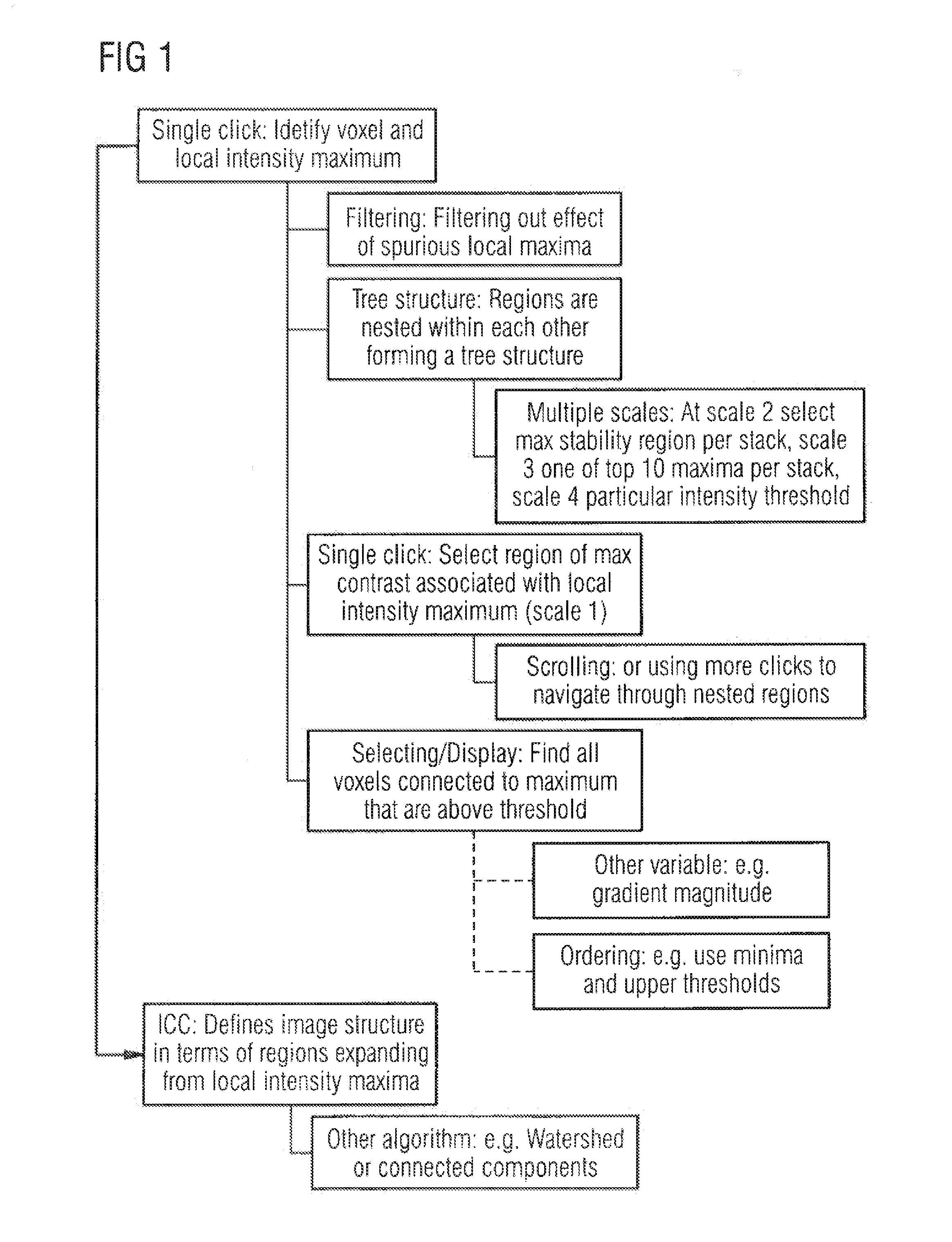
[0040]ICC—Iterative Connected Component algorithm

[0041]Threshold—a particular intensity value within an image, often above or below which all pixels or voxels are accepted for a process or algorithm.

[0042]In embodiments, the invention addresses the above noted problems by providing a PERCIST-specific reading workflow with minimal user input for ROI po...


PUM


Abstract

In methods and an apparatus for analyzing a selected region of interest in medical image data of a subject, an initial image data set is obtained, and the initial image data set is filtered to generate a filtered data set. The filtering includes computing, for each voxel of the initial image data set, a value of intensity for a standardized volume of interest centered on that voxel. A user selection of a region of interest in the initial image data set is registered, and from the filtered image data set a value of intensity for the selected region of interest is computed.

Description

BACKGROUND OF THE INVENTION[0001]1. Field of the Invention[0002]This invention is directed to methods and apparatus for analyzing a selected region of interest in medical image data of a subject.[0003]2. Description of the Prior Art[0004]In the medical imaging field, several imaging schemes are known. For example PET (Positron Emission Tomography) is a method for imaging a subject in 3D using an injected radio-active substance which is processed in the body, typically resulting in an image indicating one or more biological functions.[0005]The definition of regions or volumes of interest (ROI / VOI) is a typical precursor to quantitative analysis of medical images, such as nuclear medicine emission images (for example, PET or SPECT). Such regions may be defined around areas of high intensity which correspond to high tracer uptake (hotspots). For example, in FDG-PET images for oncology; such areas may be indicative of the presence of a tumor. Oncology physicians frequently annotate lesi...

Claims


Application Information

Patent Timeline
Patent Type & Authority Applications(United States)
IPC IPC(8): G06K9/00
CPCG06T7/0012G06T2207/10104G06T2207/30056G06T2207/20072G06T2207/20104G06T2207/10108G06T2207/30096
Inventor DECLERCK, JEROMEKADIR, TIMORKELLY, MATTHEW DAVID
Owner SIEMENS MEDICAL SOLUTIONS USA INC
Why Eureka
Try Eureka