Radiography apparatus

a technology of radiography and apparatus, applied in the field of radiography apparatus, can solve the problems of limited radiation dose that passes the outside of the actual region of interest during radiography, and achieve the effects of reducing the dose of radiation irradiating the subject, efficient slice image acquisition, and efficient slice image obtaining

Inactive Publication Date: 2011-03-31
FUJIFILM CORP
View PDF29 Cites 16 Cited by
  • Summary
  • Abstract
  • Description
  • Claims
  • Application Information

AI Technical Summary

Benefits of technology

[0033]According to the present invention, when a slice image is obtained, body thickness information about a subject is obtained, and a slice image obtainment condition representing a range in which the slice image is obtained is set based on the body thickness information. Further, the slice image is obtained based on the set slice image obtainment condition. Therefore, it is possible to efficiently set the slice image obtainment condition that is appropriate for the body thickness of the subject. Hence, it is possible to efficiently obtain the slice image.
[0034]Here, pre-shot radiography is performed to set the radiography condition in obtainment of a slice image. Further, when the pre-shot radiography is plain radiography, the pre-shot radiography is necessary to diagnose a patient. Therefore, the pre-shot radiography is always performed before obtainment of the slice image. When the slice image is obtained after pre-shot radiography, it is possible to set a slice image obtainment condition, without irradiating the subject with unnecessary radiation (additional or excessive radiation), by obtaining body thickness information about the subject based on the radiography condition in the pre-shot radiography.
[0035]A slice image may be obtained optionally after pre-shot radiography. As the slice image is obtained optionally, especially when the pre-shot radiography is plain radiography, if diagnosis is possible by using only a pre-shot image obtained in the pre-shot radiography, obtainment of the slice image may be omitted. Consequently, it is possible to reduce the dose of radiation irradiating the subject.
[0036]Radiography by the radiation source located at a position at which pre-shot radiography was performed may be skipped in obtainment of the slice image, and the pre-shot image may be used as a radiographic image obtained by using the radiation source located at the position at which pre-shot radiography was performed. Consequently, in obtainment of the slice image, radiography by the radiation source located at the position at which pre-shot radiography was performed may be omitted. Hence, it is possible to reduce the dose of radiation irradiating the subject in obtainment of the slice image.
[0037]In this case, the movement of the subject may be detected. When the subject has moved by a distance exceeding a predetermined value from the time of pre-shot radiography to the time of obtainment of the slice image, radiography by the radiation source located at the position at which pre-shot radiography was performed may be performed in obtainment of the slice image. Consequently, it is possible to prevent a drop in the image quality caused by a shift in the position of the subject between the radiographic image obtained by using the radiation source located at the position at which pre-shot radiography was performed and radiographic images obtained by using the radiation source located at different positions.
[0038]Further, past radiography conditions about a plurality of subjects may be stored in a storage means, and the past radiography conditions stored in the storage means may be referred to. If a past radiography condition about the subject to be radiographed is stored in the storage means, the slice image obtainment condition may be set based on the past radiography condition about the subject to be radiographed. Consequently, it is possible to obtain the slice image by setting the same target of radiography as that of the past radiography. Therefore, it is possible to accurately observe the progress or course of the condition of a patient by using slice images.

Problems solved by technology

Accordingly, the dose of radiation that passes the outside of the actual region of interest during radiography is limited.

Method used


Image

  • Radiography apparatus
  • Radiography apparatus
  • Radiography apparatus

Examples


first embodiment

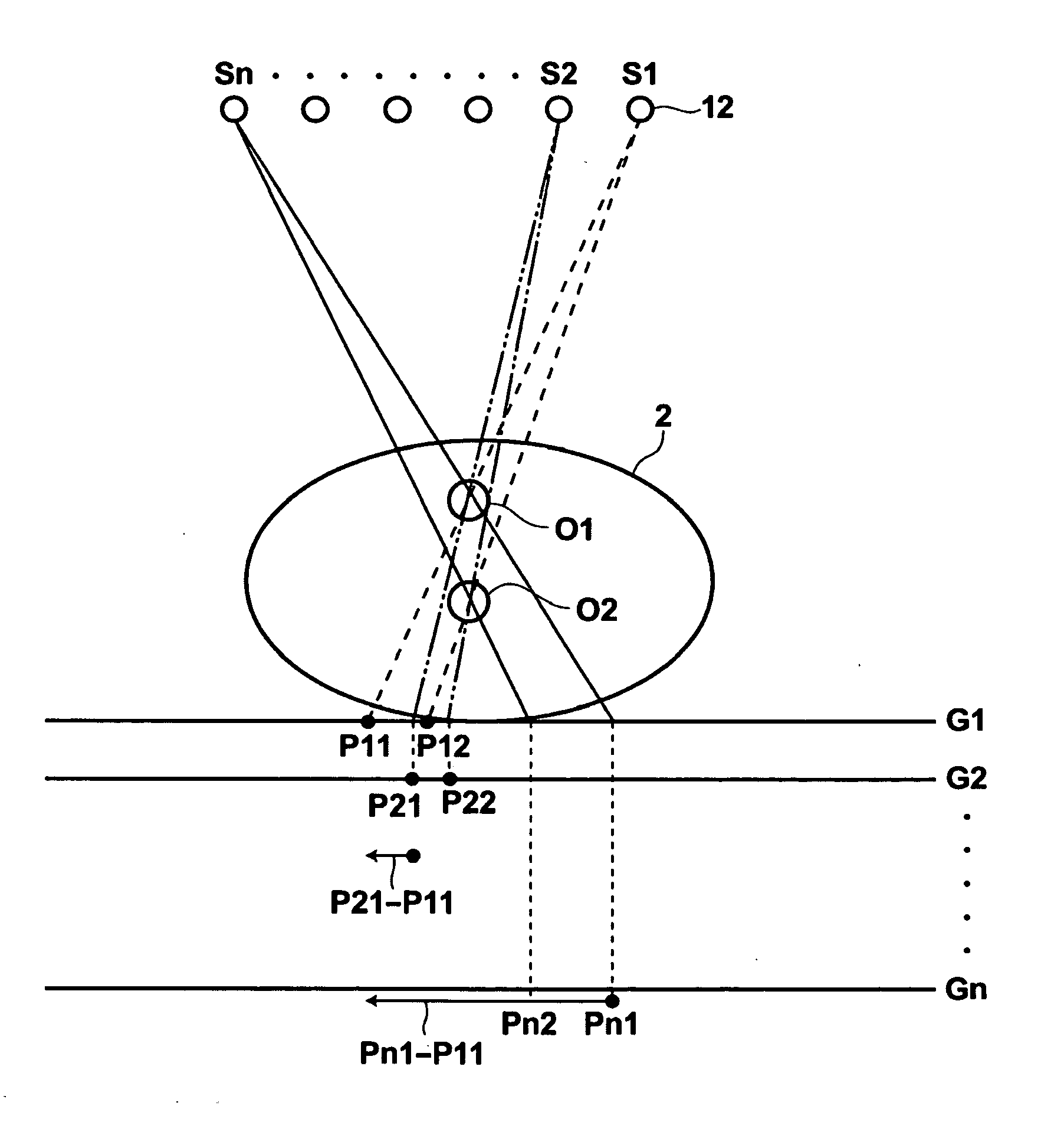
[0070]Therefore, in the first embodiment, the storage unit 26 stores a table in which the relationship of the body thickness with the position of the center plane of slice planes on which slice images are reconstructed and the reconstruction range is regulated. FIG. 4 is a table in which the relationship of the body thickness with the position of the center plane of slice planes on which slice images are reconstructed and the reconstruction range is regulated. As illustrated in FIG. 4, Table T1 correlates a thick body thickness to 15 cm as the position of the center plane, and 20 cm as the reconstruction range. Table T1 correlates a standard body thickness to 10 cm as the position of the center plane, and 10 cm as the reconstruction range. Further, Table T1 correlates a thin body thickness to 5 cm as the position of the center plane, and 5 cm as the position of the reconstruction range. Here, the position of the center plane is represented by a distance from the top board 4 of the r...

second embodiment

[0095]As described above, the slice image obtainment condition is set based on the body thickness information also in the Therefore, it is possible to efficiently set the slice image obtainment condition based on the body thickness of the subject 2. Hence, it is possible to efficiently obtain the slice image.

[0096]Next, a third embodiment of the present invention will be described. FIG. 13 is a schematic diagram illustrating the configuration of an X-ray radiography apparatus for performing tomosynthesis radiography to which a radiography apparatus according to the third embodiment of the present invention has been applied. In the third embodiment, the same reference numerals are assigned to the same elements as the elements of the first embodiment, and detailed descriptions of the elements are omitted. An X-ray radiography apparatus 10B of the third embodiment differs from the first embodiment in that the body thickness information about the subject 2 is obtained by a sensor 42, s...

third embodiment

[0099]As described above, the slice image obtainment condition is set based on the body thickness information also in the Therefore, it is possible to efficiently set the slice image obtainment condition based on the body thickness of the subject 2. Hence, it is possible to efficiently obtain the slice image.

[0100]In the third embodiment, the sensor, such as the ultrasonic sensor, is used. However, it is not necessary that the sensor is used. Any device or method may be adopted as long as a body thickness of the subject 2 is measured. For example, the body thickness information about the subject 2 may be obtained based on an image of the subject 2 imaged by using a camera or the like. In the first through third embodiments, only the X-ray tube 12 is moved. Needless to say, the present invention may be applied to a case in which both of the X-ray tube 12 and the detector 14 are moved in such a manner to be synchronized with each other.

[0101]In the first through third embodiments, to...


PUM


Abstract

When a subject is irradiated with radiation, and the radiation that has passed through the subject is detected, body thickness information about the subject is obtained. Further, a slice image obtainment condition representing a range in which a slice image is obtained in the subject is set based on the body thickness information. Further, the slice image is obtained based on the slice image obtainment condition.

Description

BACKGROUND OF THE INVENTION[0001]1. Field of the Invention[0002]The present invention relates to a radiography apparatus for performing radiography to generate slice images of a subject.[0003]2. Description of the Related Art[0004]In recent years, tomosynthesis radiography was proposed to observe affected regions of patients in more detail by a radiography apparatus using X-rays. In tomosynthesis radiography, radiography is performed by moving an X-ray tube to different positions, and by irradiating a subject by the X-ray tube at different angles from the different positions. Further, images obtained by radiography are added to obtain an image in which a desired cross section of the subject is emphasized. In tomosynthesis radiography, the X-ray tube is moved parallel to an X-ray detector, or in such a manner to draw a circle or an ellipse or oval, based on the characteristic of a radiography apparatus and the kind of slice images (tomograms) to be obtained. The subject is radiograph...

Claims


Application Information

Patent Timeline
Patent Type & Authority Applications(United States)
IPC IPC(8): A61B6/03
CPCA61B6/032A61B6/06A61B6/544A61B6/542A61B6/469
Inventor AKAHORI, SADATOENOMOTO, JUNKANAGAWA, EIICHIOHTA, YASUNORIIDA, NORIAKI
Owner FUJIFILM CORP
Why Eureka
Try Eureka