Apparatus and method for laparoscopic port site suture

a port site and suture technology, applied in the field of medical science, can solve the problems of high risk of port site hernia, cumbersome suture method, and surgeons being unable to get their fingers into the incision,

Inactive Publication Date: 2011-05-12
A H BEELEY
View PDF23 Cites 52 Cited by
  • Summary
  • Abstract
  • Description
  • Claims
  • Application Information

AI Technical Summary

Problems solved by technology

The suture method is cumbersome and difficult because the incisions are usually small and deep.
Thus, a surgeon typically is unable to get his fingers into the incision to perform the suturing procedure.
Moreover, when ports having diameters of 1 cm or more, especially in the lower part of the abdomen (below the level of the umbilicus), there is a high risk of developing a hernia through the port site (port site hernia).
Further, a port site hernia will require a further operation at a later date to repair it and this may be a very difficult procedure.
Enlargement of the incisions will result in an increased discharge of gas from the abdomen and therefore in a reduction of the working and viewing space.
This creates a series of inconveniences when suturing the patient.
Upon release of the gas, the abdominal wall lies in contact with the intra abdominal organs such as the bowel so that the risk of picking up the bowel wall during the suture procedure of the incisions is greatly increased.

Method used


Image

  • Apparatus and method for laparoscopic port site suture
  • Apparatus and method for laparoscopic port site suture
  • Apparatus and method for laparoscopic port site suture

Examples


first embodiment

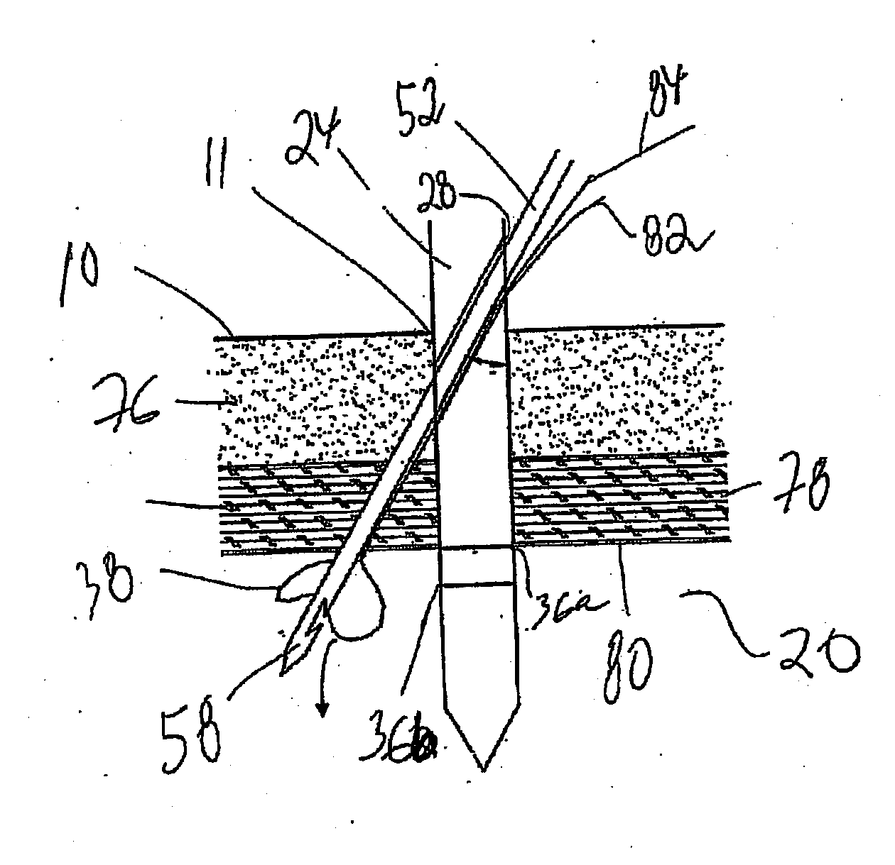
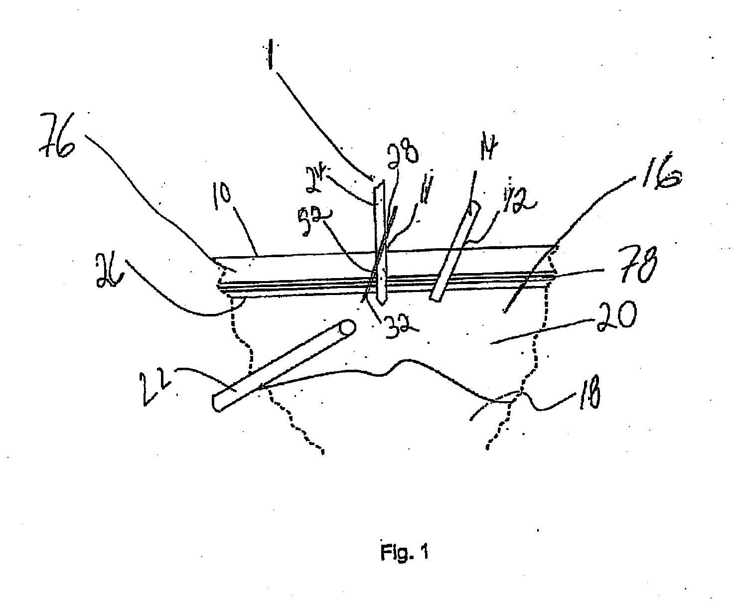
[0100]FIG. 1 is a schematic cross-sectional view of a section of the abdomen of a patient undergoing laparoscopic surgery. In the surgical procedure, a first incision 11 is made in the abdominal wall 10 of the patient, which subsequently needs to be closed. Apparatus 1 according to the invention is used in the procedure for closing the incision 11. As part of the laparoscopic surgical procedure second and third incisions perforate the abdominal wall for insertion of a port 14 and telescope 22. The port 14 facilitates delivery of instruments such as forceps (not shown) into the abdomen 16 for the procedure. Organs 18 within the abdomen are separated from the abdominal wall 10 by a space 20. Space 20 provides the working space required to perform the suture procedure and the viewing space so that the telescope 22 may provide images of the procedure within the abdomen 16. It should be noted that FIG. 1 is schematic only and the location and disposition of the apparatus 1, port 14 and t...

second embodiment

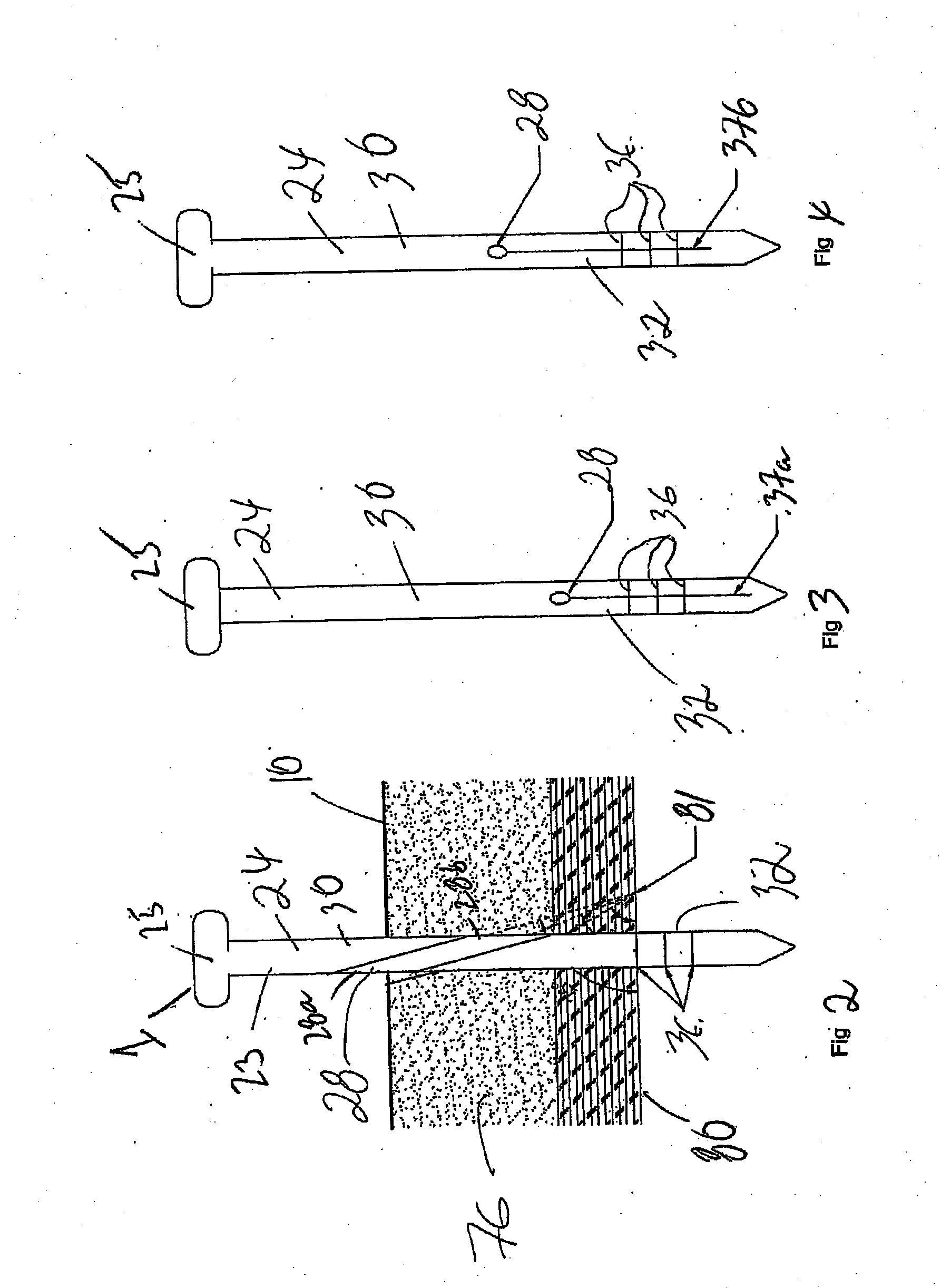
[0115]FIGS. 5 to 7 show an apparatus 2 according to the invention. In the arrangement shown in FIGS. 5 to 7, the apparatus 2 comprises a body 24 is similar to the body 24 previously described and shown in FIGS. 2 to 4, and so similar reference numerals are used to identify similar parts.

[0116]The body 24 of apparatus 2 according to the second embodiment of the invention comprises a plurality of passages 28 traversing the body 24. In the arrangement shown there are three passages 28 comprising first passage 29, second passage 30 and third passage 31. The passages 29, 30 and 31 are oriented at different angles with respect to each other. As shown in FIG. 5, passages 30 and 31 traverse the body 24 at steeper angles than passage 29. However, the passages 29, 30 and 31 are arranged such that the distal end of the needle 52 (traversing any of the passages 28, 29 and 31) arrives adjacent the body 24 at a same location 81 on the peritoneum 80, as depicted in FIG. 5. For example, a suitable ...

third embodiment

[0121]the invention (which is not shown) a trocar is configured to incorporate the passage(s) 28 and also the marks 36 and 37. The trocar may be supplied with a port. With such an arrangement, the passage(s) and the marks 36 are located at the distal end of the trocar. This enables the trocar, supplied with the port, to be used for suturing its incisions (instead of a separate body 24). Trocars are regularly used to introduce ports in abdominal walls. Thus, with this embodiment it is possible to suture the incision in which the ports are inserted with the same trocar used to insert the port. In this way it is not necessary to have a separate surgical instrument, such as body 24, to suture the port site. Passage(s) 28 and marks 36 and 27 may be included in any type of trocar. Alternatively, the trocar may include a single passage and / or any of marks 36 or 37.

[0122]In FIG. 9 there is shown a dilator 70 used for increasing the size of an incision for extraction of, for example, a large...


PUM


Abstract

A surgical device for guiding a needle during suturing of an incision comprising a body for insertion in the incision having a first passage to conduct an end of the needle below a location surrounding the incision, wherein a distal portion of the body comprises means for indicating the location to which the needle will be conducted. Methods for suturing an incision using the surgical device are also provided.

Description

FIELD OF THE INVENTION[0001]The present invention relates to the medical field and more particularly to apparatus and methods applied during surgical procedures for suturing incisionsBACKGROUND ART[0002]Invasive surgical procedures which involves the cutting of one or more incisions to access a biological cavity are widespread. The size of different types of incisions may vary depending on the type of surgical procedure being performed. For example, it might be desirable or required to perform only small incisions in order to access a biological cavity. A patient's body may be punctured in order to insert, for example, a telescope into the patient's body, to view the inside of a biological cavity. Also, a small incision may be made to insert in the biological cavity a tube that delivers gas (usually CO2) in order to insufflate the cavity thus providing a viewing space for the telescope and a working space for surgical instruments provided via other small incisions.[0003]As an exampl...

Claims


Application Information

Patent Timeline
Patent Type & Authority Applications(United States)
IPC IPC(8): A61B17/04A61M29/00
CPCA61B17/0057A61B2017/00637A61B2019/4836A61M29/00A61B2017/00663A61B2090/0807A61B17/0482A61B2017/06042A61B2090/0811A61B17/062
Inventor BEELEY, ANTONY HUGH
Owner A H BEELEY
Why Eureka
Try Eureka