Surgical access port with embedded imaging device

a surgical access port and imaging device technology, applied in the field of endoscopic devices, can solve the problems of reducing the risk of damaging abdominal organs, affecting the use of patients,

Inactive Publication Date: 2011-06-30
PSIP LLC
View PDF99 Cites 125 Cited by
  • Summary
  • Abstract
  • Description
  • Claims
  • Application Information

AI Technical Summary

Problems solved by technology

Such observation reduces the risk of damaging abdominal organs beneath the point of insertion, such as may occur when the trocar is pushed too far into the abdominal cavity.
This would then result in the loss of visualization of the surgical field, which interrupts the surgical procedure and interrupts the use of the surgical instruments until the surgical field can again be visualized with the laparoscope.
In addition, sometimes during the course of a surgery an endoscopist or surgeon determined that the view through the laparoscope is not optimal for safe manipulation of the instruments, and it is necessary to withdraw the laparoscope from the primary port and insert it through one of the ancillary ports in order to provide visualization of the surgical field from a more appropriate angle.
This also interrupts the surgical procedure and increase risk to the patient.
However, because laparoscopes are relatively long and heavy, a surgeon or an assistant must have one hand occupied with the laparoscope at all times unless it is attached to a robotic arm.
Furthermore, laparoscopes require sterilization between uses, and using more than one laparoscope for a procedure would result in significant additional expense for sterilization.
Because laparoscopes are very expensive, using more than one laparoscope for a surgical procedure would require a hospital or surgical facility to make a substantial additional investment to have extra laparoscopes on hand, which also requires increases maintenance and sterilization expenses.

Method used


Image

  • Surgical access port with embedded imaging device
  • Surgical access port with embedded imaging device
  • Surgical access port with embedded imaging device

Examples


Embodiment Construction

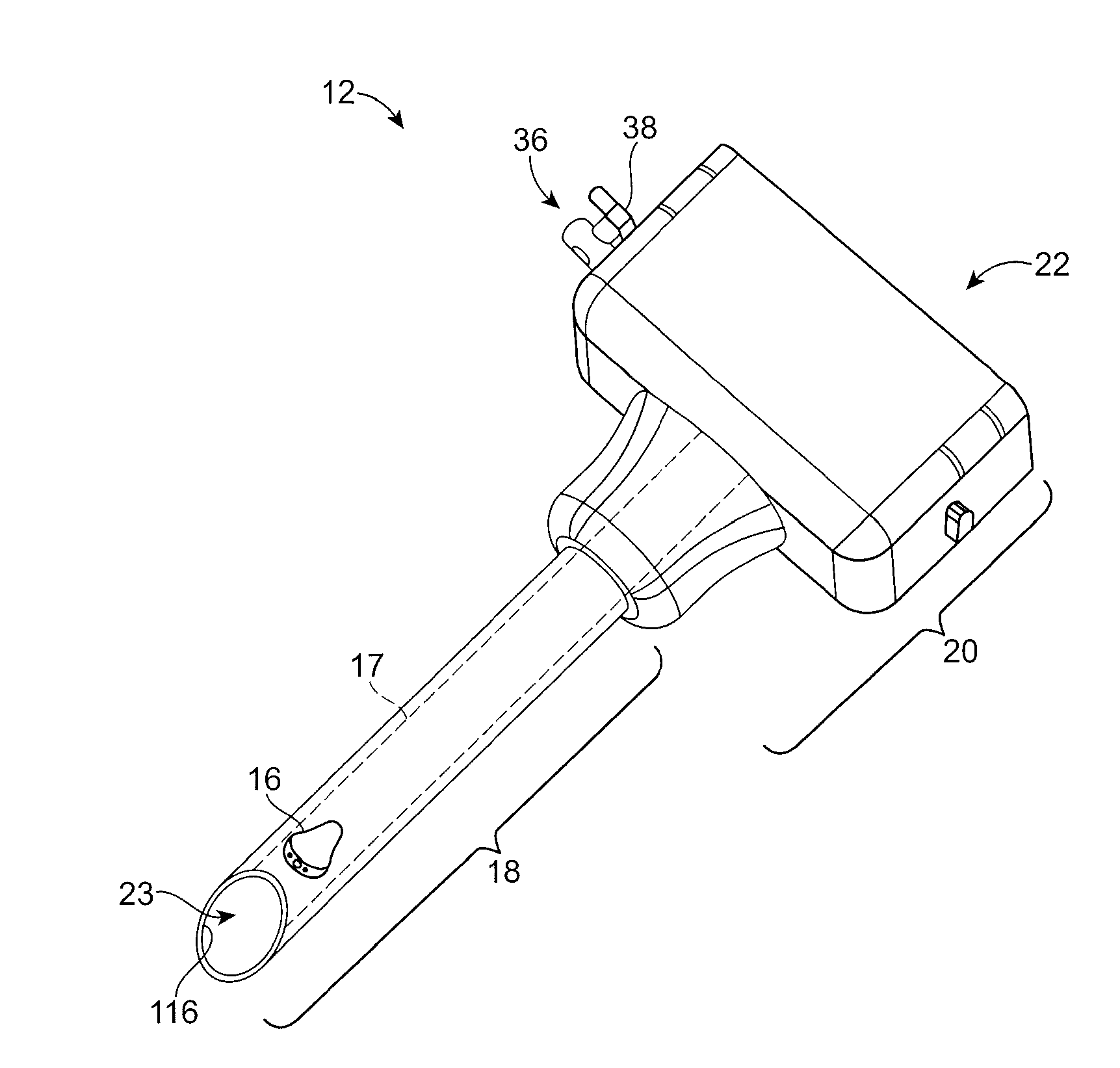
[0051]The term “endoscopic surgery” is a broad term that includes many varieties of surgeries such as laparoscopy. The scope of the present invention includes various types of endoscopic procedures, including laparoscopic surgery and other minimally invasive forms of surgery. The present invention also applies to any type of surgery that makes use of a trocar or cannula or similar devices.

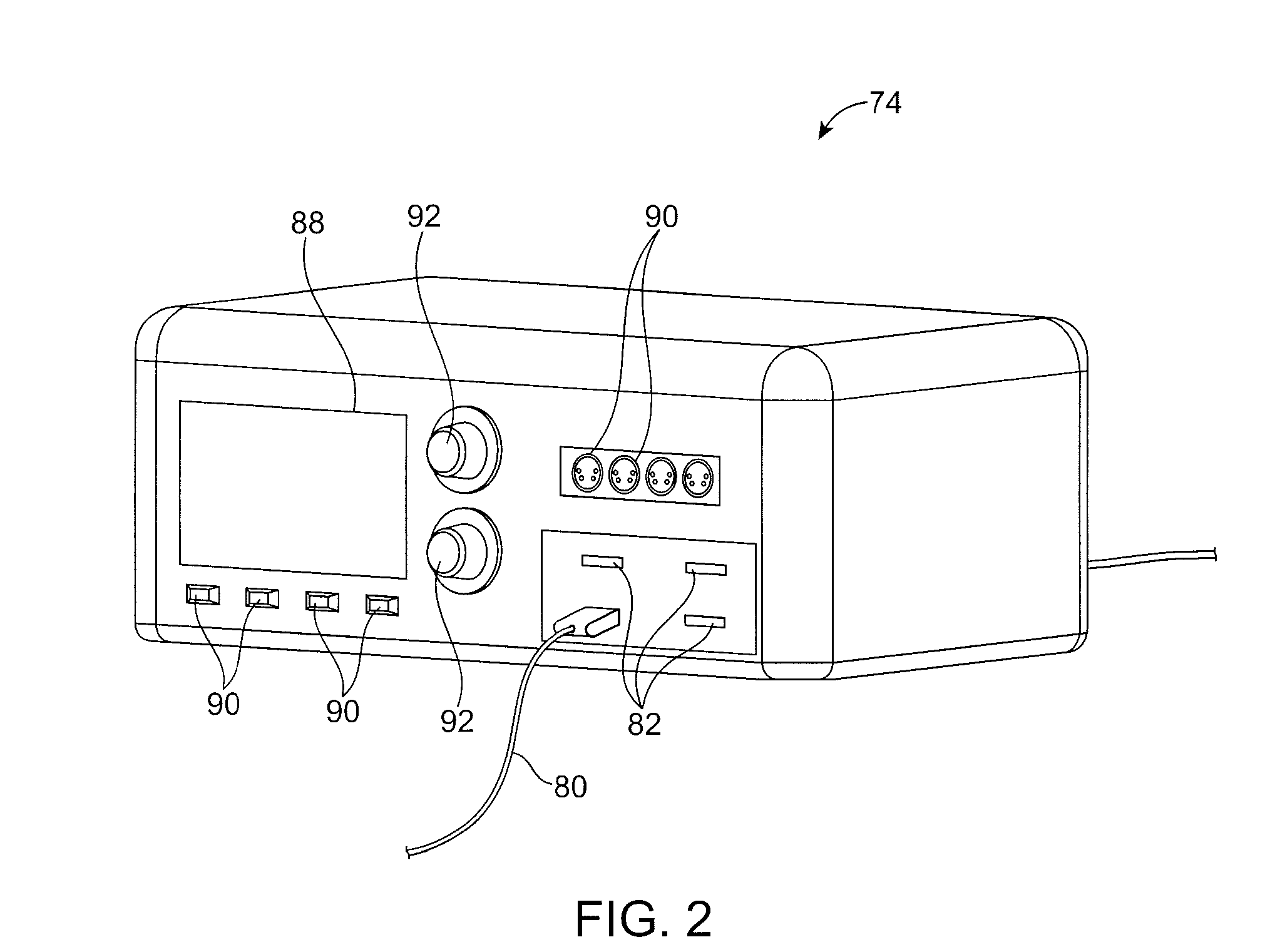
[0052]In an embodiment of the present invention, a trocar is inserted into a cannula until it snaps into place as the projections on the trocar handle engage with complementary grooves on the corresponding sections of the cannula handle. An integrated imaging device, such as a camera, forms part of the cannula, trocar, or both. Corresponding electrical cabling for the camera is connected to connectors on an external control box and the cannula handle. The camera is powered on through the control box, and the control box begins to process the images captured by the camera and displays them on a moni...


PUM


Abstract

Disclosed is a disposable access port for use in endoscopic procedures, including laparoscopic procedures. The access port includes a cannula with an embedded camera in communication with an external control box. In operation, a trocar is combined with the access port to facilitate insertion of the access port into an anatomical site. Prior to insertion, the camera is pushed inside the cannula, where it remains during insertion. The trocar is removed after the access port has been inserted to allow surgical instruments to access the anatomical site. During removal of the trocar, a portion of the trocar urges the camera out of the cannula, thereby allowing visualization of the anatomical site. The camera can be fixedly or adjustably mounted on the port. A camera may also be mounted on the trocar. The trocar may include irrigation and suction channels to facilitate a clear view of the anatomical site.

Description

CLAIM OF PRIORITY[0001]This application is a divisional of and claims the benefit of priority under 35 U.S.C. § 120 to U.S. patent application Ser. No. 11 / 834,540, filed on Aug. 6, 2007, which claims the benefit of priority under 35 U.S.C. § 119(e) to U.S. Provisional Patent Application Ser. No. 60 / 835,543, filed Aug. 4, 2006, the benefit of priority of each of which is claimed hereby, and each of which is incorporated by reference herein in its entirety.BACKGROUND OF THE INVENTION[0002]1. Field of the Invention[0003]The present invention relates generally to the field of endoscopic devices, and more particularly, to laparoscopic surgical devices including laparoscopic ports which provide minimally invasive access to the abdominal cavity.[0004]2. Description of the State of the Art[0005]Endoscopic surgery has become the new standard for surgical procedures. A specific type of endoscopic surgery, laparoscopic surgery, has become the preferred method for surgeries involving the organs...

Claims


Application Information

Patent Timeline
Patent Type & Authority Applications(United States)
IPC IPC(8): A61B1/32A61B1/045
CPCA61B1/00154A61B1/05A61B1/3132A61B17/3417A61B2019/5217A61B19/5225A61B2017/00221A61B2017/3454A61B17/3421A61B2090/3614A61B90/37
Inventor BAYER, LEXHIGGINS, JACK
Owner PSIP LLC
Why Eureka
Try Eureka