Visceral organ suspension device for endoscopic surgery

A suspension device and endoscope technology, applied in the field of medical devices, can solve the problems of poor liver feasibility, liver subcapsular hematoma, high price, etc., and achieve the effect of increasing the contact area, clear exposure, and convenient operation

Active Publication Date: 2020-07-10
THE FIRST AFFILIATED HOSPITAL OF ZHENGZHOU UNIV
View PDF13 Cites 4 Cited by
  • Summary
  • Abstract
  • Description
  • Claims
  • Application Information

AI Technical Summary

Problems solved by technology

The assistant can add a special puncture hole in the abdominal wall, and use fan-shaped forceps to pull the left lobe of the liver forward to the right to expose the stomach and duodenum. However, this method requires an additional puncture hole and occupies one arm of the assistant. steadily revealed
It is also possible to suture the liver tissue with silk thread, and pass both ends of the silk thread out of the abdominal cavity to suspend and fix the liver on the anterior abdominal wall. Although this method can save one puncture hole, long-term pulling of the silk thread will damage the liver parenchyma and its tissues. The internal blood vessels and bile ducts play a cutting role, causing liver damage
Commercial liver retractors can also be used, but this device not only requires a special puncture hole, but also is expensive and inconvenient to operate. In addition, the metal retractor compresses the liver for a long time may cause liver subcapsular hematoma, cause additional liver damage
[0005] At present, several patents have designed liver suspension devices. Patent document CN203436352U discloses a laparoscopic liver suspen...

Method used


Image

  • Visceral organ suspension device for endoscopic surgery
  • Visceral organ suspension device for endoscopic surgery
  • Visceral organ suspension device for endoscopic surgery

Examples


Embodiment 1

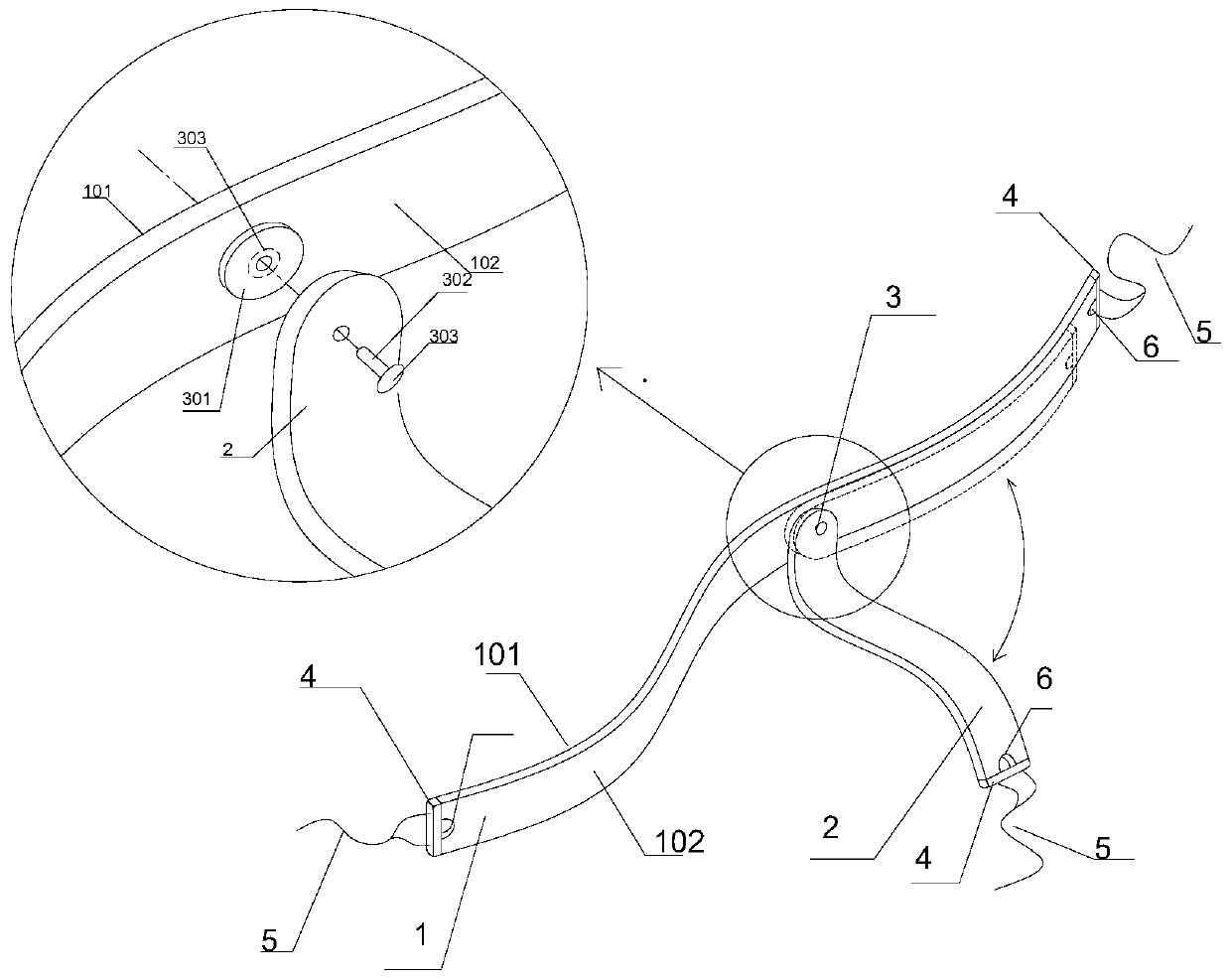
[0034] A liver suspension device used in laparoscopic surgery, such as figure 1 As shown, its structure mainly includes an upper suspension belt 1 and a lower suspension belt 2. The upper suspension belt 1 is a soft strip-shaped plastic film with a length of 20 cm and a width of 1 cm. One side is a suspension contact surface 101 and the other side is a pivotal joint surface 102 . The lower suspension belt 2 is a soft strip-shaped plastic film with a length of 8 cm and a width of 1 cm. One end has a pivot hole and is pivotally connected to the pivotal surface 102 of the upper suspension belt 1 by the rotating connection part 3, and the other end is free. The lower suspension belt 2 can rotate 360 ​​degrees around the pivot point on a plane parallel to the upper suspension belt 1, so that the angle between the upper suspension belt 1 and the lower suspension belt 2 can be opened and closed. Both ends of the upper suspension belt 1 and the free end of the lower suspension belt 2...

Embodiment 2

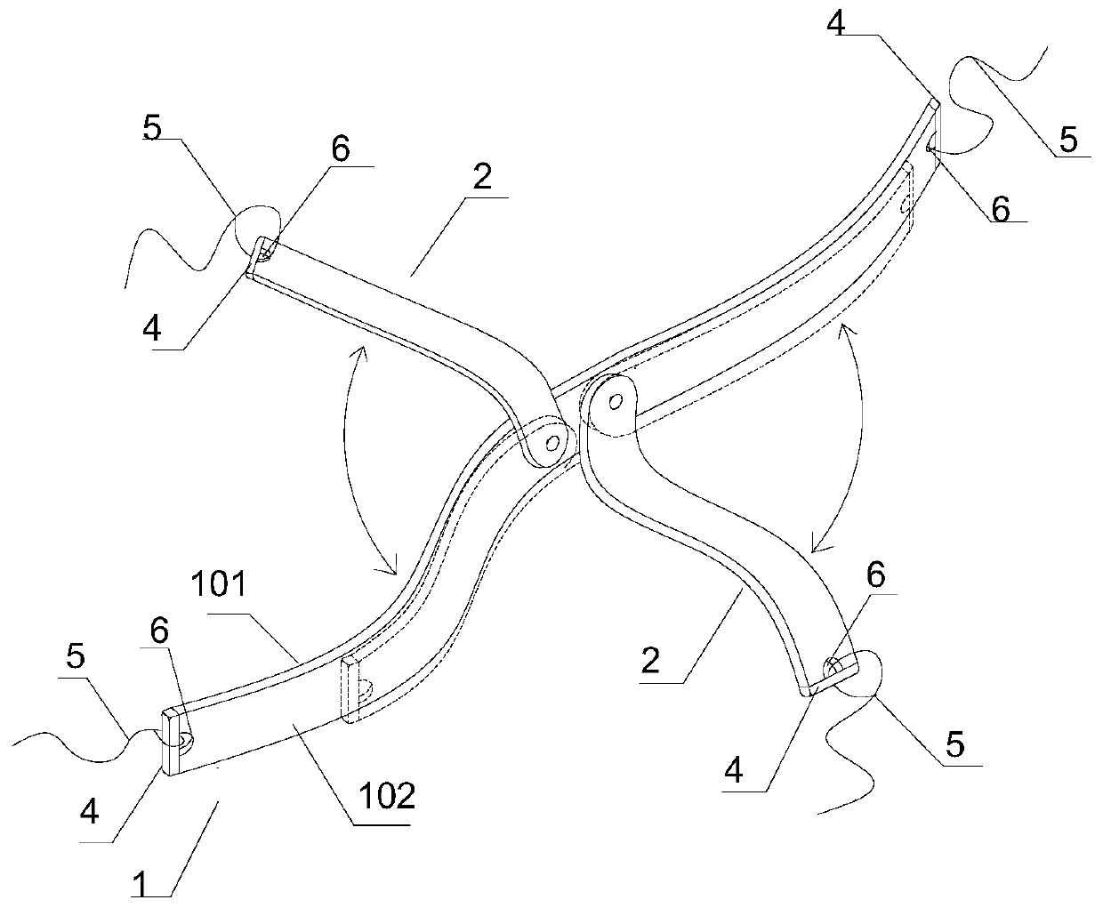
[0038] A lobar suspension device for thoracoscopic surgery, such as figure 2 As shown, its structure mainly includes an upper suspension belt 1 and two lower suspension belts 2 . The upper suspension belt 1 is a soft strip-shaped plastic film with a length of 15-20 cm and a width of 1 cm. One side is a suspension contact surface 101 and the other side is a pivotal joint surface 102 . Each lower suspension belt 2 is a soft strip-shaped plastic film with a length of 6-8 cm and a width of 1 cm. Embodiment 1 is the same), and the other end is free. Each lower suspension belt 2 can rotate around its pivot point in a plane parallel to the upper suspension belt 1 . Both ends of the upper suspension belt 1 and the free end of each lower suspension belt 2 are provided with a support side strip 4 made of elastic memory resin material, and a reserved through hole 6 is opened on the plastic film inside the support side strip 4 .

[0039] When using the lung lobe suspension device of ...

Embodiment 3

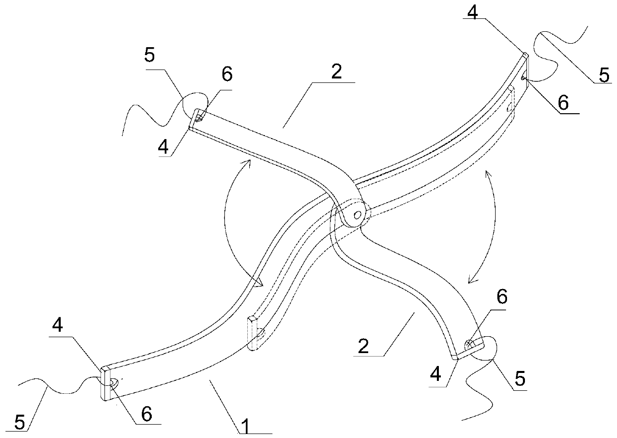
[0041] An organ or tissue suspension device for endoscopic surgery, its overall structure is similar to the suspension device described in Embodiment 2, the difference is that, as image 3 As shown, the two lower suspension belts 2 and the upper suspension belt 1 are stacked and pivoted at the same position through the same rotating connection part 3 .

[0042] Its usage method can refer to embodiment 2.


PUM


Abstract

The invention provides a visceral organ suspension device for endoscopic surgery, which structurally comprises a basic suspension belt formed by arranging elastic edgings at two ends of a flexible belt-shaped film along the width direction and used for being arranged along the long diameter direction of a visceral organ so as to suspend a visceral organ main body; an additional hanging belt is composed of one or more flexible belt-shaped thin films; one end of each flexible belt-shaped film is pivoted with the basic suspension belt, and the other end of each flexible belt-shaped film is free and is provided with an elastic edge strip along the width direction; the additional hanging belt is used for hanging other parts of the viscera beyond the hanging range of the basic hanging belt; preformed holes are formed in the two ends of the basic hanging belt and the free end of each additional hanging belt and used for being connected with an in-vitro fixing facility after a traction line penetrates through the preformed holes. The device provided by the invention can continuously and stably pull large tissues such as liver and lung lobes, efficiently and clearly reveal the operation view field, slightly presses the pulled tissues, cannot cause damage to the tissues or organs, can be repeatedly used in the same operation, and is simple in structure and convenient to operate.

Description

technical field [0001] The invention relates to the field of medical instruments, in particular to an organ suspension device for endoscopic surgery. Background technique [0002] Endoscopic surgery, including laparoscopy and thoracoscopy, has the advantages of less trauma and low risk, and has been increasingly used in clinical practice. Examples include laparoscopic gastric and duodenal surgery, thoracoscopic esophagectomy or lobectomy, laparoscopic rectal cancer resection, and more. In order to make this type of minimally invasive endoscopic surgery go smoothly, it is often necessary to suspend nearby organs or tissues that block the line of sight and hinder the operation, so as to fully reveal the surgical field of view. [0003] There are densely distributed important blood vessels, bile ducts, and lymphatic vessels around the stomach and duodenum. When performing laparoscopic total gastrectomy, partial gastrectomy, or duodenal-related operations, the surgeon must alwa...

Claims


Application Information

Patent Timeline
IPC IPC(8): A61B17/02
CPCA61B17/02A61B17/00234
Inventor 葛鹏磊吴阳党晓卫张弓高志强乔师师
Owner THE FIRST AFFILIATED HOSPITAL OF ZHENGZHOU UNIV
Why Eureka
Try Eureka