Microcalcification enhancement from digital mammograms

a digital mammogram and microcalcification technology, applied in image enhancement, image analysis, instruments, etc., can solve the problems of low accuracy, low accuracy, and limited number of orientations, and achieve the effect of enhancing microcalcifications

Inactive Publication Date: 2011-09-15
THREE PALM SOFTWARE
View PDF3 Cites 10 Cited by
  • Summary
  • Abstract
  • Description
  • Claims
  • Application Information

AI Technical Summary

Benefits of technology

[0010] To solve the previously existing problems identified in the BACKGROUND OF THE INVENTION, this invention processes digital mammograms by first partitioning and mapping the image into three homogeneous areas: the breast glandular tissue area, the fat tissue area and the dense tissue area (including pectoral muscle). So a single enhancement filter can be used for each of the density

Problems solved by technology

This is computationally expensive.
The limited number of orientations also may not well characterize the complex edge shape of the microcalcifications.
The derivative methods are, by nature, affected by noise.
In addition, the inherent inhomogeneity of breast tissue as seen in mammography images often interferes with this enhancement process, and so decreases the segmentation quality.

Method used


Image

  • Microcalcification enhancement from digital mammograms
  • Microcalcification enhancement from digital mammograms
  • Microcalcification enhancement from digital mammograms

Examples


Embodiment Construction

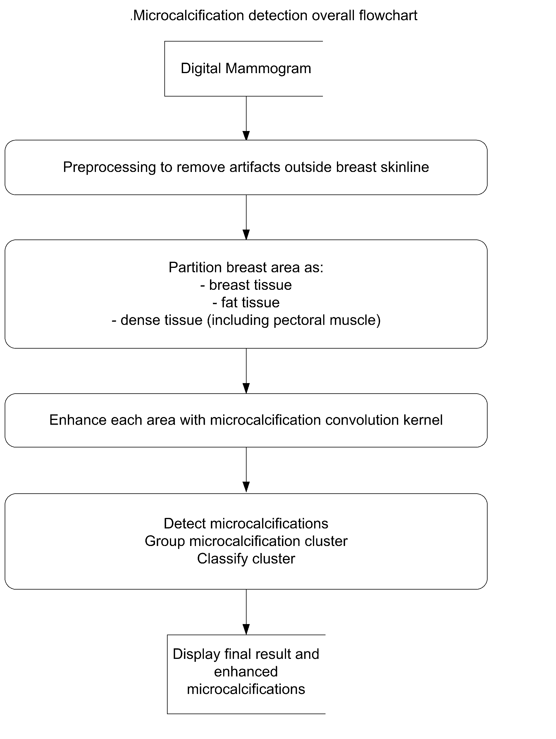
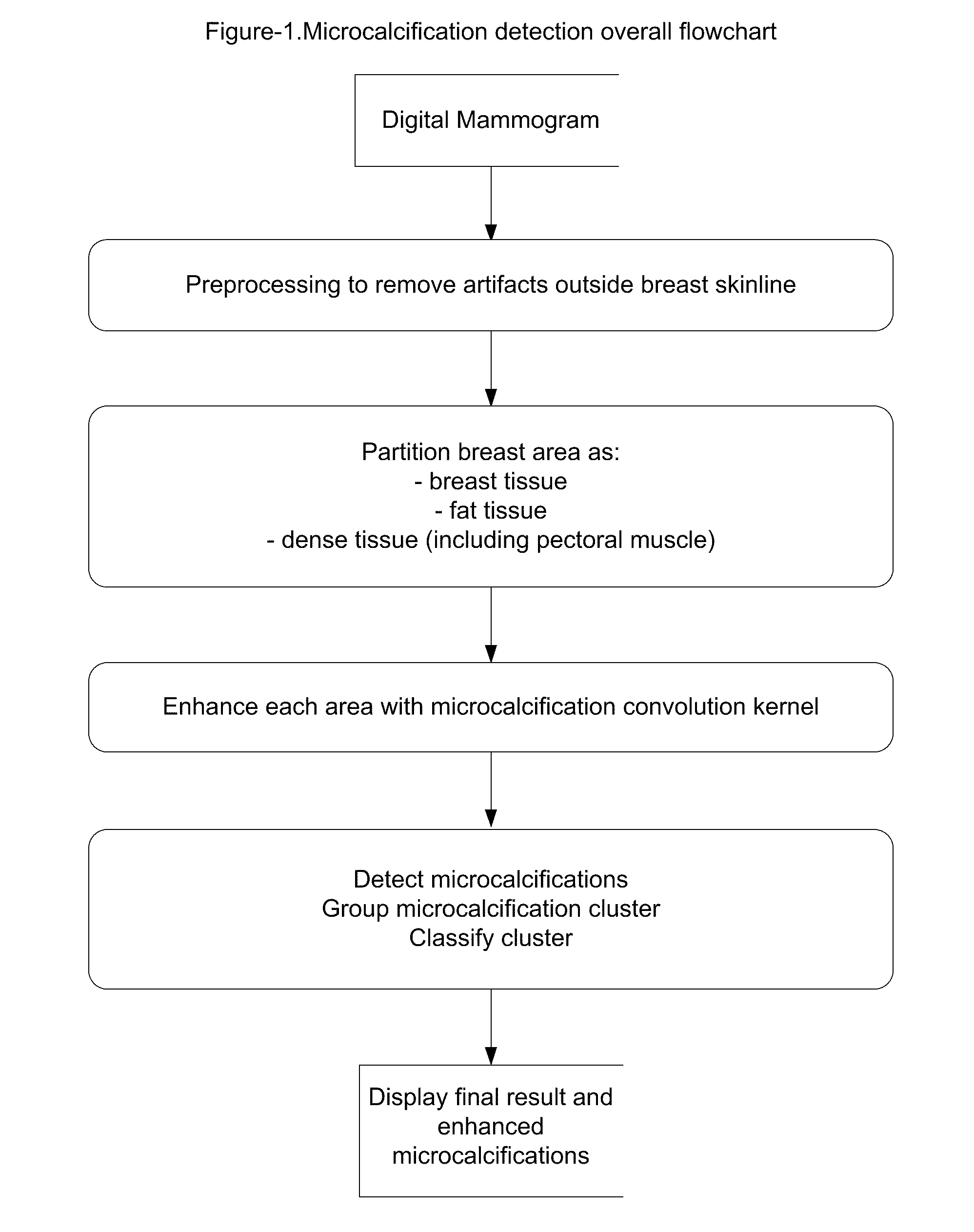
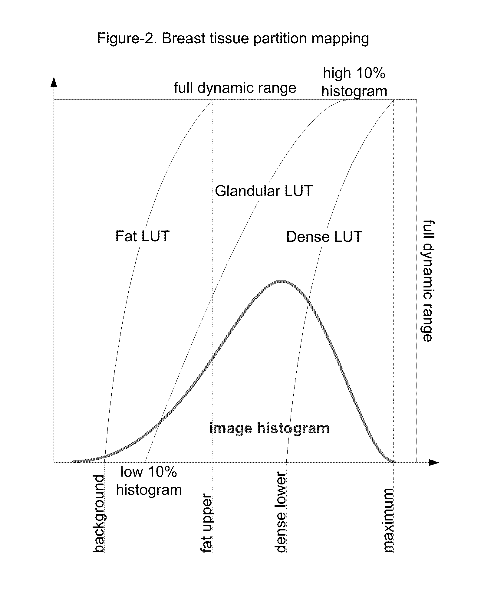
[0015] Early detection of breast cancer is the goal of mammography screening. With the rapid transition from film to digital acquisition and reading, more radiologists can benefit from advanced image processing and computational intelligence techniques when they are applied to this task. The method and system in this invention will be used as either a “second read” or a “concurrent read” tool for digital mammography screening—ultimately, will be used as a “communicative read” tool for radiologists. In this task, enhancement of microcalcifications is the initial step for a CAD server or a diagnosis review workstation. As shown in FIG. 1, the microcalcification detection system includes steps of preprocessing to remove artifacts outside breast tissue area; partitioning the breast area as breast glandular tissue area, fat tissue area and dense tissue area which each area is in a sub-range of full pixel dynamic range; remapping and enhancing each area using a filter with a convolution k...


PUM


Abstract

The present invention provides a method for enhancing microcalcifications for computer-aided lesion detection, review and diagnosis. The method includes two steps: partitioning of the breast tissue area and filtering with a convolution kernel. The partitioning process delineates: breast glandular tissue area; fat tissue sub-area and dense tissue sub-area. The 2D or 3D convolution kernels are designed to highlight small spot regions of rapid intensity changes on 2D mammograms or 3D tomosynthesis mammography images. The size of such a kernel is calculated based on the resolution of the mammographic images that are produced from each manufacturer's digital radiography device.

Description

CROSS-REFERENCE TO RELATED APPLICATIONS U.S. Patents Documents [0001] U.S. Pat. No. 5,365,429 January 1993 “Computer detection of microcalcifications in mammograms”[0002] U.S. Pat. No. 6,075,878 June 2000 “Method for determining an optimally weighted wavelet transform based on supervised training for detection of microcalcifications in digital mammograms”[0003] U.S. Pat. No. 6,137,898 November 1998 “Gabor filtering for improved microcalcification detection in digital mammograms”STATEMENT REGARDING FEDERALLY SPONSORED RESEARCH OR DEVELOPMENT [0004] Not Applicable. REFERENCE TO SEQUENCE LISTING, A TABLE, OR A COMPUTER PROGRAM LISTING COMPACT DISC APPENDIX [0005] Not Applicable. BACKGROUND OF THE INVENTION [0006] The present invention relates generally to the field of medical imaging analysis. Particularly, the present invention relates to a method and system for enhancement of microcalcifications from digital mammography images in conjunction with computer-aided detection, review and ...

Claims


Application Information

Patent Timeline
IPC IPC(8): G06K9/00
CPCG06T5/20G06T7/0012G06T2207/30096G06T2207/30068G06T7/0081G06T7/11
Inventor ZHANG, HEIDIHEFFERNAN, PATRICK
Owner THREE PALM SOFTWARE
Why Eureka
Try Eureka