Medical image diagnosis apparatus and image data processing apparatus

a diagnostic apparatus and image technology, applied in the field of medical image diagnosis apparatus and image data processing apparatus, can solve the problems of difficult to decide the most appropriate treatment for the tumor, and the prognostic factor of the vascular invasion data adjacent to the tumor, and achieve the effect of improving the diagnostic accuracy and accuracy of the tumor

Active Publication Date: 2012-02-02
TOSHIBA MEDICAL SYST CORP
View PDF2 Cites 12 Cited by
  • Summary
  • Abstract
  • Description
  • Claims
  • Application Information

AI Technical Summary

Benefits of technology

[0009]To solve the above-noted problems and drawbacks, the embodiments consistent with the present invention provide a medical image diagnosis apparatus, an image data processing apparatus that can generate and display efficacious therapy assist data for a tumor therapy and a method for controlling display of the therapy assist data of a patient, based on volume data acquired from therapy target organs.

Problems solved by technology

Thus, the proposed differential diagnosis for a tumor cannot obtain the prognostic factor of vascular invasion data adjacent the tumor.
Consequently, it has been very difficult to decide the most appropriate therapy for the tumor.

Method used


Image

  • Medical image diagnosis apparatus and image data processing apparatus
  • Medical image diagnosis apparatus and image data processing apparatus
  • Medical image diagnosis apparatus and image data processing apparatus

Examples


Embodiment Construction

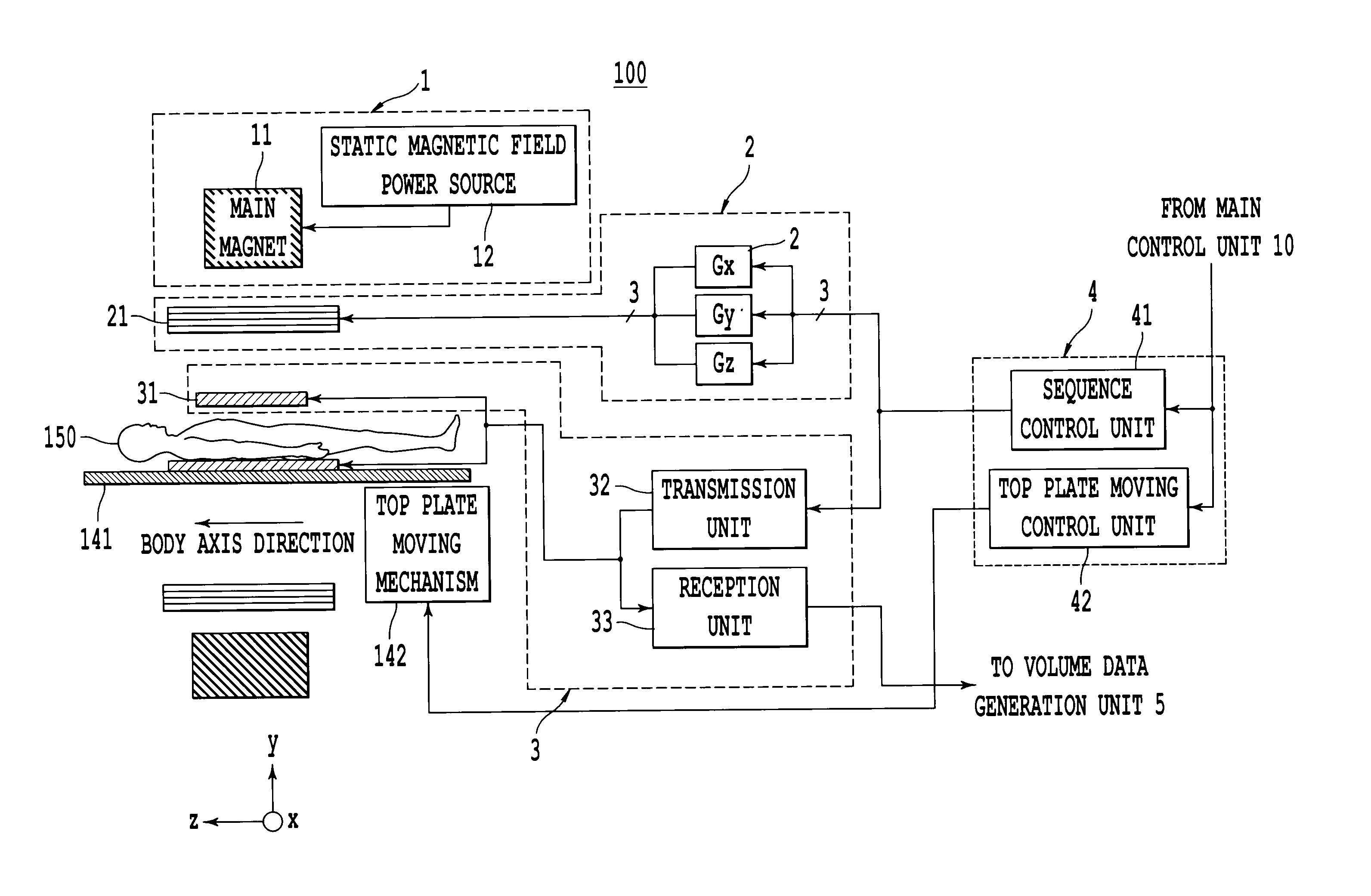
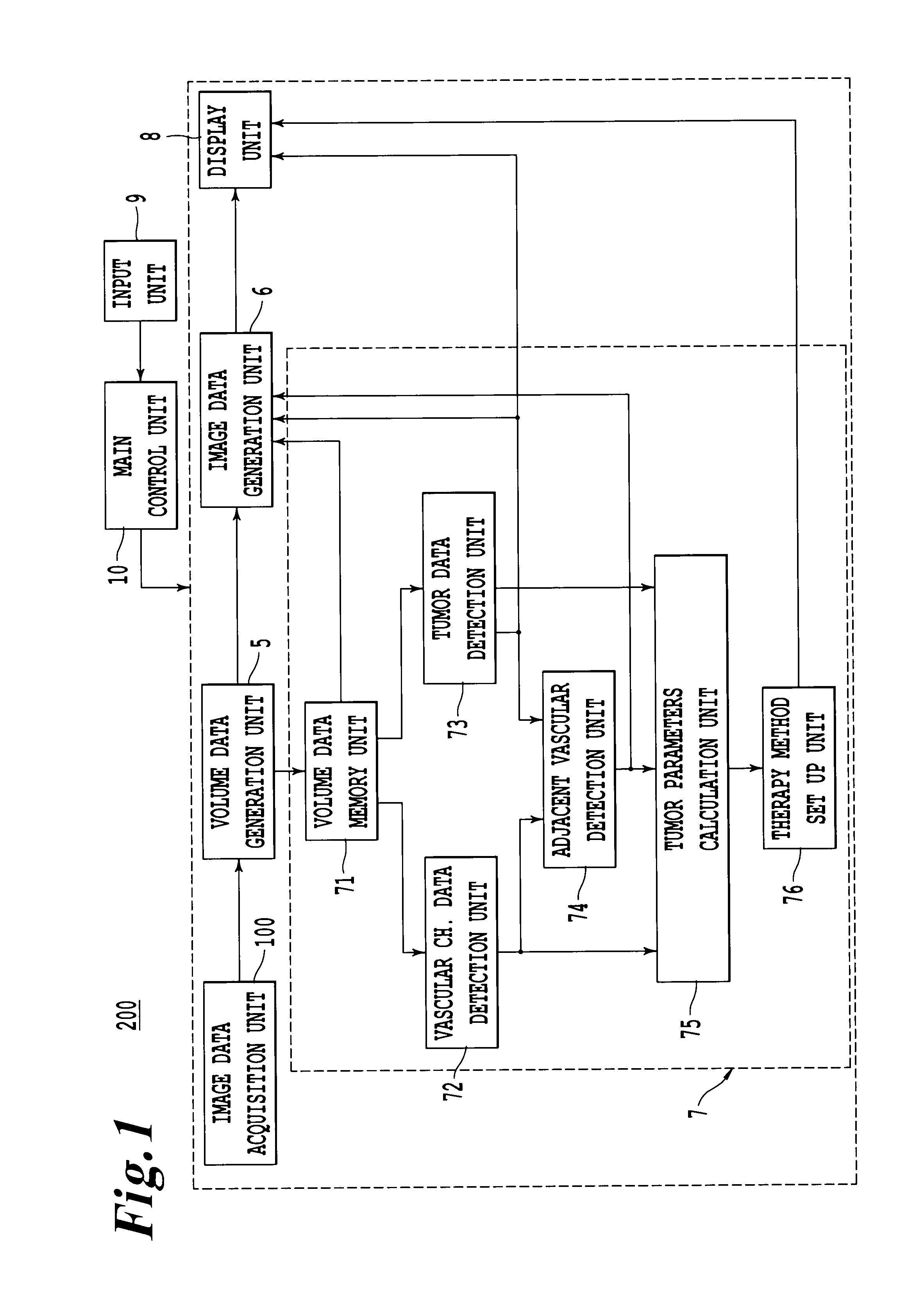
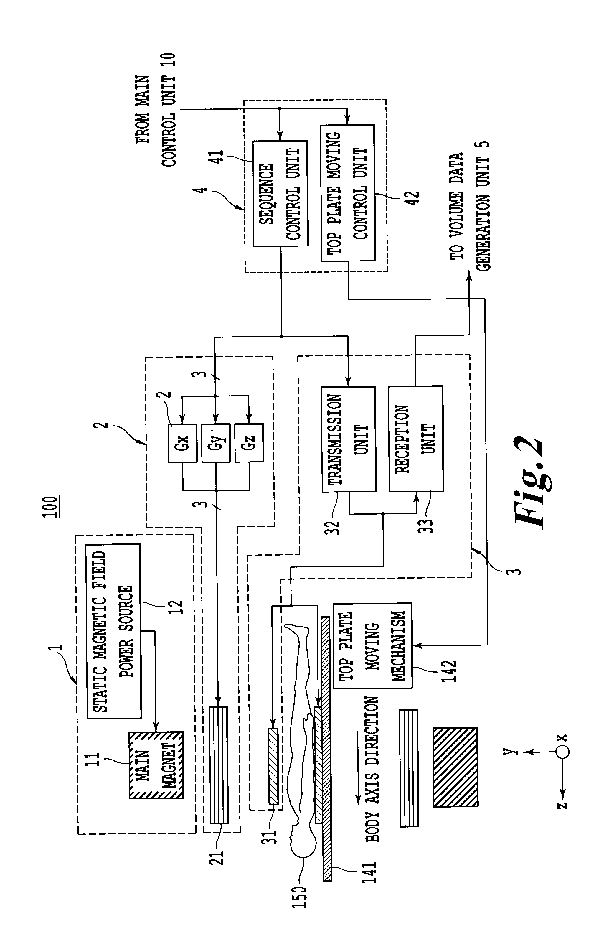
[0046]A medical image diagnosis apparatus according to one embodiment of the present invention detects tumor candidate regions by selecting tumor enhanced phases among extensive volume data acquired in time series through an MR imaging for covering a whole therapy target organ in a patient (hereinafter simply referred to as “extensive volume data”). The image diagnosis apparatus, further detects adjacent vascular regions to the tumor candidate regions by selecting vascular enhanced phases based on the extensive volume data. Further, the image diagnosis apparatus calculates various tumor parameters based on sizes of the detected tumor candidate regions and position data of the detected adjacent vascular regions for setting up recommended therapy policies for the tumors. The recommended therapy policy data and narrow scope volume data for covering the tumor candidate regions and the adjacent vascular regions (hereinafter simply referred to as “narrow scope volume data”) are displayed ...


PUM


Abstract

A medical image diagnosis apparatus and an image data processing apparatus that diagnoses vascular invasion status of a tumor and decides the most appropriate therapy method for the tumor by extracting tumor candidate regions and vascular regions based on extensive volume data derived from covering a therapy target organ, and specifying an adjacent vascular region existing within a prescribed scope from a gravity center of a tumor. The tumor candidate regions and vascular regions are generated as 3D image data or MPR image data for submitting a recommended therapy method for the tumor by adding branch data of the adjacent vascular.

Description

CROSS-REFERENCE TO RELATED APPLICATIONS[0001]This application claims the benefit of priority under 35 U.S.C. §119(a) from Japanese Patent Application No. 2010-118572 filed May 25, 2010, the contents of which are expressly incorporated herein by reference in their entirety.BACKGROUND OF THE INVENTION[0002]A. Field of the Invention[0003]Embodiments consistent with the present invention relate to a medical image diagnosis apparatus and an image data processing apparatus that can generate and display efficacious therapy assist data for a tumor (cancer) therapy.[0004]B. Background of the Invention[0005]Recently, with developments of a living body data detection unit and an operation process units operating at high speeds and with high level functions, medical image diagnosis using such as an X-ray computer tomography (CT) apparatus or a magnetic resonance imaging (MRI) apparatus can acquire 3 dimensional data (volume data) in a short time. Consequently, such a medical diagnosis can perfo...

Claims


Application Information

Patent Timeline
Patent Type & Authority Applications(United States)
IPC IPC(8): A61B6/00
CPCG01R33/5608A61B6/5211G16H50/20
Inventor AOYAGI, KOTA
Owner TOSHIBA MEDICAL SYST CORP
Why Eureka
Try Eureka