Automated contrast enhancement for contouring

a technology of contouring and automatic enhancement, applied in image enhancement, image analysis, instruments, etc., can solve the problems of requiring significant expert knowledge to execute, tedious and time-consuming segmentation by hand, and one of the main limitations of patient throughput in clinical workflow

Active Publication Date: 2012-02-09
KONINKLIJKE PHILIPS ELECTRONICS NV
View PDF12 Cites 2 Cited by
  • Summary
  • Abstract
  • Description
  • Claims
  • Application Information

AI Technical Summary

Problems solved by technology

Segmentation by hand is tedious and time-consuming, requiring significant expert knowledge to execute.
For example, for some applications like radiotherapy in the head and neck region, the segmentation step is one of the

Method used


Image

  • Automated contrast enhancement for contouring
  • Automated contrast enhancement for contouring
  • Automated contrast enhancement for contouring

Examples


Embodiment Construction

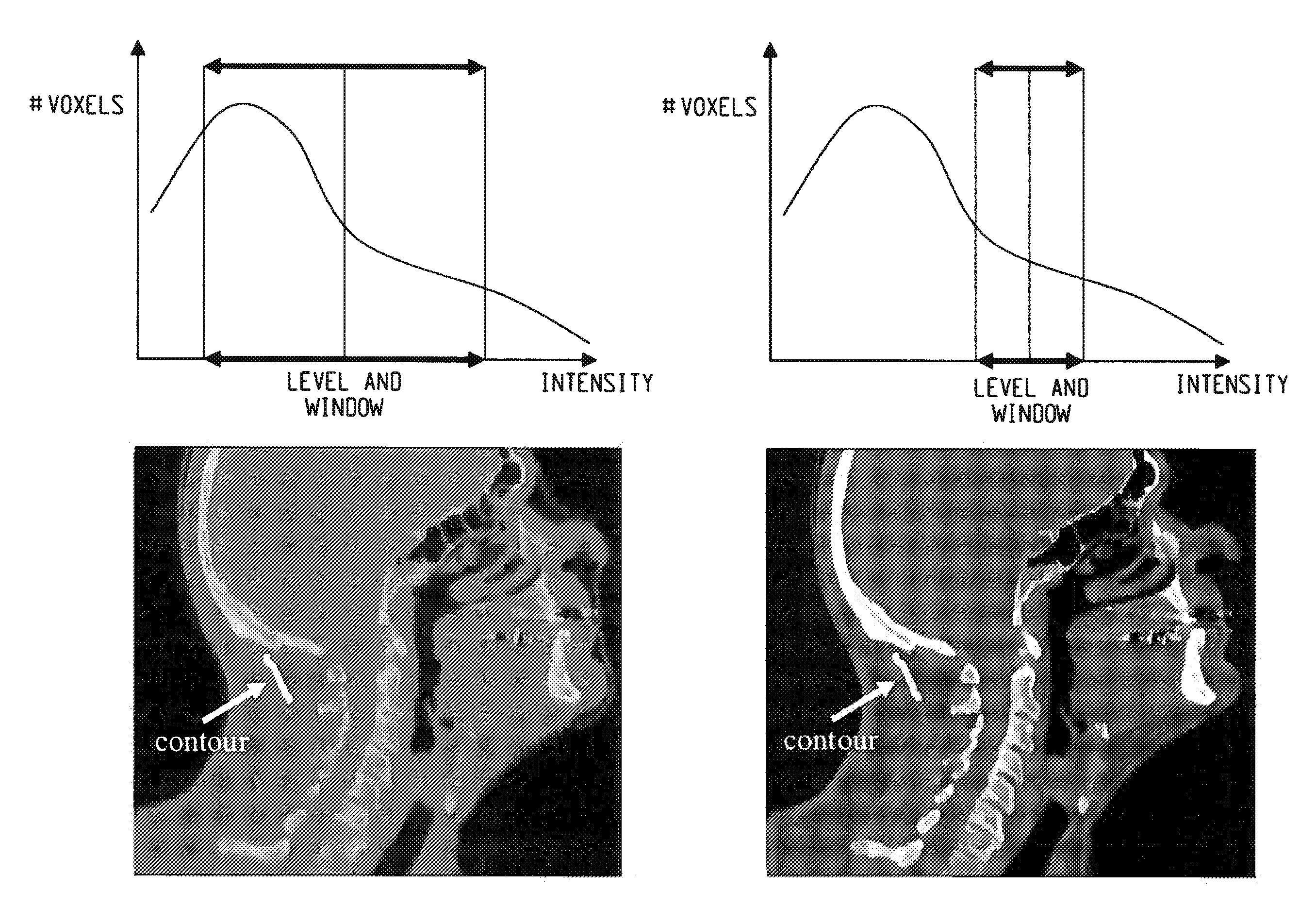
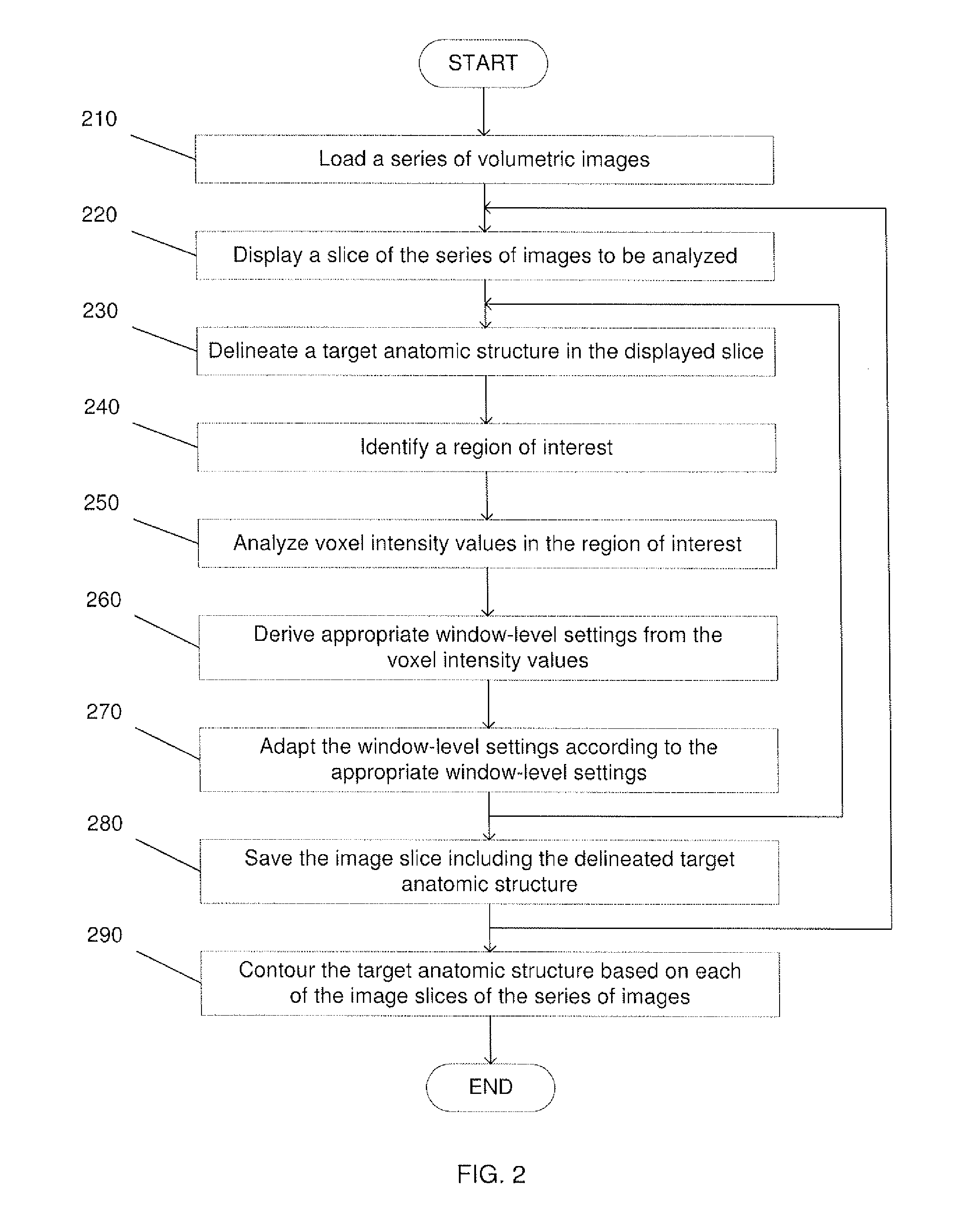
[0009]The exemplary embodiments set forth herein may be further understood with reference to the following description and the appended drawings, wherein like elements are referred to with the same reference numerals. The exemplary embodiments relate to a system and method for segmentation of a standard anatomy in volumetric images acquired from CT, MRI, etc. In particular, exemplary embodiments set forth herein describe a method for automatically adjusting image visualization in volumetric images such that a target structure may be easily distinguished from neighboring structures.

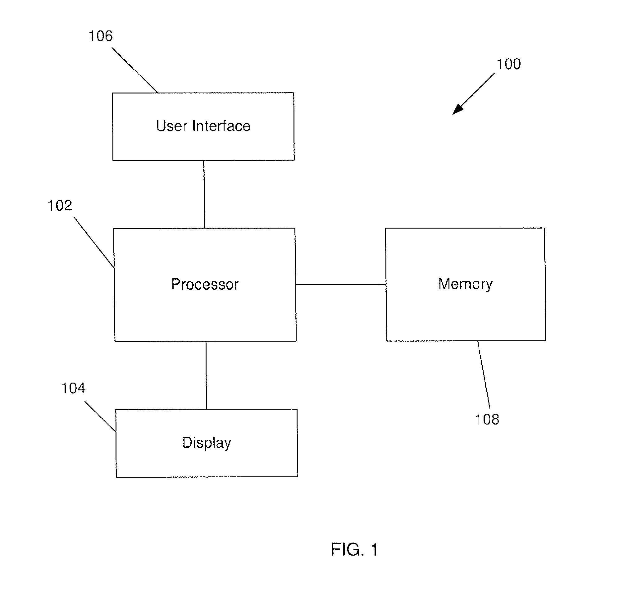
[0010]FIG. 1 shows an exemplary embodiment of a system 100 for automatically adjusting window-level settings such that a target anatomic structure is optimally visible in an image slice being analyzed. The system 100 comprises a processor 102 for mapping window-level settings to voxel intensity values, a display 104 for displaying volumetric images and a user interface 106 for drawing a target anatomic str...


PUM


Abstract

A system and method for automatic contrast enhancement for contouring. The system and method including displaying a volumetric image slice to be analyzed, receiving a delineation of a target anatomic structure in the volumetric image slice, identifying a region of interest based upon an area being delineated in the volumetric image slice, analyzing voxel intensity values in the region of interest and determining an appropriate window-level setting based on the voxel intensity values.

Description

BACKGROUND[0001]Segmentation is the process of extracting anatomic configurations from images. Many applications in medicine require segmentation of standard anatomy in volumetric images acquired from CT, MRI and other imaging devices. Clinicians, or other professionals, often use segmentation for treatment planning. For example, segmentation may be used for radiation therapy planning such that radiation treatment may be delivered, at a desired dosage, to a target tissue. Currently, segmentation can be performed manually, in which the clinician examines individual image slices and manually draws two-dimensional contours of a relevant organ in each slice. The hand-drawn contours are then combined to produce a three-dimensional representation of the relevant organ. Alternatively, the clinician may use an automatic algorithm for segmentation.[0002]Most structures, however, are still delineated manually slice-by-slice in volumetric medical datasets. Segmentation by hand is tedious and t...

Claims


Application Information

Patent Timeline
IPC IPC(8): G06T15/00G06K9/00
CPCG06T7/0083G06T2207/10072G06T5/008G06T2207/20104G06T2207/30004G06T2207/20012G06T7/12G16H30/40G16H40/63
Inventor SCHULZ, HEINRICHBYSTROV, DANIEL
Owner KONINKLIJKE PHILIPS ELECTRONICS NV
Why Eureka
Try Eureka