Method and system for characterization and mapping of tissue lesions

a tissue lesions and mapping technology, applied in the field of tissue lesions characterization and mapping, can solve the problems of invasive cancer and metastases, the possibility of successful therapy is dramatically diminished, and the conventional clinical process of optical examination has very limited capabilities in detecting cancerous and pre-cancerous tissue lesions

Inactive Publication Date: 2005-04-28
KREOS CAPITAL V UK LTD
View PDF30 Cites 77 Cited by
  • Summary
  • Abstract
  • Description
  • Claims
  • Application Information

AI Technical Summary

Benefits of technology

[0033] In a further embodiment, the step of filtering the light source comprises a plurality of optical filters and a mechanism for selecting the filter that is interposed to the tissue illumination optical path, thus enabling the tuning of the center wavelength and the spectral width of the light illuminating the tissue.
[0040] In yet another embodiment, the image or the images which are determined for the mapping of the grade of the alterations in biochemical and / or functional characteristics of tissue can be overimposed onto the color or black and white image of the same area of tissue under examination displayed on the monitor, so that abnormal tissue areas are highlighted and their borders are demarcated, facilitating the selection of a representative area for taking a biopsy sample, the selective surgical removal of the abnormal area and the evaluation of the accuracy in selecting and removing the appropriate section of the tissue.
[0046] In a further embodiment, in the case of microscopes used in clinical diagnostic examinations, surgical microscopes and colposcopes, comprise an articulated arm onto which the optical head is attached, which optical head comprises an objective lens, focusing optics, a mechanism for selecting the magnification, an eyepiece, a mount for attaching a camera, an illuminator and two linear polarizers, where the two linear polarizers are attached, one at a point along the optical path of the illuminating light beam and the other at a point along the optical path of the rays that form the image of the tissue, with the capability of rotating the polarization planes of these light polarizing optical elements, so that when these planes are perpendicular to each other, the contribution of the tissue's surface reflection to the formed image is eliminated.
[0047] In another embodiment, in the case of endoscopy, the endoscope may comprise optical means for transferring light from the light source onto the tissue surface and for collecting and transferring along almost the same axis and focusing the rays that form the image of the tissue, and two linear polarizers, where the two linear polarizers are attached, one at a point along the optical path of the illuminating light beam and the other at a point along the optical path of the rays that form the image of the tissue, with the capability of rotating the polarization planes of these light polarizing optical elements, so that when these planes are perpendicular to each other, the contribution of the tissue's surface reflection to the formed by the endoscope image is eliminated.
[0048] In another embodiment, in the case of microscopes used in clinical diagnostic examinations, surgical microscopes and colposcopes, may additionally comprise a reflective objective lens, where the reflective objective replaces the refractive one, which reflective objective is devised so that in the central part of its optical front aperture the second reflection mirror is located, and in the rear part (non-reflective) of this mirror, illumination means are attached from which light is emitted toward the object, so that with or without illumination beam zooming and focusing optics the central ray of the emitted light cone is coaxial, with the central ray of the light beam that enters the imaging lens, and with the aid of zooming and focusing optics of illumination beam that may be adjusted simultaneously and automatically with the mechanism for varying the magnification of the optical imaging system, the illuminated area and the field-of-view of the imaging system, are varying simultaneously and proportionally, so that any decrease in image brightness caused by increasing the magnification, is compensated with the simultaneous zooming and focusing of the illumination beam.

Problems solved by technology

In the opposite case the lesion can progress in depth, resulting in the development of invasive cancer and metastases.
At this stage, the possibilities of successful therapy are dramatically diminished.
The conventional clinical process of optical examination have very limited capabilities in detecting cancerous and pre-cancerous tissue lesions.
However, biopsy sampling poses several problems, such as: a) risk for sampling errors associated with the visual limitations in detecting and localizing suspicious areas; b) biopsy can alter the natural history of the intraepithelial lesion; c) mapping and monitoring of the lesion require multiple tissue sampling, which is subjected to several risks and limitations; d) the diagnostic procedure performed with biopsy sampling and histologic evaluation is qualitative, subjective, time consuming, costly and labor intensive.
The main disadvantage of fluorescence spectroscopy is that the existing biochemical modifications associated with the progress of the disease are not manifested in a direct way as modifications in the measured fluorescence spectra.
In other words the spectra carry convoluted information for several components and therefore it is difficult assess alterations in tissue features of diagnostic importance; and b) The spectra are broad due to the fact that a large number of tissue components are optically excited and contribute to the captured optical signal.
As a result the spectra do not carry specific information for the pathologic alterations and thus they are of limited diagnostic value.
The last has the capability of providing more analytical information, it requires however complex instrumentation and ideal experimental conditions, which substantially hinder their clinical use.
It is generally known that tissues are characterized by the lack of spatial homogeneity and consequently the spectral analysis of distributed spatial points is insufficient for the characterization of their status.
However, due to the insignificance of the spectral differences between normal and pathologic tissue, which is in general the case, inspection in narrow spectral bands does not allow the highlighting of these characteristics and even more so, the identification and staging of the pathologic area.
Furthermore, in several cases of early pathologic conditions, the phenomenon of temporary staining after administering the agent, is short-lasting and thus the examiner is not able to detect the provoked alterations and even more so, to assess their intensity and extent.
In other cases, the staining of the tissue progresses very slowly, with the consequence of patient discomfort and creation of problems for the examiner in assessing the intensity and extent of the alterations, since they are continuously changing.
The above have as direct consequence, the downgrading of the diagnostic value of these diagnostic procedures and thus its usefulness is limited to facilitate the localization of suspected areas for obtaining biopsy samples.
The main disadvantage of these techniques is that they provide point information, which is inadequate for the analysis of the spatially non-homogenous tissue.
These techniques (imaging and non-imaging) however, provide information of limited diagnostic value, due to the fact that the structural tissue alterations, which are accompanying the development of the disease, are not manifested as significant and characteristic alterations on the measured spectra.
Consequently, the captured spectral information cannot be directly correlated with the tissue pathology, a fact which limits the clinical usefulness of these techniques.
Nevertheless they provide limited information for the in vivo identification and staging of the disease.
Nevertheless, the experimental method employed in the published paper is characterized by quite a few disadvantages, such as: The imaging monochromator requires time for changing the imaging wavelength and as a consequence it is inappropriate for multispectral imaging and analysis of dynamic phenomena.
The lack of data modeling and parametric analysis of the characteristics of the phenomenon kinetics in any spatial point of the area of interest restrict the usefulness of the method in experimental studies and hinder its clinical implementation.
The optics used for the imaging of the area of interest are of general purpose and are not comply with the special technical requirements for the clinical implementation of the method.
Clinical implementation of the presented system is also hindered by the fact that it does not integrate appropriate means for ensuring the stability of the relative position between the tissue surface and image capturing module, during the snapshot imaging procedure.
This reduces substantially the precision in the calculation of the curves in any spatial point, that express the kinetics of marker-tissue interaction.

Method used


Image

  • Method and system for characterization and mapping of tissue lesions
  • Method and system for characterization and mapping of tissue lesions
  • Method and system for characterization and mapping of tissue lesions

Examples


Embodiment Construction

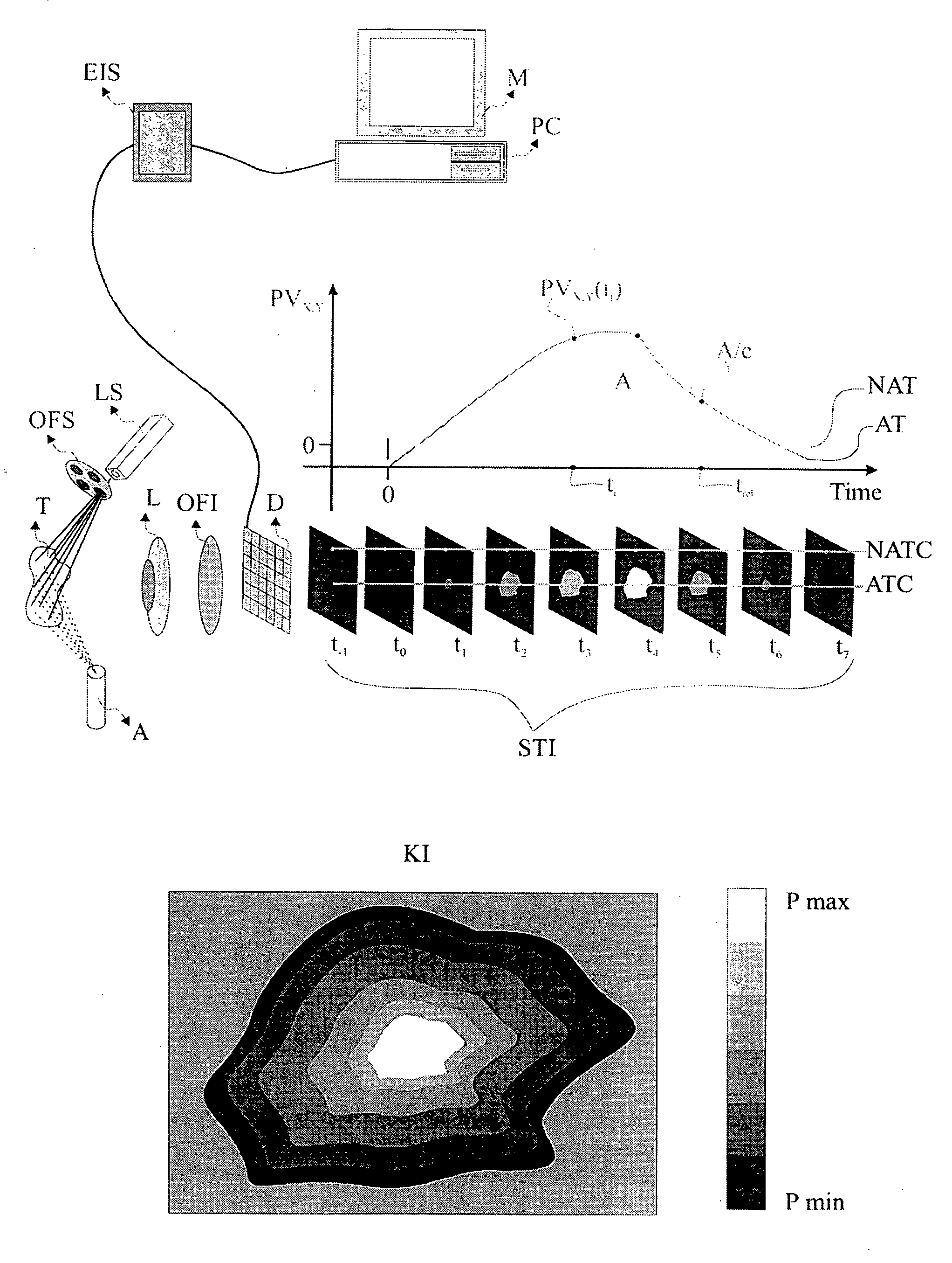
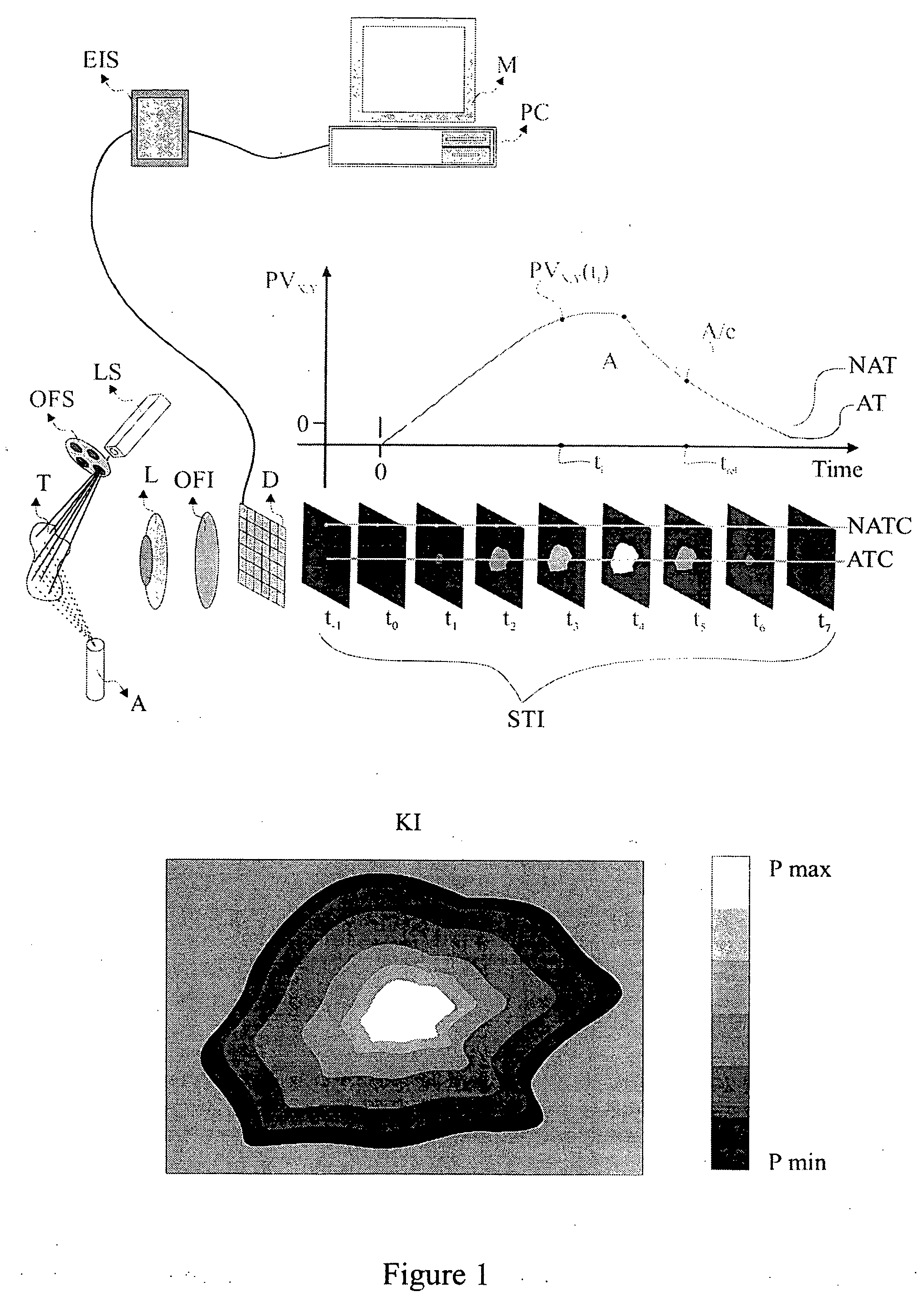
[0057] The present invention is directed to a method and system for the in-vivo, non-invasive detection and mapping of the biochemical and or functional alterations of tissue, e.g., tissue within a subject. Upon selection of the appropriate agent which enhances the optical contrast between normal and pathologic tissue (depending on the tissue's pathology), this agent is administered, e.g., topically to the tissue. In FIG. 1, the tissue (T), is sprayed using an atomizer (A), which contains the agent, e.g., acetic acid. At the same time, the tissue is illuminated with a source that emits light at a specific spectral band, depending on the optical characteristics of both the agent and the tissue. Illumination and selection of the spectral characteristics of the incident to the tissue light can be performed with the aid of a light source (LS) and a mechanism for selecting optical filters (OFS). Of course there are several other methods for illuminating the tissue and for selecting the s...


PUM

PropertyMeasurementUnit
timeaaaaaaaaaa
volumeaaaaaaaaaa
depthaaaaaaaaaa

Abstract

The present invention provides a method and an apparatus for the in vivo, non-invasive, early detection of alterations and mapping of the grade of these alterations, caused in the biochemical and / or in the functional characteristics of epithelial tissues during the development of tissue atypias, dysplasias, neoplasias and cancers. The method is based, at least in part, on the simultaneous measurement of the spatial, temporal and spectral alterations in the characteristics of the light that is re-emitted from the tissue under examination, as a result of a combined tissue excitation with light and special chemical agents. The topical or systematic administration of these agents result in an evanescent contrast enhancement between normal and abnormal areas of tissue. The apparatus enables the capturing of temporally successive imaging in one or more spectral bands simultaneously. Based on the measured data, the characteristic curves that express the agent-tissue interaction kinetics, as well as numerical parameters derived from these data, are determined in any spatial point of the examined area. Mapping and characterization of the lesion, are based on these parameters.

Description

RELATED APPLICATIONS [0001] This application is a continuation application of application Ser. No. 10 / 346,338, filed on Jan. 16, 2003, pending, which is a continuation of Ser. No. 09 / 739,089, filed on Dec. 15, 2000, pending, which in turn claims priority to Greek National Application Serial No. 20000 / 00102 filed on Mar. 28, 2000 and U.S. provisional Application No. 60 / ______, entitled “Method And Apparatus For Amplifying Pathological Features In Tissues”, filed on Dec. 15, 1999. The contents of all of the aforementioned application(s) are hereby incorporated by reference.FIELD OF THE INVENTION [0002] The present invention is directed to a method and apparatus for the in vivo, non invasive detection and mapping of the biochemical and / or functional pathologic alterations of human tissues. BACKGROUND OF THE INVENTION [0003] Cancer precursors signs are the so called pre-cancerous states, which are curable if they are detected at an early stage. In the opposite case the lesion can progre...

Claims


Application Information

Patent Timeline
Patent Type & Authority Applications(United States)
IPC IPC(8): G01N21/64A61B1/00A61B1/267A61B1/273A61B1/303A61B1/307A61B1/31A61B5/00A61B10/00G01N21/27G01N21/65G01N21/75G01N33/15G01N33/48
CPCA61B1/043A61B1/303A61B5/0059A61B5/0071A61B5/0075A61B5/0084A61B1/0646G01N21/21G01N21/31G01N21/6486G01N2021/6417G01N2021/6423A61B1/00186A61B5/0088
Inventor BALAS, CONSTANTINOS
Owner KREOS CAPITAL V UK LTD
Why Eureka
Try Eureka