Image interpretation report generation apparatus, method and program
a report and image technology, applied in the field of image interpretation report generation apparatus and method, can solve the problems of increasing the work load of image interpretation report input, difficult to recognize the position of each character string in the medical image, and the editing operation is extremely complicated, so as to facilitate the reference, easy to understand the medical image corresponding, and easy to refer to the attachment image
- Summary
- Abstract
- Description
- Claims
- Application Information
AI Technical Summary
Benefits of technology
Problems solved by technology
Method used
Image
Examples
first embodiment
[0072]Next, a configuration related to a medical image processing function of the first embodiment will be described.
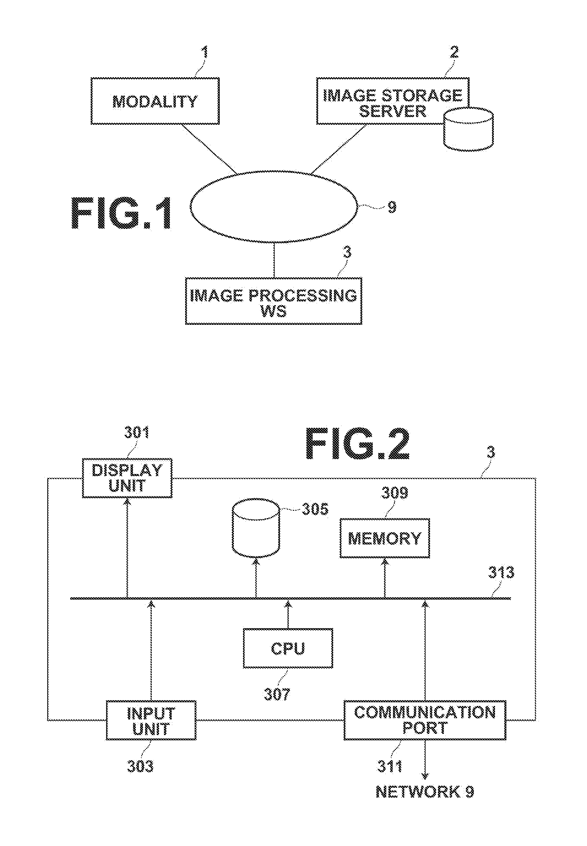
[0073]FIG. 2 is a schematic block diagram illustrating the configuration of the image processing workstation 3. As illustrated in FIG. 2, an image interpretation report generation apparatus of the first embodiment is constituted by the image processing workstation 3 including a display unit 301, such as a liquid crystal monitor, an input unit 303, a hard disk 305, a CPU 307, a memory 309, and a communication interface 311. The display unit 301 displays various kinds of information. The input unit 303 is composed of a keyboard, a mouse and the like for inputting various kinds of information. The hard disk 305 stores various programs for controlling the image interpretation report generation apparatus of the present embodiment and various kinds of data, such as image data. The various programs include an image interpretation report generation program of the present inve...
second embodiment
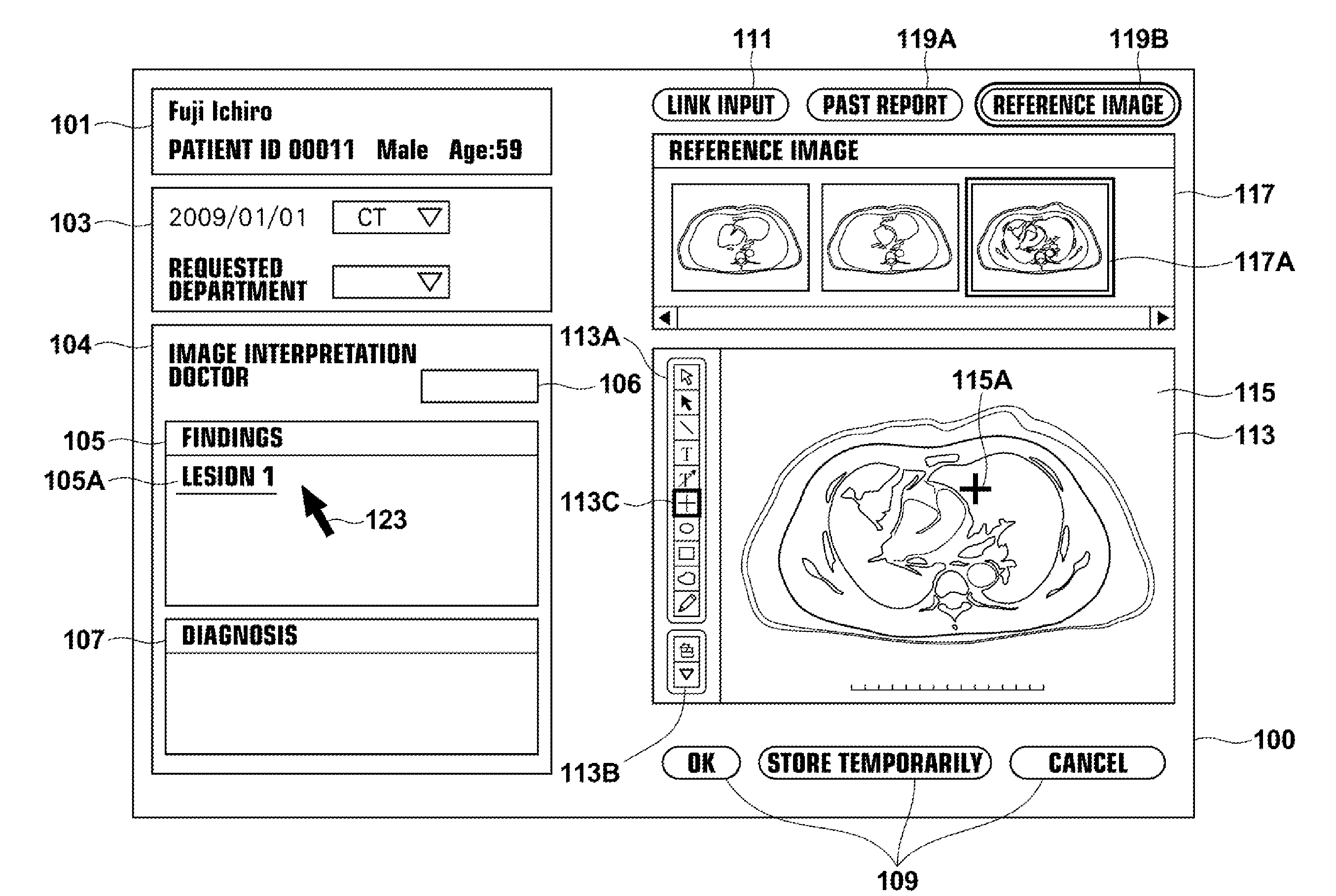
[0124]FIG. 8 is a conceptual diagram of an image interpretation report and a medical image displayed by an image interpretation report generation function of the In FIGS. 8 and 9, the composition of elements to which the same numbers as those in FIG. 5 are assigned is same as FIG. 5. Therefore, explanation of the elements will be omitted.
[0125]As illustrated in FIG. 8, in the image interpretation report generation screen, the image interpretation report display means 30 includes an attachment image display area 121 in an image interpretation report area 114, and displays, as attachment images, thumbnails 121A, 121B, and 121C together with the image interpretation report. The thumbnails 121A, 121B, and 121C are reduced medical images including the positions of lesion regions corresponding to the link characters. The thumbnail 121A of the medical image is a reduced medical image including the position 115A of the lesion region. The thumbnail 121B is a reduced medical image including ...
third embodiment
[0140]First, in a manner similar to ST101, the image interpretation report is set in a condition in which an input of link is possible (ST301). For example, as illustrated in FIG. 5, the link input button 111 may be selected by the input unit 303. Further, an automatic lesion region insertion button, or the like may be provided, and a button for making input of a link character possible, as described in the third embodiment, or the like may be provided. The buttons may be turned on to display an input screen.
[0141]Next, when the link input mode is turned on, the lesion region detection means 70 automatically detects a lesion, such as an abnormal shadow (ST302). When the lesion region detection means 70 has a function for detecting plural lesions, automatic lesion region detection is repeated until detection of lesion regions is completed by all of the lesion region detection functions (ST303 is N).
[0142]When all of lesion region detection is completed (ST303 is Y), and the position ...
PUM
 Abstract
Description
Claims
Application Information
 - R&D
- Intellectual Property
- Life Sciences
- Materials
- Tech Scout
- Unparalleled Data Quality
- Higher Quality Content
- 60% Fewer Hallucinations
Browse by: Latest US Patents, China's latest patents, Technical Efficacy Thesaurus, Application Domain, Technology Topic, Popular Technical Reports.
© 2025 PatSnap. All rights reserved.Legal|Privacy policy|Modern Slavery Act Transparency Statement|Sitemap|About US| Contact US: help@patsnap.com


