
Computer-aided diagnosis of retinal pathologies using frontal en-face views of optical coherence tomography

a technology of optical coherence tomography and computer-aided diagnosis, which is applied in the field of methods and systems for processing and representing images in ophthalmology, can solve the problems of involuntary creation of artifacts, limited number of cross-sectional images that can be obtained for evaluation and examination of the entire retina, and rendering the entire data set unreliabl

Inactive Publication Date: 2012-08-02
OPTOVUE
View PDF5 Cites 34 Cited by
  • Summary
  • Abstract
  • Description
  • Claims
  • Application Information

AI Technical Summary

Benefits of technology

"The present invention provides a method and system for computer-aided diagnosis for ophthalmology using OCT data. The method involves acquiring OCT data and obtaining an RPE fit from the data. The system includes an OCT imager and a computer that generates a set of frontal en-face images based on the RPE fit. These images are suitable for qualitative and quantitative assessment of the retina. The technical effect of this invention is to provide a more accurate and reliable tool for diagnosis and treatment of ophthalmic diseases."

Problems solved by technology

However, due to limitation of scan speed in imaging device based on time-domain technology (TD-OCT), only a very limited number of cross-sectional images can be obtained for evaluation and examination of the entire retina.
One of the challenges posed by the current technological advances in imaging techniques is the efficient and meaningful processing and presentation of the massive amounts of data collected at ever increasing imaging rates.
One common problem with this type of en-face image processing technique and other volume rendering techniques is the appearance of artifacts created by the involuntary motion of the subject's eye while a data set is being collected.
The motion introduces relative displacements of the collected images so that salient physical features appear discontinuous in the resulting 3D data set, rendering the entire data set unreliable.
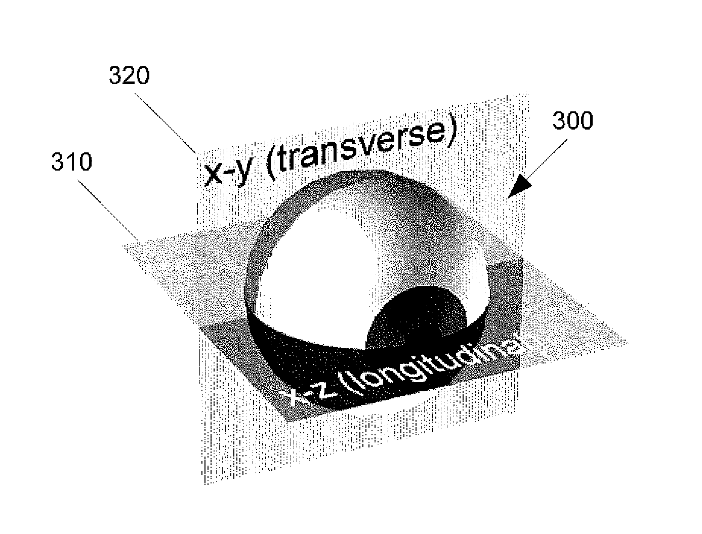
Another challenge that commonly occurs in the processing of OCT images is the central focus on reliable and reproducible layer segmentation in the B-scan (X-Z) images.
However, it becomes very unreliable, and in some cases impossible, to segment various layers accurately where there are significant layer profile alternations.

Method used


Image

  • Computer-aided diagnosis of retinal pathologies using frontal en-face views of optical coherence tomography
  • Computer-aided diagnosis of retinal pathologies using frontal en-face views of optical coherence tomography
  • Computer-aided diagnosis of retinal pathologies using frontal en-face views of optical coherence tomography

Examples


Embodiment Construction

[0026]Optical Coherence Tomography (OCT) technology has been commonly used in the medical industry to obtain information-rich content in three-dimensional (3D) data sets. OCT can be used to provide imaging for catheter probes during surgery. In the dental- industry, OCT has been used to guide dental procedures. In the field of ophthalmology, OCT is capable of generating precise and high resolution 3D data sets that can be used to detect and monitor different eye diseases in the cornea and the retina. A new data presentation scheme and design, tailored to retrieve the most commonly used and expected information from these massive 3D data sets, can further expand the application of OCT technology for different clinical application and further enhance the quality and information-richness of 3D data set obtained by OCT technologies.

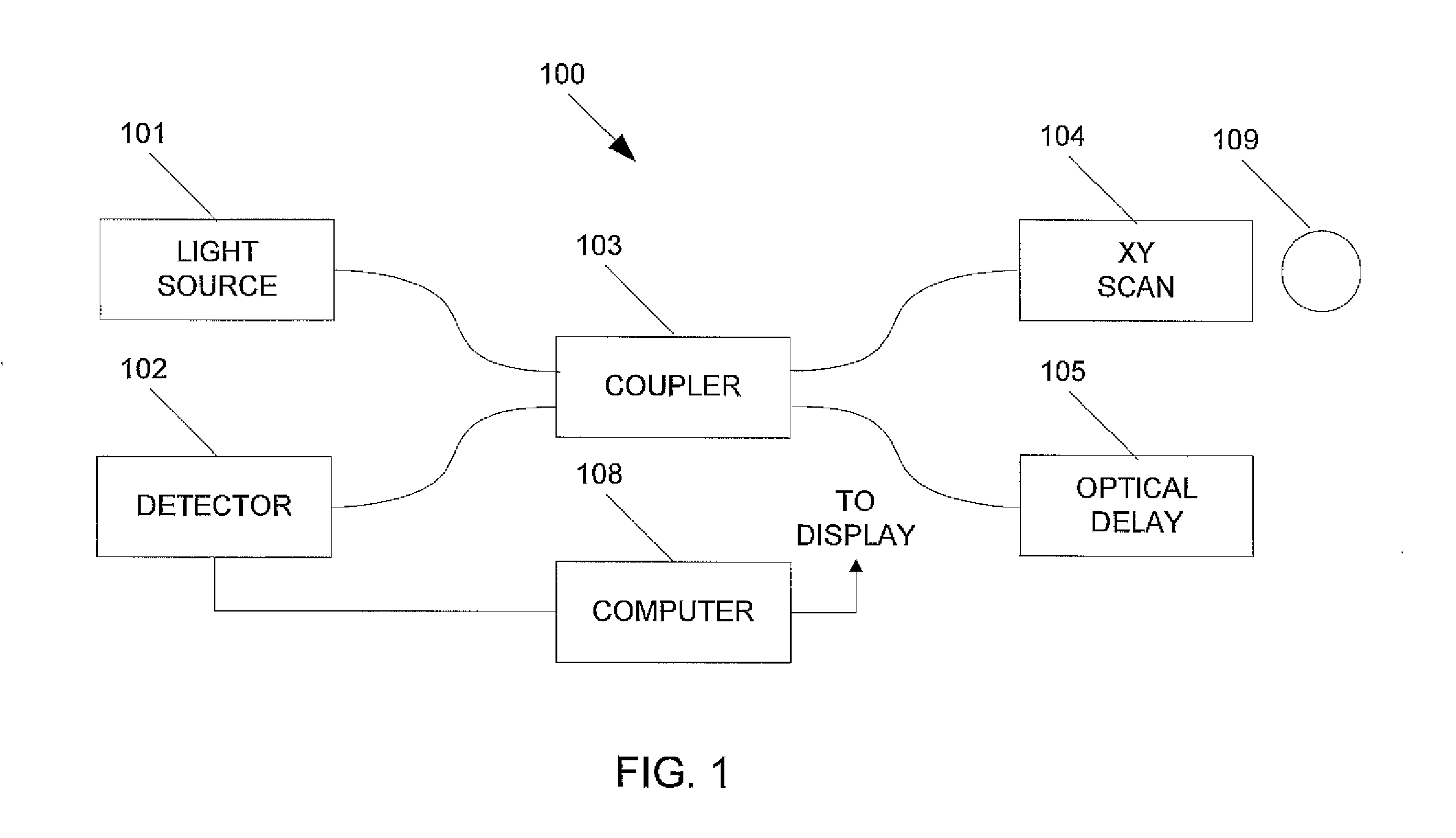
[0027]FIG. 1 illustrates an example of an OCT imager 100 that can be utilized in processing and presenting an OCT data set according to some embodiments of t...


PUM


Abstract

A system and methods of computer-aided diagnosis for ophthalmology are described that includes acquiring OCT data, determining an RPE fit from the OCT data, and displaying en face images based on the RPE fit.

Description

RELATED APPLICATIONS[0001]This application claims priority to U.S. Provisional Application No. 61 / 437,449, filed on Jan. 28, 2011, which is herein incorporated by reference in its entirety.BACKGROUND[0002]1. Field of the Invention[0003]The embodiments described herein relate generally to methods and systems for processing and representing images in ophthalmology for diagnosis and treatment of diseases or any other physiological conditions.[0004]2. Description of Related Art[0005]Optical Coherence Tomography (OCT) is an optical signal and processing technique that captures three-dimensional (3D) data sets with micrometer resolution. This OCT imaging modality has been commonly used for non-invasive imaging of object of interest, such as retina of the human eye, over the past 15 years. A cross sectional retinal image as a result of an OCT scan allows users and clinicians to evaluate various kinds of ocular pathologies in the field of ophthalmology. However, due to limitation of scan sp...

Claims


Application Information

Patent Timeline
Patent Type & Authority Applications(United States)
IPC IPC(8): A61B3/14
CPCA61B3/102A61B3/1225G06T7/0012G06T2207/10101G06T2207/30041
Inventor WEI, JAYLUMBROSO, BRUNOJANG, BENDAVIS, JOHN
Owner OPTOVUE
Learn More